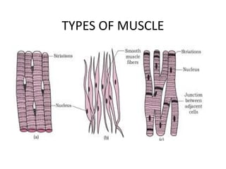
Types of MuscularTissue
• The three types of muscular tissue-skeletal,
cardiac, and smooth.
• Although the different types of muscular
tissue share some properties, they differ from
one another in their microscopic anatomy,
location, and how they are controlled by the
nervous and endocrine systems.
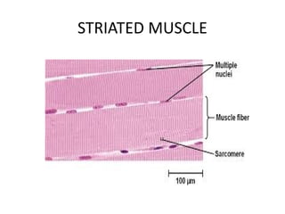
Skeletal muscle
• Skeletalmuscle tissue is so named because most
skeletal musles move bones of the skeleton. (A few
skeletal muscles attach to and move the skin or other
skeletal muscles.)
• Skeletal muscle tissue is striated: Alternating light and
dark protein bands (striations) are seen when the
tissue is examined with a microscope .
• Skeletal muscle tissue works mainly in a voluntary
manner.
• Its activity can be consciously controlled by neurons
(nerve cells) that are part of the somatic (voluntary)
division of the nervous system.
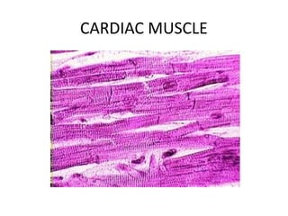
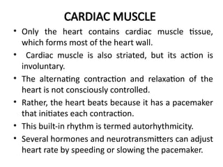
CARDIAC MUSCLE
• Onlythe heart contains cardiac muscle tissue,
which forms most of the heart wall.
• Cardiac muscle is also striated, but its action is
involuntary.
• The alternating contraction and relaxation of the
heart is not consciously controlled.
• Rather, the heart beats because it has a pacemaker
that initiates each contraction.
• This built-in rhythm is termed autorhythmicity.
• Several hormones and neurotransmitters can adjust
heart rate by speeding or slowing the pacemaker.
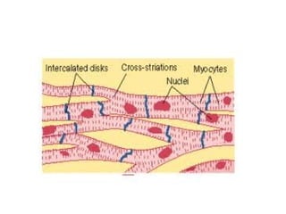
SMOOTH MUSCLE
• Smoothmuscle tissue is located in the walls of hollow
internal structures, such as blood vessels, airways, and most
organs in the abdominopelvic cavity.
• It is also found in the skin, attached to hair follicles.
• Under a microscope, it lacks the striations of skeletal and
cardiac muscle tissue. For this reason, it looks nonstriated,
which is why it is referred to as smooth.
• The action of smooth muscle is usually involuntary, and
some smooth muscle tissue, such as the muscles that propel
food through the gastrointestinal tract, has autorhythmicity.
• Both cardiac muscle and smooth muscle are regulated by
neurons that are part of the autonomic (involuntary)
division of the nervous system and by hormones released by
endocrine glands.
10.
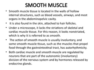
CHARACTER STRIATED NONSTRIATED CARDIAC
Shape Cylindrical Spindle Branched
Occurrence Associated
with
skeleton
Associated
with internal
organs
Exclusively found
in the heart
Nuclei Multinucleat
ed
Uninucleated Uninucleated
Striations Present Absent Present
Sarcoplasmic
reticulum
Well
developed
Poorly
developed
Poorly developed
Action Voluntary Involuntary Involuntary
Fatigue/Non
Fatigue
Fatigue
muscle
Non-Fatigue
muscle
Non-Fatigue
muscle
11.
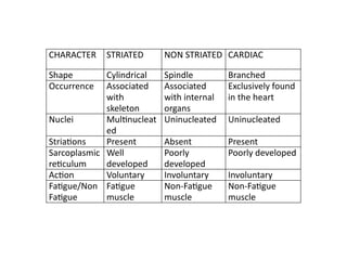
FAST MUSCLE/ TWITCH/PHASIC SLOW/TONIC
React rapidly React slowly
Move appendages,muscles of
wings,mouth parts
Surround gastrointestinal
tract.ureter etc
Have definite origin & insertion Lack definite origin & insertion
Have large fibres for great strength Small fibres
Less extensive blood vascular system extensive blood vascular system
Fewer mitochondria because
oxidative metabolism is secondary
Increased number of
mitochondria for oxidative
metabolism
Large number of glycolytic enzymes
for release of energy
fewer number of glycolytic
enzymes
Adapted for rapid & powerful activity Adapted for prolonged,continued
activity
12.
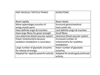
White Muscles RedMuscle
Associated with rapid
activities(jumping, flying)
Associated with slow
activities
Energy from glycolysis Energy from oxidative
phosphorylation
Abundant glycogen Abundant
myoglobin,mitochondria,fat
Subject to fatigue soon Capable of sustained
contraction
13.
Functions of MuscularTissue
1. Producing body movements: Movements of the whole
body such as walking and running, and localized
movements such as grasping a pencil, keyboarding, or
nodding the head as a result of muscular contractions, rely
on the integrated functioning of skeletal muscles, bones,
and joints.
2. Stabilizing body positions:
Skeletal muscle contractions stabilize joints and help
maintain body positions, such as standing or sitting.
Postural muscles contract continuously when you are
awake; for example, sustained contractions of your neck
muscles hold your head upright .
14.
3. Storing andmoving substances within the body:
• Storage is accomplished by sustained contractions of ringlike bands
of smooth muscle called sphincters, which prevent outflow of the
contents of a hollow organ.
• Temporary storage of food in the stomach or urine in the urinary
bladder is possible because smooth muscle sphincters close off the
outlets of these organs.
• Contraction and relaxation of smooth muscle in the walls of blood
vessels help adjust blood vessel diameter and thus regulate the rate
of blood flow.
• Smooth muscle contractions also move food and substances such as
bile and enzymes through the gastrointestinal tract, push gametes
(sperm and oocytes) through the passageways of the reproductive
systems, and propel urine through the urinary system.
• Skeletal muscle contractions promote the flow of lymph and aid the
return of blood in veins to the heart.
15.
4. Generating heat:
•As muscular tissue contracts, it produces
heat, a process known as thermogenesis.
• Much of the heat generated by muscle is used
to maintain normal body temperature.
• Involuntary contractions of skeletal muscles,
known as shivering, can increase the rate of
heat production.
16.
Properties of MuscularTissue Muscular tissue
1. Electrical excitability,a property of both
muscle and nerve cells is the ability to
respond to certain stimuli by producing
electrical signals called action potentials
(impulses). Action potentials in muscles are
referred to as muscle action potentials.
2. Contractility is the ability of muscular tissue
to contract forcefully when stimulated by an
action potetial.
17.
2. Extensibility isthe ability of muscular tissue to
stretch, within limits, without being damaged.
Normally, smooth muscle is subject to the
greatest amount of stretching. For example, each
time your stomach fills with food, the smooth
muscle in the wall is stretched, Cardiac muscle
also is stretched each time the heart fills with
blood,
3. Elasticity (e-las-TIS-i-tē) is the ability of muscular
tissue to return to its original length and shape
after contraction or extension.
18.
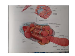
SKELETAL MUSCLE TISSUE
•Skeletal muscles is composed of hundreds to
thousands of cells, which are called muscle
fibers because of their elongated shapes.
• Thus, muscle cell and muscle fiber are two
terms for the same structure.
• Skeletal muscle also contains connective
tissues surrounding muscle fibers.
• The subcutaneous layer or hypodermis, which
separates muscle from skin is composed of
areolar connective tissue and adipose tissue.
19.
• It providesa pathway for nerves, blood vessels,
and lymphatic vessels to enter and exit muscles.
• Fascia is a dense sheet or broad band of irregular
connective tissue that lines the body wall and
limbs and supports and surrounds muscles and
other organs of the body.
• Three layers of connective tissue extend from the
fascia to protect and strengthen skeletal muscle .
• The outermost layer of dense, irregular
connective tissue, encircling the entire muscle, is
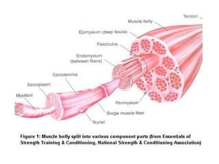
the epimysium .
20.
• Perimysium isalso a layer of dense, irregular
connective tissue, but it surrounds groups of 10 to
100 or more muscle fibers, separating them into
bundles called fascicles.
• Many fascicles are large enough to be seen with
the naked eye.
• Endomysium penetrates the interior of each
fascicle and separates individual muscle fibers
from one another.
• The epimysium, perimysium, and endomysium all
are continuous with the connective tissue that
attaches skeletal muscle to other structures, such
as bone or another muscle.
24.
• A skeletalmuscle refers to multiple bundles
(fascicles) of cells joined together.
• Muscle fibers are cylindrical and have more than
one nucleus.
• Muscle fibers are in turn composed of myofibrils.
• The myofibrils are composed of actin and myosin
filaments, repeated in units called sarcomeres,
which are the basic functional units of the muscle
fiber.
• The sarcomere is responsible for the striated
appearance of skeletal muscle and forms the basic
machinery necessary for muscle contraction.
25.
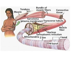
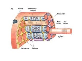
• The cellmembrane is called the sarcolemma with the
cytoplasm known as the sarcoplasm. In the sarcoplasm
are the myofibrils.
• The myofibrils are long protein bundles about 1
micrometer in diameter each containing myofilaments.
Between the myofibrils are the mitochondria.
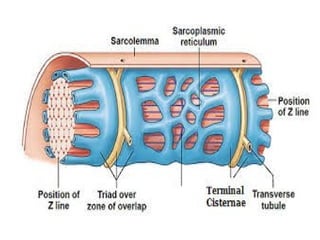
• The muscle fiber contains sarcoplasmic reticulum.
• The sarcoplasmic reticulum surrounds the myofibrils and
holds a reserve of the calcium ions needed to cause a
muscle contraction.
28.
• Periodically, ithas dilated end sacs known as
terminal cisternae.
• In between two terminal cisternae is a tubular
infolding called a transverse tubule (T tubule).
• T tubules are the pathways for action potentials
to signal the sarcoplasmic reticulum to release
calcium, causing a muscle contraction.
• Together, two terminal cisternae and a
transverse tubule form a triad.
29.
• The principalcytoplasmic proteins are myosin
and actin (also known as "thick" and "thin"
filaments, respectively) which are arranged in
a repeating unit called a sarcomere.
• The interaction of myosin and actin is
responsible for muscle contraction.
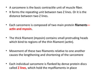
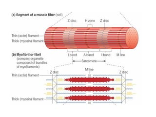
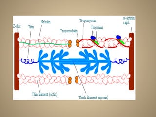
• A sarcomereis the basic contractile unit of muscle fiber.
• It forms the repeating unit between two Z lines. Or it is the
distance between two Z lines.
• Each sarcomere is composed of two main protein filaments—
actin and myosin.
• The thick filament (myosin) contains small protruding heads
which bind to regions of the thin filament (actin).
• Movement of these two filaments relative to one another
causes the lengthening and shortening of the sarcomere
• Each individual sarcomere is flanked by dense protein discs
called Z lines, which hold the myofilaments in place
32.
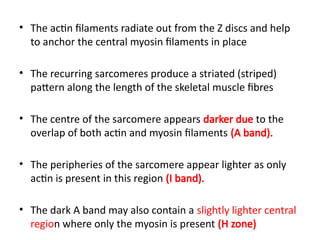
• The actinfilaments radiate out from the Z discs and help
to anchor the central myosin filaments in place
• The recurring sarcomeres produce a striated (striped)
pattern along the length of the skeletal muscle fibres
• The centre of the sarcomere appears darker due to the
overlap of both actin and myosin filaments (A band).
• The peripheries of the sarcomere appear lighter as only
actin is present in this region (I band).
• The dark A band may also contain a slightly lighter central
region where only the myosin is present (H zone)
34.
Muscle Proteins
There arethree kinds of proteins:
1. Contractile proteins : Proteins which generate
force during contraction.
2. Regulatory proteins: Proteins which switch the
contraction process off and on.
3. Structural proteins: Keep the thick and thin
filaments in proper alignment, give the
myofibrils elasticity,link myofibrils to
sarcolemma.
35.
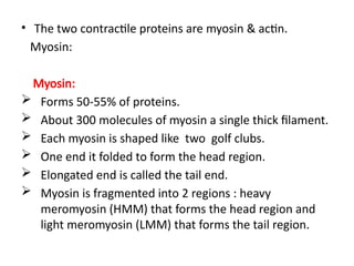
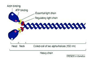
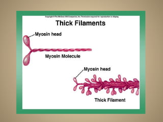
• The twocontractile proteins are myosin & actin.
Myosin:
Myosin:
Forms 50-55% of proteins.
About 300 molecules of myosin a single thick filament.
Each myosin is shaped like two golf clubs.
One end it folded to form the head region.
Elongated end is called the tail end.
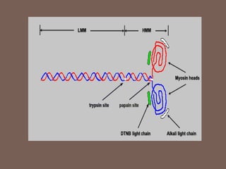
Myosin is fragmented into 2 regions : heavy
meromyosin (HMM) that forms the head region and
light meromyosin (LMM) that forms the tail region.
39.
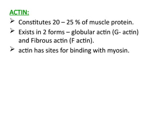
ACTIN:
Constitutes 20– 25 % of muscle protein.
Exists in 2 forms – globular actin (G- actin)
and Fibrous actin (F actin).
actin has sites for binding with myosin.
41.
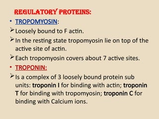
Regulatory proteins:
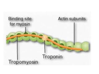
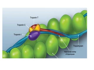
• TROPOMYOSIN:
Looselybound to F actin.
In the resting state tropomyosin lie on top of the
active site of actin.
Each tropomyosin covers about 7 active sites.
• TROPONIN:
Is a complex of 3 loosely bound protein sub
units: troponin I for binding with actin; troponin
T for binding with tropomyosin; troponin C for
binding with Calcium ions.
45.
Structural Proteins
1. Titin: stuctural protein that connects Z disc to M
line of sarcomere,stabilizing thick filament position.
2. α actinin : binds to actin molecules of thin filament
and to titin.
3. Myomesin : molecules of myomesin form M line.
4. Nebulin: long non elastic protein wrapped around
the entire length of each thin filament. It anchors
the thin filament to the Z disc and regulates the
length of thin filaments.
5. Dystrophin: links thin filaments to the sarcolemma.
46.
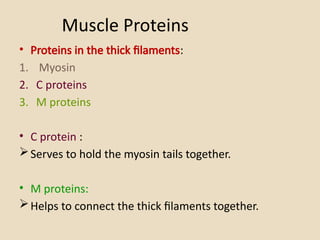
Muscle Proteins
• Proteinsin the thick filaments:
1. Myosin
2. C proteins
3. M proteins
• C protein :
Serves to hold the myosin tails together.
• M proteins:
Helps to connect the thick filaments together.
47.
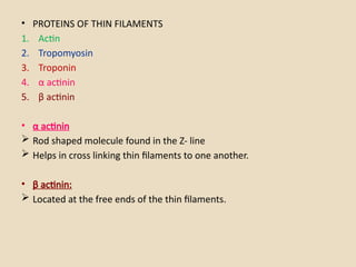
• PROTEINS OFTHIN FILAMENTS
1. Actin
2. Tropomyosin
3. Troponin
4. α actinin
5. β actinin
• α actinin
Rod shaped molecule found in the Z- line
Helps in cross linking thin filaments to one another.
• β actinin:
Located at the free ends of the thin filaments.