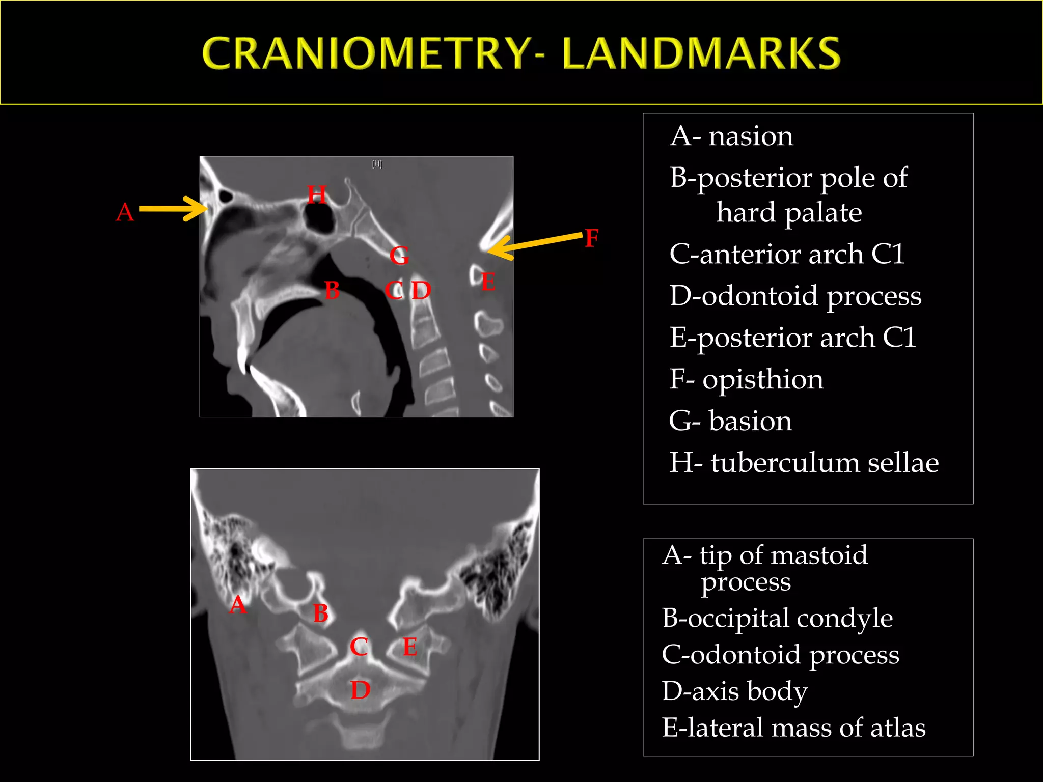
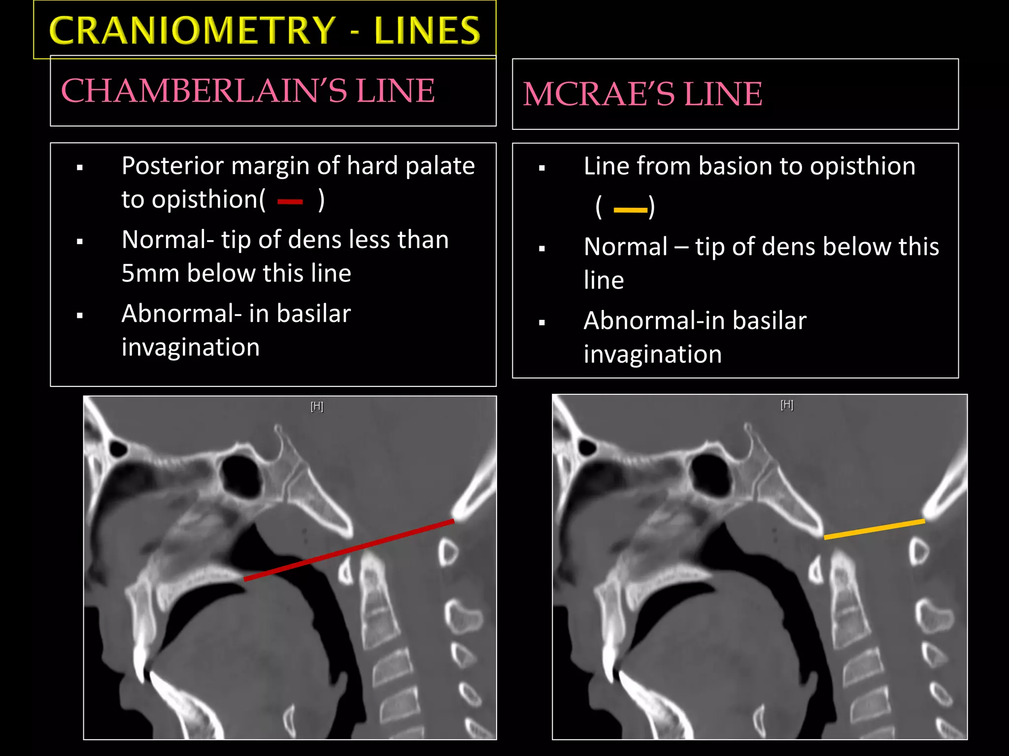
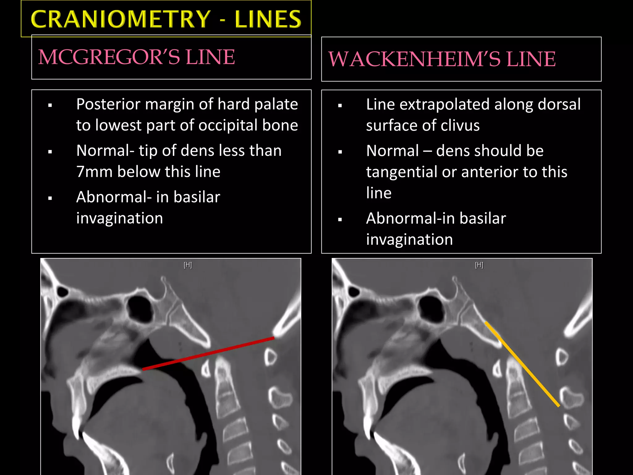
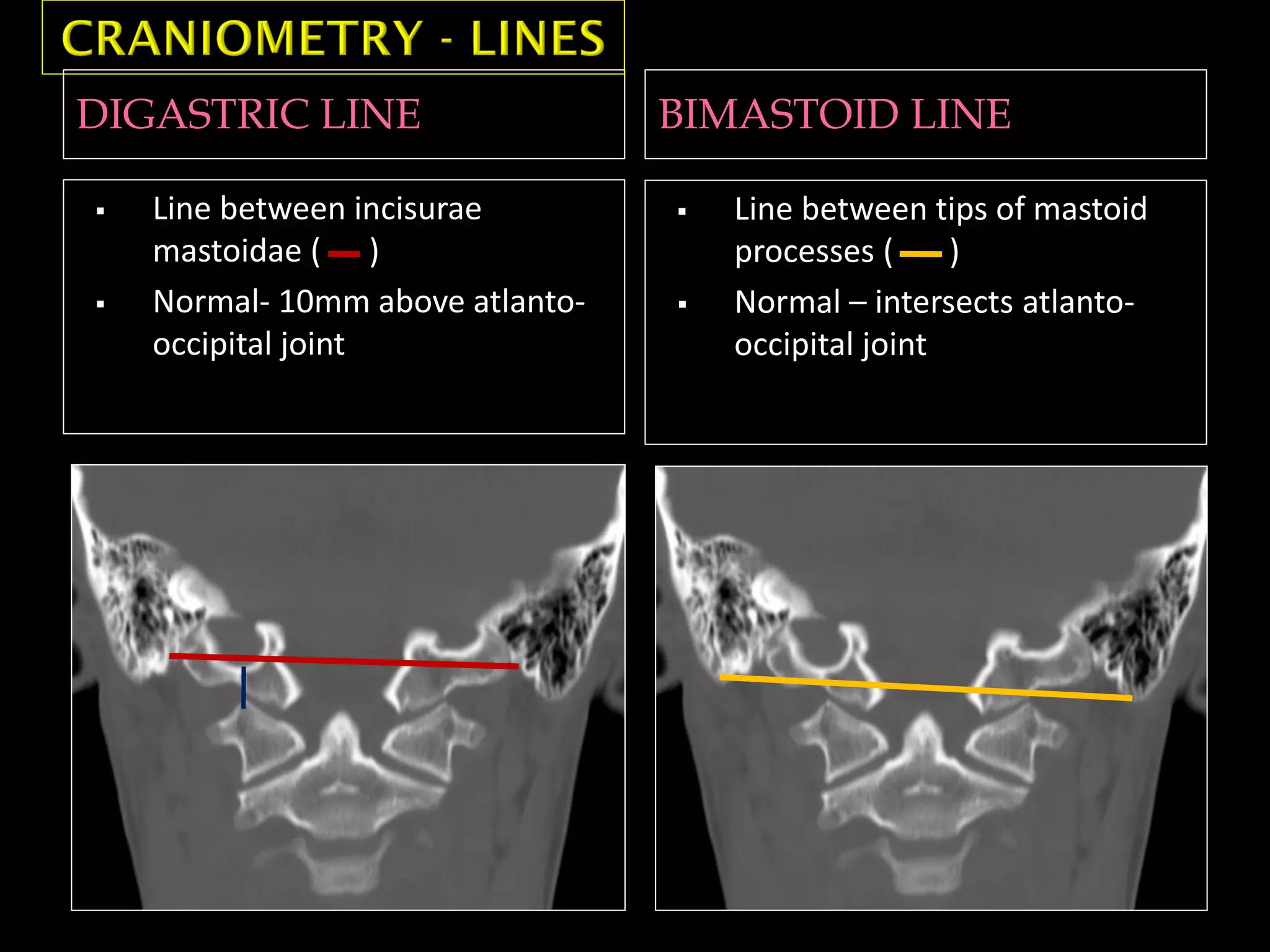
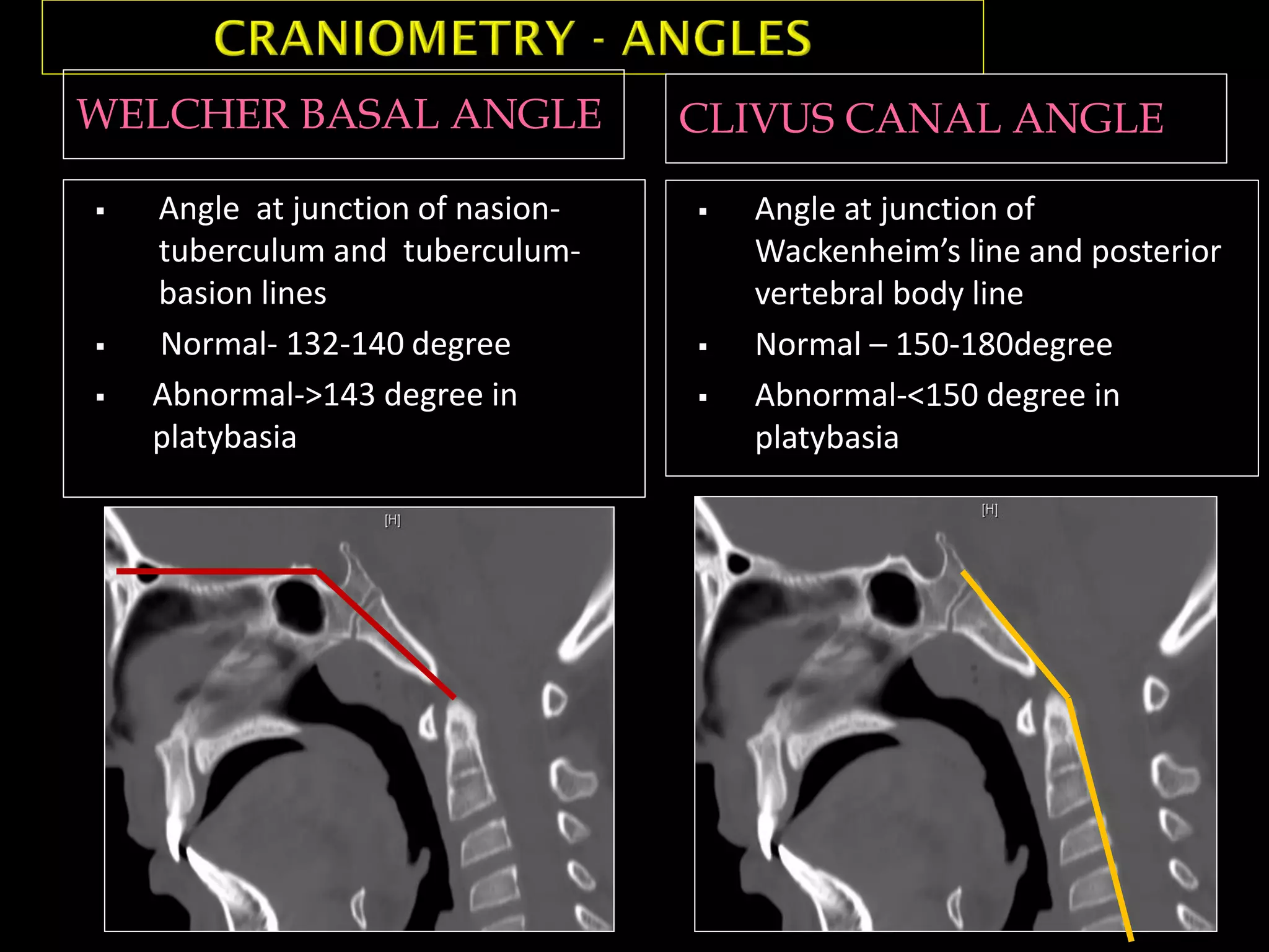
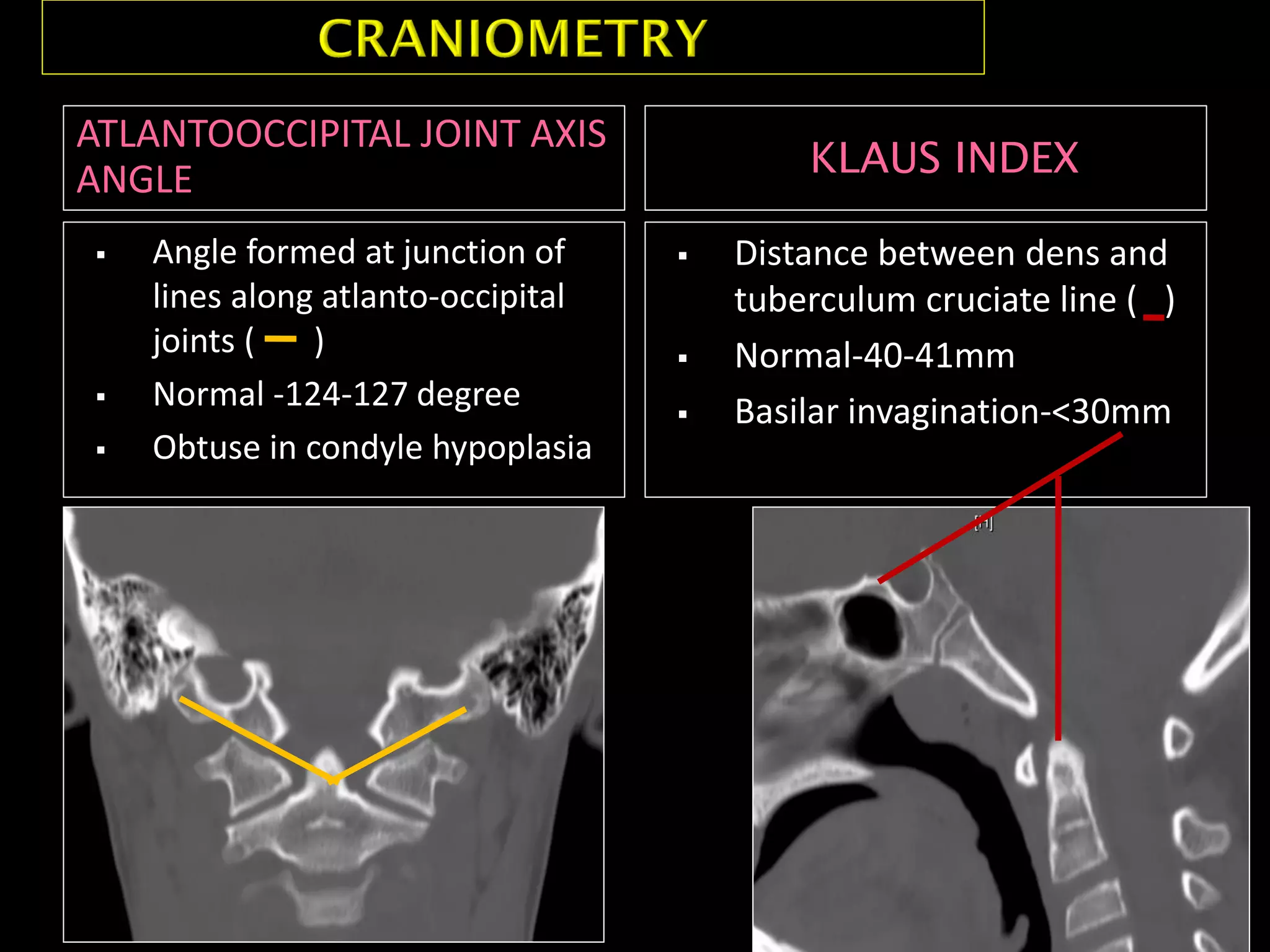
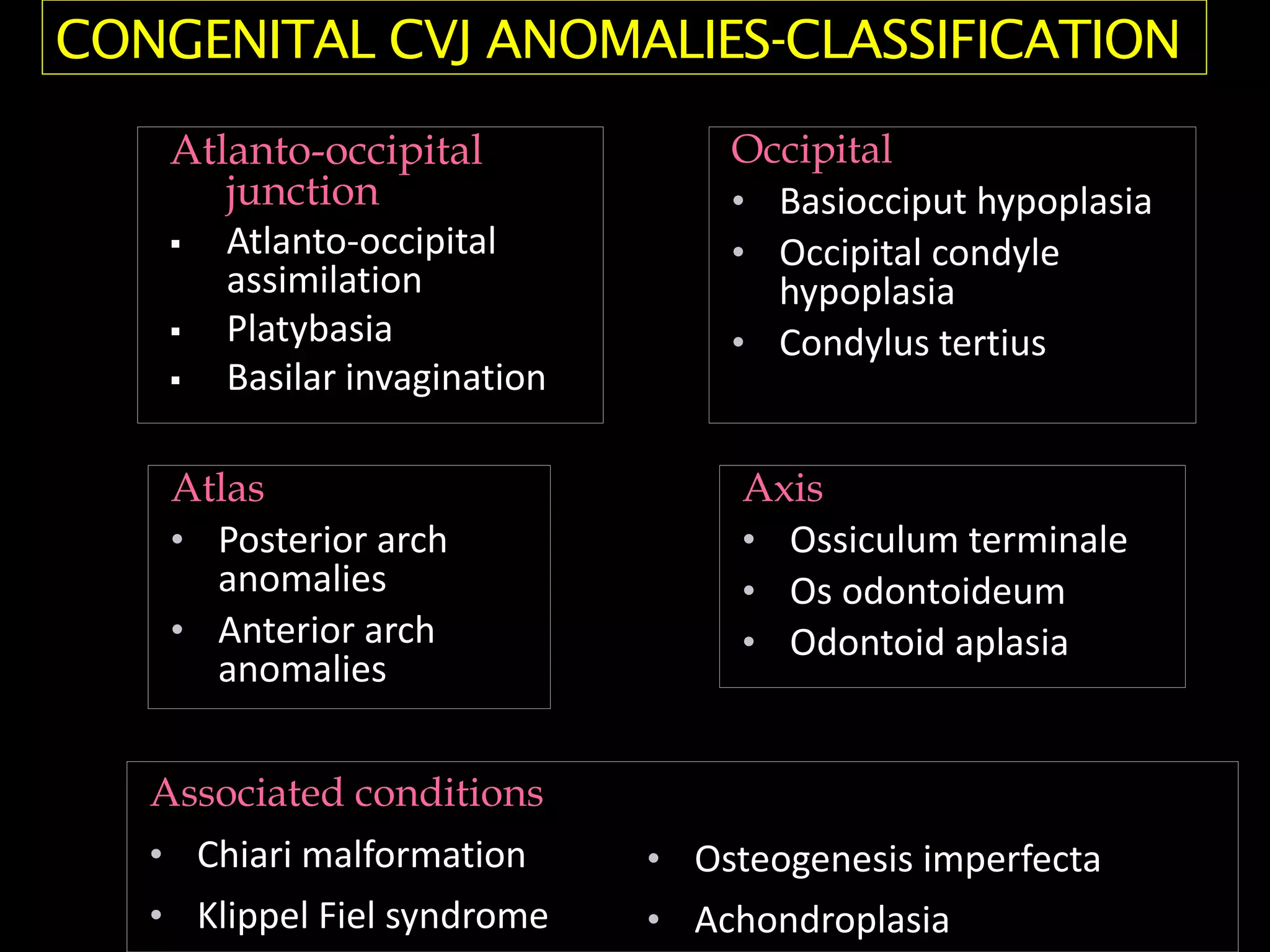
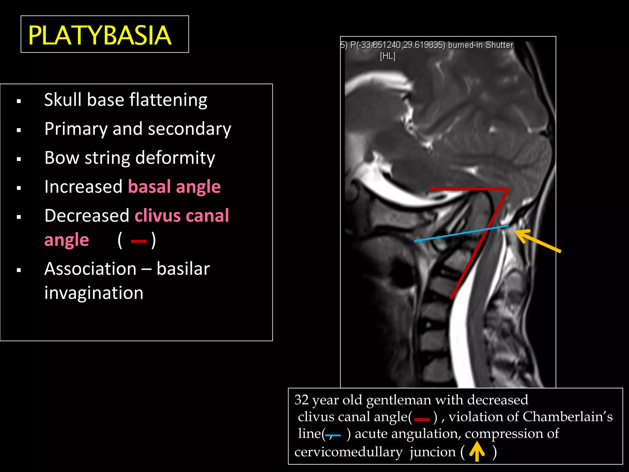
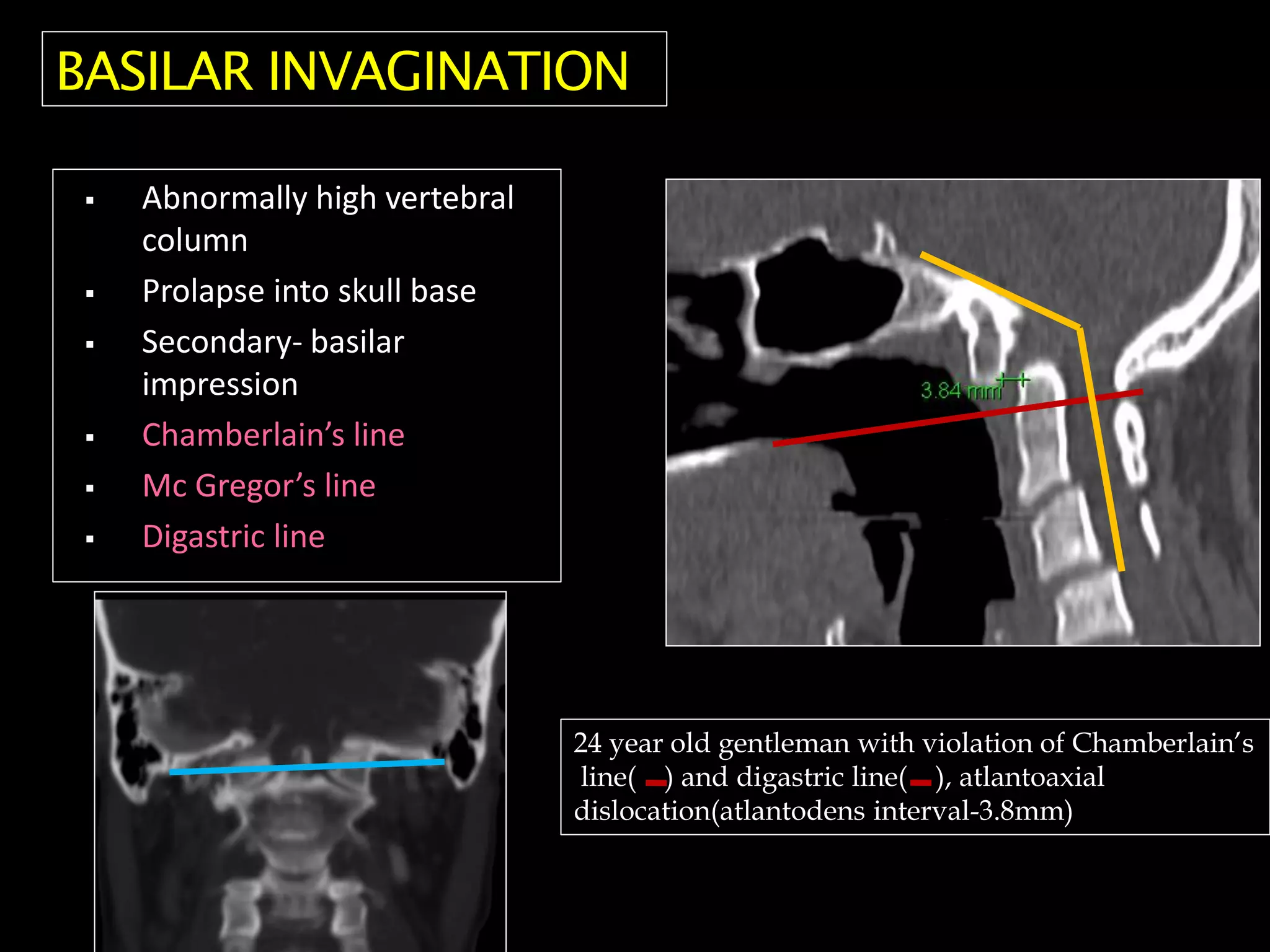
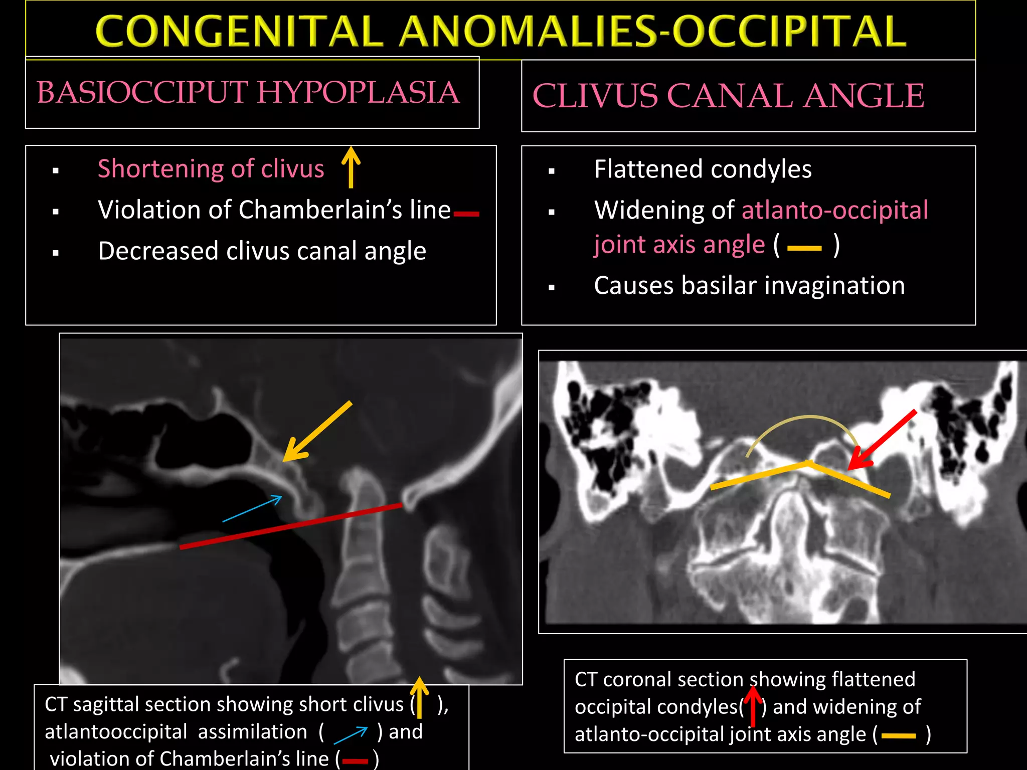
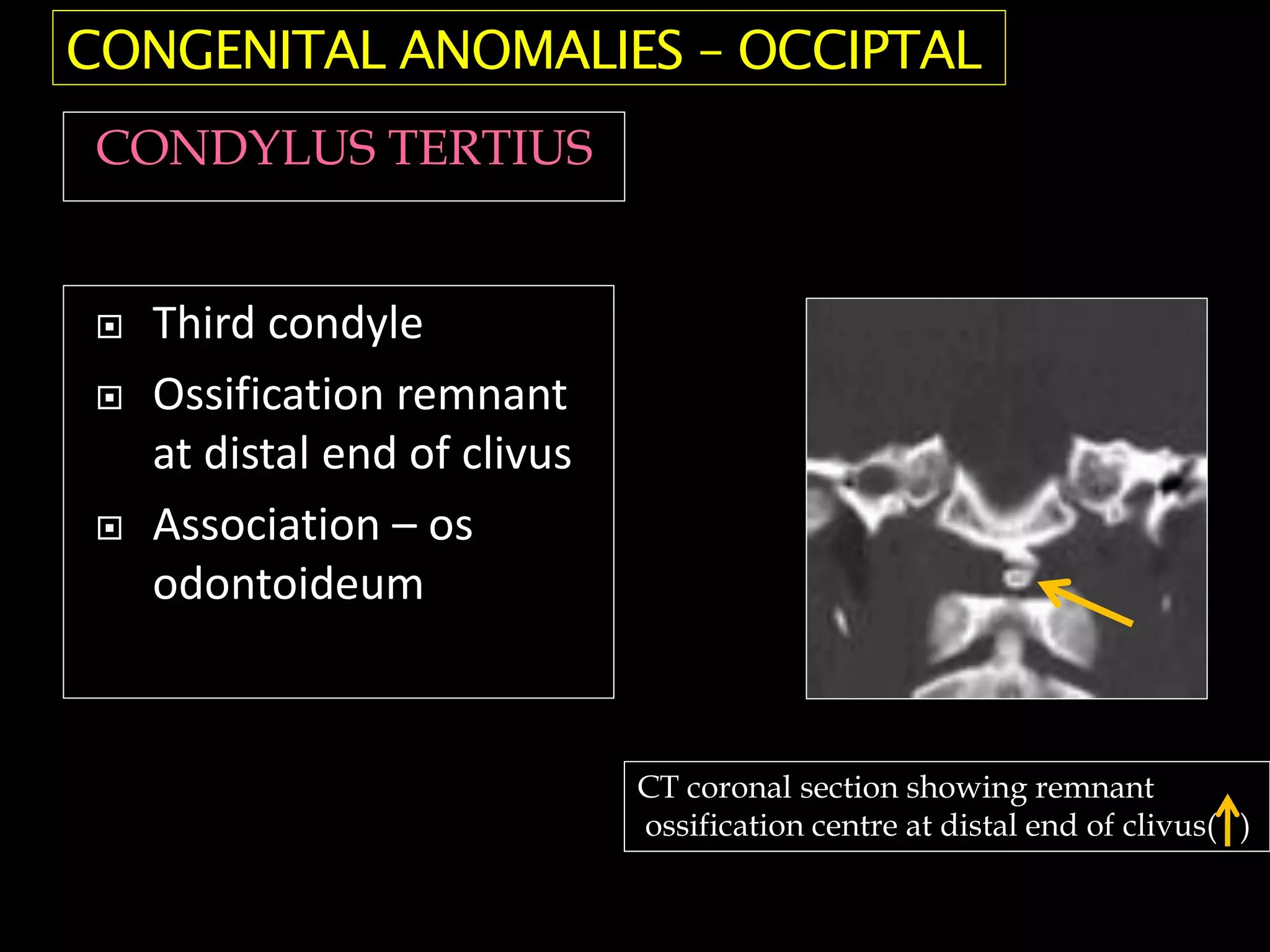
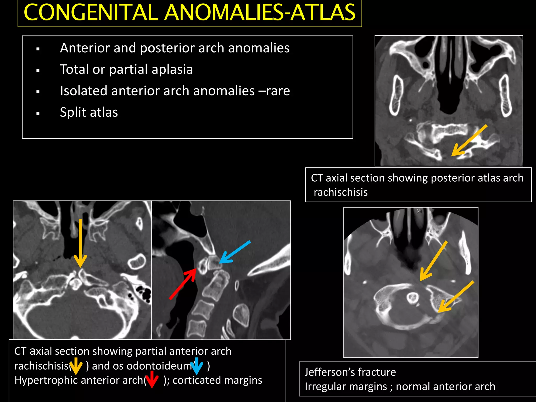
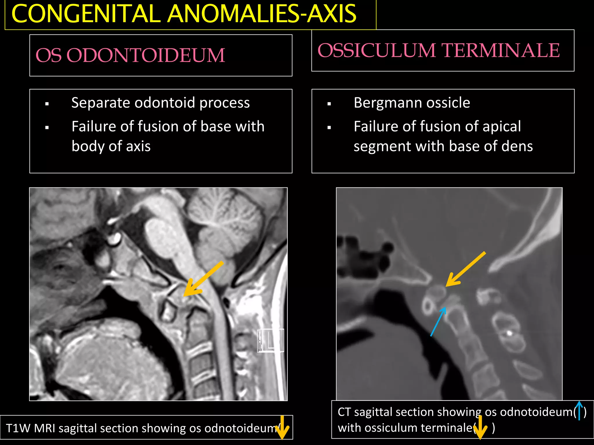
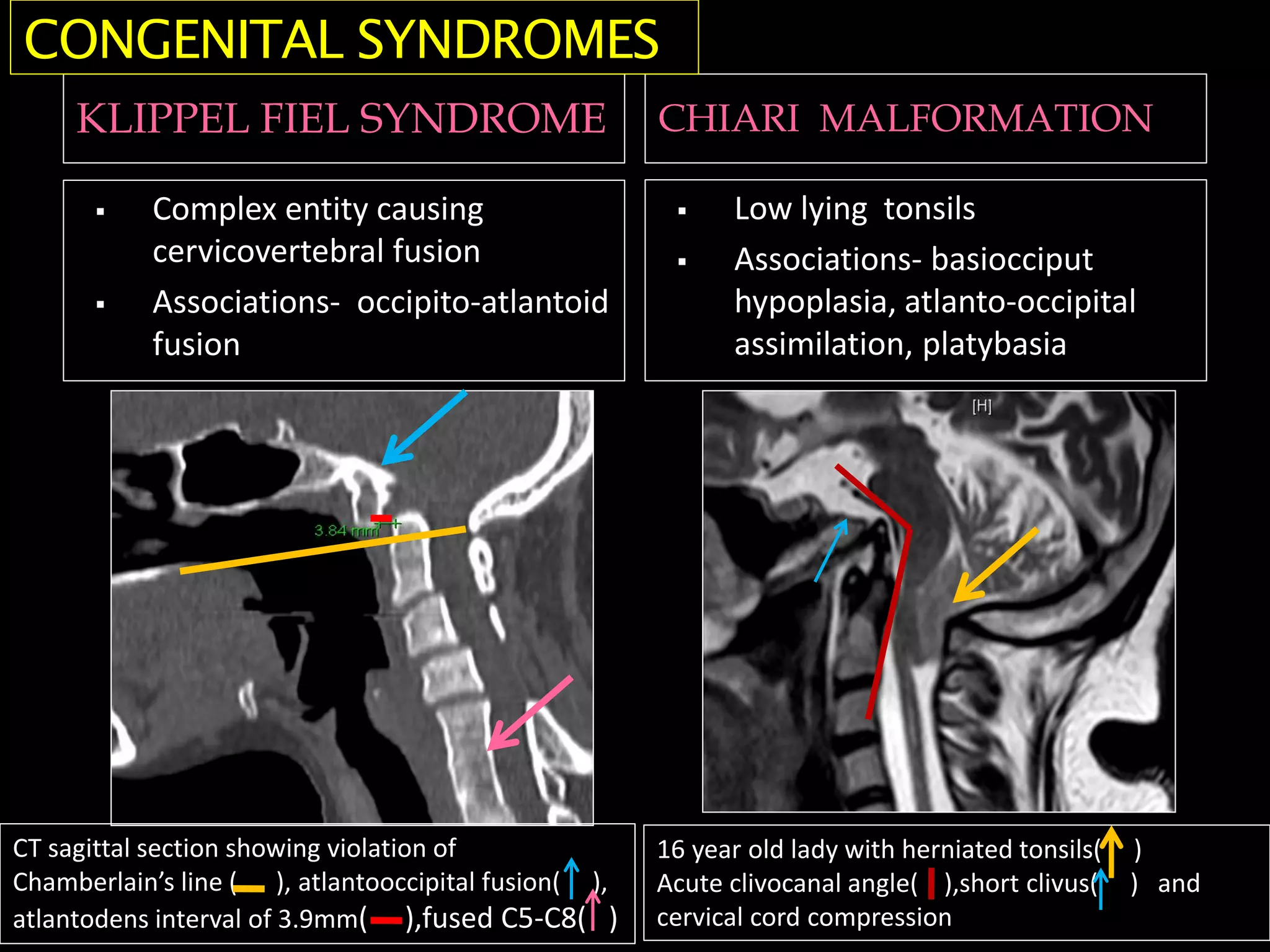
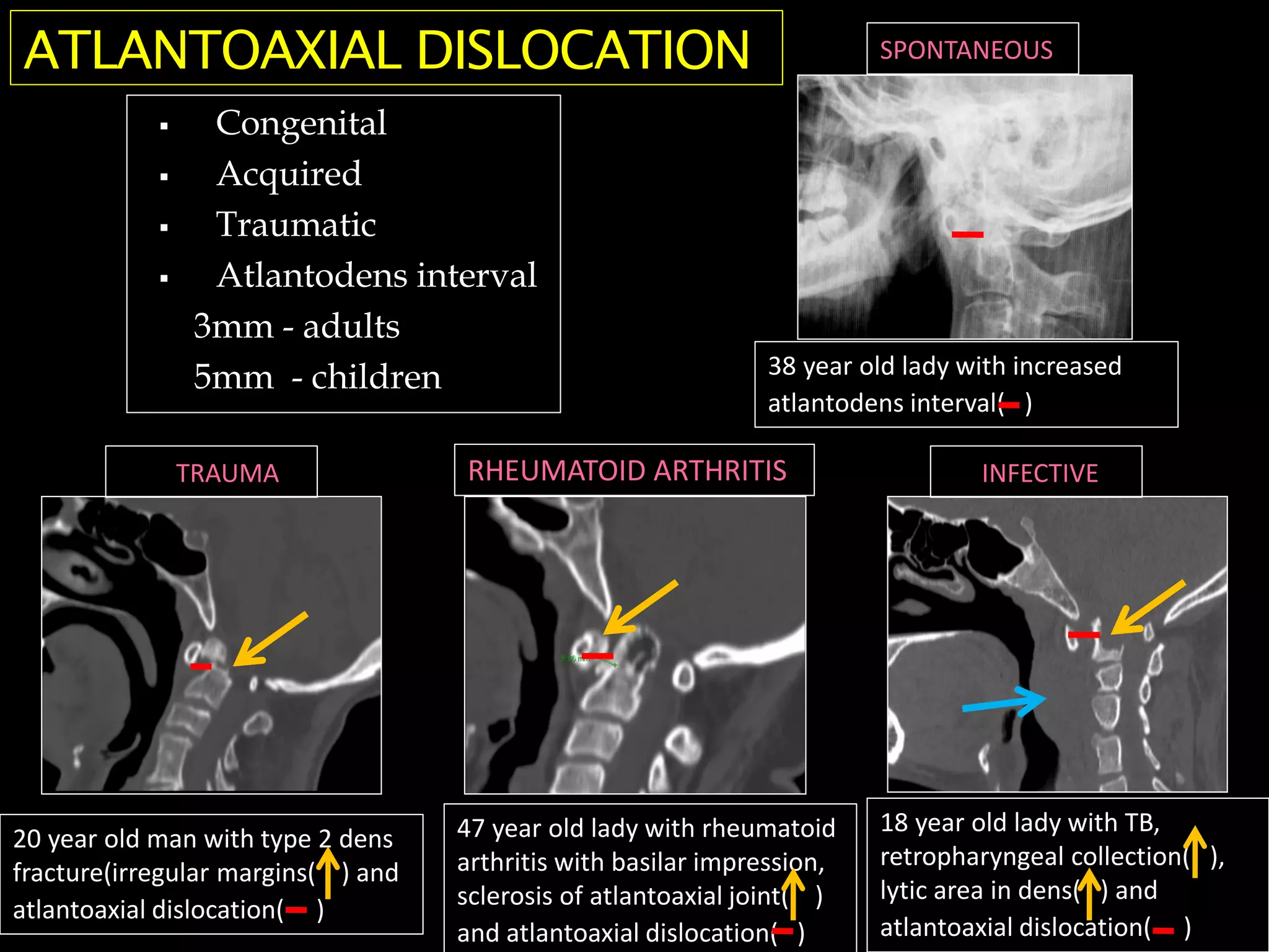
Abstract ID: IRIA -1251
 Knowledge of CVJ anomalies is important as it contains
vital structures including cervicomedullary junction
ď‚§ To illustrate the various methods of craniometry
used in diagnosing CVJ anomalies
Aims and objectives
Materials and methods
ď‚§ Multimodality radiological assessment of CVJ
anomalies along with radiological findings in
commonly encountered congenital CVJ anomalies
is illustrated
A- nasion
B-posterior pole of
hard palate
C-anterior arch C1
D-odontoid process
E-posterior arch C1
F- opisthion
G- basion
H- tuberculum sellae
A
B
C D E
F
G
H
A
B
C
D
E
A- tip of mastoid
process
B-occipital condyle
C-odontoid process
D-axis body
E-lateral mass of atlas
CHAMBERLAIN’S LINE MCRAE’S LINE
ď‚§ Posterior margin of hard palate
to opisthion( )
ď‚§ Normal- tip of dens less than
5mm below this line
ď‚§ Abnormal- in basilar
invagination
ď‚§ Line from basion to opisthion
( )
 Normal – tip of dens below this
line
ď‚§ Abnormal-in basilar
invagination
MCGREGOR’S LINE WACKENHEIM’S LINE
ď‚§ Posterior margin of hard palate
to lowest part of occipital bone
ď‚§ Normal- tip of dens less than
7mm below this line
ď‚§ Abnormal- in basilar
invagination
ď‚§ Line extrapolated along dorsal
surface of clivus
 Normal – dens should be
tangential or anterior to this
line
ď‚§ Abnormal-in basilar
invagination
DIGASTRIC LINE BIMASTOID LINE
ď‚§ Line between incisurae
mastoidae ( )
ď‚§ Normal- 10mm above atlanto-
occipital joint
ď‚§ Line between tips of mastoid
processes ( )
 Normal – intersects atlanto-
occipital joint
WELCHER BASAL ANGLE CLIVUS CANAL ANGLE
ď‚§ Angle at junction of nasion-
tuberculum and tuberculum-
basion lines
ď‚§ Normal- 132-140 degree
ď‚§ Abnormal->143 degree in
platybasia
ď‚§ Angle at junction of
Wackenheim’s line and posterior
vertebral body line
 Normal – 150-180degree
ď‚§ Abnormal-<150 degree in
platybasia
ATLANTOOCCIPITAL JOINT AXIS
ANGLE KLAUS INDEX
ď‚§ Angle formed at junction of
lines along atlanto-occipital
joints ( )
ď‚§ Normal -124-127 degree
ď‚§ Obtuse in condyle hypoplasia
ď‚§ Distance between dens and
tuberculum cruciate line ( )
ď‚§ Normal-40-41mm
ď‚§ Basilar invagination-<30mm
 Chamberlain’s line (palato-
occipital line)
 Palato–suboccipital line
(McGregor line)
ď‚§ Foramen magnum line (McRae
line)
ď‚§ Height of the posterior cranial
fossa(Klaus Index)
 Wackenhein’s clival canal line
 Bull’s angle (Atlanto-palatal
angle)
ď‚§ Atlanto-temporo-
mandibularindex (Fischgold)
• Bimastoid line (Fischgold&
Metzer)
• Bidigastric line (Fischgold&
Metzer)
• Condylar angle (Schmidt &
Fischer)
• Basal angle (Welcher)
• Boogard’s angle
Atlanto-occipital
junction
ď‚§ Atlanto-occipital
assimilation
ď‚§ Platybasia
ď‚§ Basilar invagination
Occipital
• Basiocciput hypoplasia
• Occipital condyle
hypoplasia
• Condylus tertius
Atlas
• Posterior arch
anomalies
• Anterior arch
anomalies
Axis
• Ossiculum terminale
• Os odontoideum
• Odontoid aplasia
Associated conditions
• Chiari malformation
• Klippel Fiel syndrome
• Osteogenesis imperfecta
• Achondroplasia
CONGENITAL CVJ ANOMALIES-CLASSIFICATION
ď‚§ Failure of segmentation of
C1 and skull base
 Association – C2-C3
fusion, atlantoaxial
subluxation
CT coronal section showing complete atlanto-
occipital assimilation on right side and incomplete
atlanto-occipital assimilation on left side( )
CONGENITAL ANOMALIES-ATLANTO-OCCIPITAL
ATLANTOOCCIPITAL
ASSIMILATION
CT sagittal section showing complete atlanto-occipital
assimilation( ),short clivus( ),violation of Chamberlain’s
line( )-basilar invagination and atlantoaxial dislocation( )
ď‚§ Skull base flattening
ď‚§ Primary and secondary
ď‚§ Bow string deformity
ď‚§ Increased basal angle
ď‚§ Decreased clivus canal
angle ( )
 Association – basilar
invagination
32 year old gentleman with decreased
clivus canal angle( ) , violation of Chamberlain’s
line( , ) acute angulation, compression of
cervicomedullary juncion ( )
PLATYBASIA
ď‚§ Abnormally high vertebral
column
ď‚§ Prolapse into skull base
ď‚§ Secondary- basilar
impression
 Chamberlain’s line
 Mc Gregor’s line
ď‚§ Digastric line
24 year old gentleman with violation of Chamberlain’s
line( ) and digastric line( ), atlantoaxial
dislocation(atlantodens interval-3.8mm)
BASILAR INVAGINATION
BASIOCCIPUT HYPOPLASIA CLIVUS CANAL ANGLE
ď‚§ Shortening of clivus
 Violation of Chamberlain’s line
ď‚§ Decreased clivus canal angle
ď‚§ Flattened condyles
ď‚§ Widening of atlanto-occipital
joint axis angle ( )
ď‚§ Causes basilar invagination
CT sagittal section showing short clivus ( ),
atlantooccipital assimilation ( ) and
violation of Chamberlain’s line ( )
CT coronal section showing flattened
occipital condyles( ) and widening of
atlanto-occipital joint axis angle ( )
 Third condyle
 Ossification remnant
at distal end of clivus
 Association – os
odontoideum
CT coronal section showing remnant
ossification centre at distal end of clivus( )
CONGENITAL ANOMALIES – OCCIPTAL
CONDYLUS TERTIUS
ď‚§ Anterior and posterior arch anomalies
ď‚§ Total or partial aplasia
 Isolated anterior arch anomalies –rare
ď‚§ Split atlas
CT axial section showing posterior atlas arch
rachischisis
CT axial section showing partial anterior arch
rachischisis( ) and os odontoideum( )
Hypertrophic anterior arch( ); corticated margins
Jefferson’s fracture
Irregular margins ; normal anterior arch
CONGENITAL ANOMALIES-ATLAS
OS ODONTOIDEUM OSSICULUM TERMINALE
ď‚§ Separate odontoid process
ď‚§ Failure of fusion of base with
body of axis
ď‚§ Bergmann ossicle
ď‚§ Failure of fusion of apical
segment with base of dens
CONGENITAL ANOMALIES-AXIS
T1W MRI sagittal section showing os odnotoideum(
CT sagittal section showing os odnotoideum( )
with ossiculum terminale( )
KLIPPEL FIEL SYNDROME CHIARI MALFORMATION
ď‚§ Complex entity causing
cervicovertebral fusion
ď‚§ Associations- occipito-atlantoid
fusion
ď‚§ Low lying tonsils
ď‚§ Associations- basiocciput
hypoplasia, atlanto-occipital
assimilation, platybasia
CONGENITAL SYNDROMES
16 year old lady with herniated tonsils( )
Acute clivocanal angle( ),short clivus( ) and
cervical cord compression
CT sagittal section showing violation of
Chamberlain’s line ( ), atlantooccipital fusion( ),
atlantodens interval of 3.9mm( ),fused C5-C8( )
ď‚§ Congenital
ď‚§ Acquired
ď‚§ Traumatic
ď‚§ Atlantodens interval
3mm - adults
5mm - children
ATLANTOAXIAL DISLOCATION
20 year old man with type 2 dens
fracture(irregular margins( ) and
atlantoaxial dislocation( )
47 year old lady with rheumatoid
arthritis with basilar impression,
sclerosis of atlantoaxial joint( )
and atlantoaxial dislocation( )
18 year old lady with TB,
retropharyngeal collection( ),
lytic area in dens( ) and
atlantoaxial dislocation( )
38 year old lady with increased
atlantodens interval( )
SPONTANEOUS
INFECTIVE
RHEUMATOID ARTHRITIS
TRAUMA
ď‚§ Understanding of the important land marks and
accurate assessment of the lines and angles is
crucial in the evaluation of craniovertebral
junction anomalies
CONCLUSION
REFERENCES
• Wendy etal, Craniovertebral junction:Normal craniometry and
congenital anomalies; Radiographics:1994:14:225-277
• Goel A,Basilar invagination,Chiari malformation,syringomyelia:a
review,Neurology India, 2009(3):235-246
• Tassanawipas etal, magnetic resonance imaging study of the
craniocervical junction, J Orth surg, 2005:13(3):228-231
• Harris J, The cervicocranium:its radiographic assessment,
Radiology 2001;218:337-351

CVJ lines and angles.pdf

  • 1.
  • 2.
     Knowledge ofCVJ anomalies is important as it contains vital structures including cervicomedullary junction  To illustrate the various methods of craniometry used in diagnosing CVJ anomalies Aims and objectives Materials and methods  Multimodality radiological assessment of CVJ anomalies along with radiological findings in commonly encountered congenital CVJ anomalies is illustrated
  • 3.
    A- nasion B-posterior poleof hard palate C-anterior arch C1 D-odontoid process E-posterior arch C1 F- opisthion G- basion H- tuberculum sellae A B C D E F G H A B C D E A- tip of mastoid process B-occipital condyle C-odontoid process D-axis body E-lateral mass of atlas
  • 4.
    CHAMBERLAIN’S LINE MCRAE’SLINE  Posterior margin of hard palate to opisthion( )  Normal- tip of dens less than 5mm below this line  Abnormal- in basilar invagination  Line from basion to opisthion ( )  Normal – tip of dens below this line  Abnormal-in basilar invagination
  • 5.
    MCGREGOR’S LINE WACKENHEIM’SLINE  Posterior margin of hard palate to lowest part of occipital bone  Normal- tip of dens less than 7mm below this line  Abnormal- in basilar invagination  Line extrapolated along dorsal surface of clivus  Normal – dens should be tangential or anterior to this line  Abnormal-in basilar invagination
  • 6.
    DIGASTRIC LINE BIMASTOIDLINE  Line between incisurae mastoidae ( )  Normal- 10mm above atlanto- occipital joint  Line between tips of mastoid processes ( )  Normal – intersects atlanto- occipital joint
  • 7.
    WELCHER BASAL ANGLECLIVUS CANAL ANGLE  Angle at junction of nasion- tuberculum and tuberculum- basion lines  Normal- 132-140 degree  Abnormal->143 degree in platybasia  Angle at junction of Wackenheim’s line and posterior vertebral body line  Normal – 150-180degree  Abnormal-<150 degree in platybasia
  • 8.
    ATLANTOOCCIPITAL JOINT AXIS ANGLEKLAUS INDEX ď‚§ Angle formed at junction of lines along atlanto-occipital joints ( ) ď‚§ Normal -124-127 degree ď‚§ Obtuse in condyle hypoplasia ď‚§ Distance between dens and tuberculum cruciate line ( ) ď‚§ Normal-40-41mm ď‚§ Basilar invagination-<30mm
  • 9.
     Chamberlain’s line(palato- occipital line)  Palato–suboccipital line (McGregor line)  Foramen magnum line (McRae line)  Height of the posterior cranial fossa(Klaus Index)  Wackenhein’s clival canal line  Bull’s angle (Atlanto-palatal angle)  Atlanto-temporo- mandibularindex (Fischgold) • Bimastoid line (Fischgold& Metzer) • Bidigastric line (Fischgold& Metzer) • Condylar angle (Schmidt & Fischer) • Basal angle (Welcher) • Boogard’s angle
  • 10.
    Atlanto-occipital junction  Atlanto-occipital assimilation  Platybasia Basilar invagination Occipital • Basiocciput hypoplasia • Occipital condyle hypoplasia • Condylus tertius Atlas • Posterior arch anomalies • Anterior arch anomalies Axis • Ossiculum terminale • Os odontoideum • Odontoid aplasia Associated conditions • Chiari malformation • Klippel Fiel syndrome • Osteogenesis imperfecta • Achondroplasia CONGENITAL CVJ ANOMALIES-CLASSIFICATION
  • 11.
     Failure ofsegmentation of C1 and skull base  Association – C2-C3 fusion, atlantoaxial subluxation CT coronal section showing complete atlanto- occipital assimilation on right side and incomplete atlanto-occipital assimilation on left side( ) CONGENITAL ANOMALIES-ATLANTO-OCCIPITAL ATLANTOOCCIPITAL ASSIMILATION CT sagittal section showing complete atlanto-occipital assimilation( ),short clivus( ),violation of Chamberlain’s line( )-basilar invagination and atlantoaxial dislocation( )
  • 12.
     Skull baseflattening  Primary and secondary  Bow string deformity  Increased basal angle  Decreased clivus canal angle ( )  Association – basilar invagination 32 year old gentleman with decreased clivus canal angle( ) , violation of Chamberlain’s line( , ) acute angulation, compression of cervicomedullary juncion ( ) PLATYBASIA
  • 13.
     Abnormally highvertebral column  Prolapse into skull base  Secondary- basilar impression  Chamberlain’s line  Mc Gregor’s line  Digastric line 24 year old gentleman with violation of Chamberlain’s line( ) and digastric line( ), atlantoaxial dislocation(atlantodens interval-3.8mm) BASILAR INVAGINATION
  • 14.
    BASIOCCIPUT HYPOPLASIA CLIVUSCANAL ANGLE  Shortening of clivus  Violation of Chamberlain’s line  Decreased clivus canal angle  Flattened condyles  Widening of atlanto-occipital joint axis angle ( )  Causes basilar invagination CT sagittal section showing short clivus ( ), atlantooccipital assimilation ( ) and violation of Chamberlain’s line ( ) CT coronal section showing flattened occipital condyles( ) and widening of atlanto-occipital joint axis angle ( )
  • 15.
     Third condyle Ossification remnant at distal end of clivus  Association – os odontoideum CT coronal section showing remnant ossification centre at distal end of clivus( ) CONGENITAL ANOMALIES – OCCIPTAL CONDYLUS TERTIUS
  • 16.
     Anterior andposterior arch anomalies  Total or partial aplasia  Isolated anterior arch anomalies –rare  Split atlas CT axial section showing posterior atlas arch rachischisis CT axial section showing partial anterior arch rachischisis( ) and os odontoideum( ) Hypertrophic anterior arch( ); corticated margins Jefferson’s fracture Irregular margins ; normal anterior arch CONGENITAL ANOMALIES-ATLAS
  • 17.
    OS ODONTOIDEUM OSSICULUMTERMINALE ď‚§ Separate odontoid process ď‚§ Failure of fusion of base with body of axis ď‚§ Bergmann ossicle ď‚§ Failure of fusion of apical segment with base of dens CONGENITAL ANOMALIES-AXIS T1W MRI sagittal section showing os odnotoideum( CT sagittal section showing os odnotoideum( ) with ossiculum terminale( )
  • 18.
    KLIPPEL FIEL SYNDROMECHIARI MALFORMATION  Complex entity causing cervicovertebral fusion  Associations- occipito-atlantoid fusion  Low lying tonsils  Associations- basiocciput hypoplasia, atlanto-occipital assimilation, platybasia CONGENITAL SYNDROMES 16 year old lady with herniated tonsils( ) Acute clivocanal angle( ),short clivus( ) and cervical cord compression CT sagittal section showing violation of Chamberlain’s line ( ), atlantooccipital fusion( ), atlantodens interval of 3.9mm( ),fused C5-C8( )
  • 19.
    ď‚§ Congenital ď‚§ Acquired ď‚§Traumatic ď‚§ Atlantodens interval 3mm - adults 5mm - children ATLANTOAXIAL DISLOCATION 20 year old man with type 2 dens fracture(irregular margins( ) and atlantoaxial dislocation( ) 47 year old lady with rheumatoid arthritis with basilar impression, sclerosis of atlantoaxial joint( ) and atlantoaxial dislocation( ) 18 year old lady with TB, retropharyngeal collection( ), lytic area in dens( ) and atlantoaxial dislocation( ) 38 year old lady with increased atlantodens interval( ) SPONTANEOUS INFECTIVE RHEUMATOID ARTHRITIS TRAUMA
  • 20.
     Understanding ofthe important land marks and accurate assessment of the lines and angles is crucial in the evaluation of craniovertebral junction anomalies CONCLUSION REFERENCES • Wendy etal, Craniovertebral junction:Normal craniometry and congenital anomalies; Radiographics:1994:14:225-277 • Goel A,Basilar invagination,Chiari malformation,syringomyelia:a review,Neurology India, 2009(3):235-246 • Tassanawipas etal, magnetic resonance imaging study of the craniocervical junction, J Orth surg, 2005:13(3):228-231 • Harris J, The cervicocranium:its radiographic assessment, Radiology 2001;218:337-351