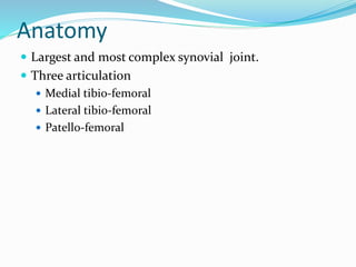
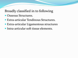
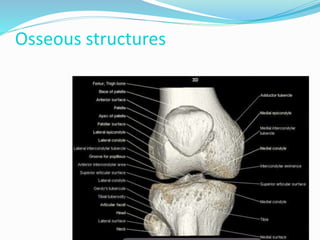
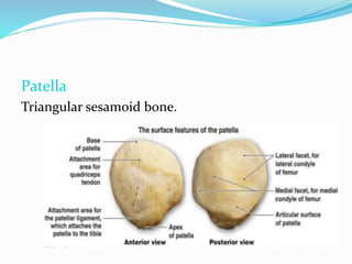
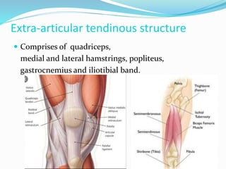
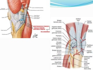
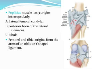
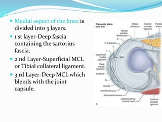
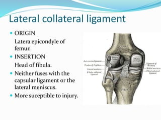
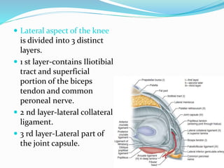
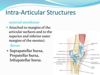
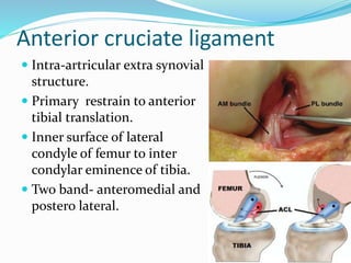
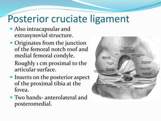
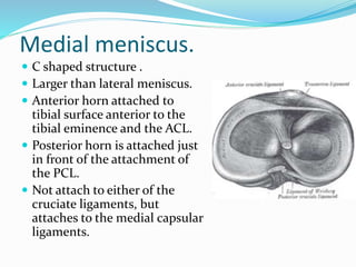
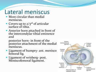
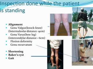
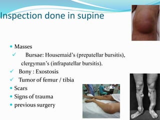
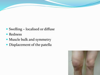
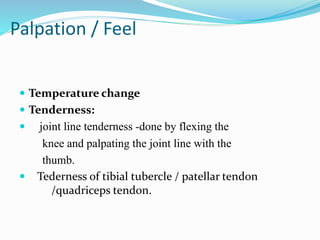
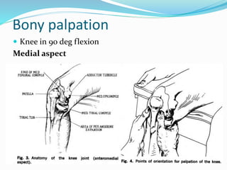
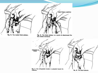
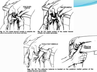
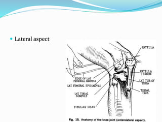
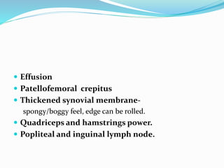
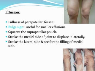
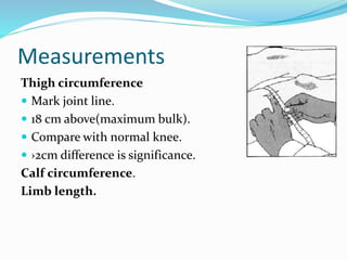
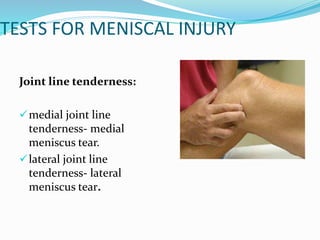
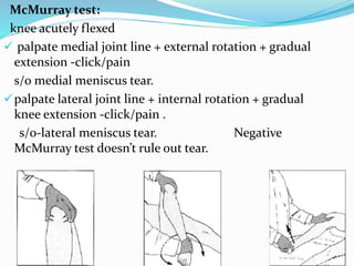
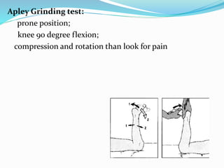
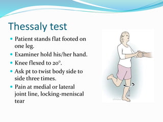
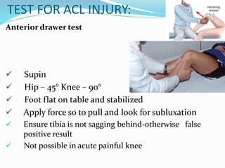
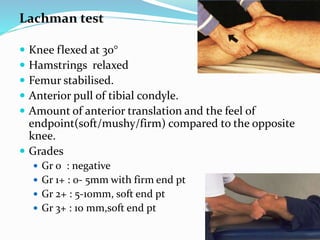
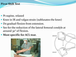
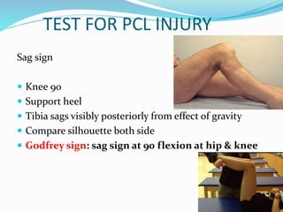
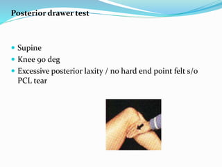
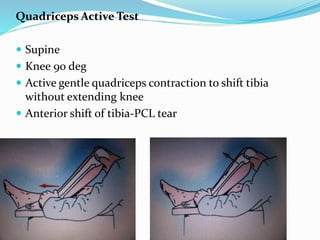
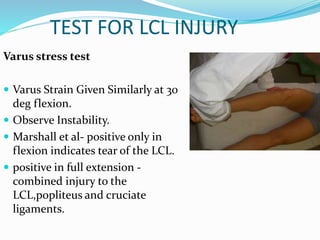
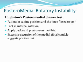
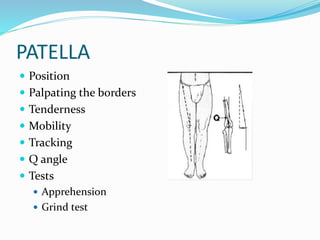
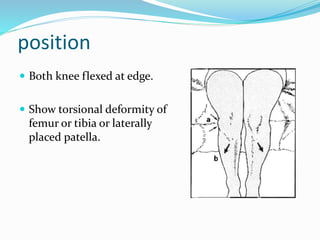
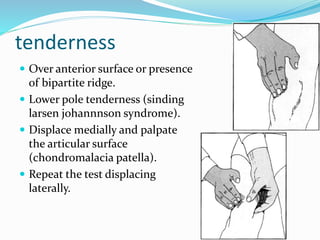
Dr Barun Patel
Examination of the knee
 Introduction
 Anatomy
 History taking
 Routine knee joint examination
 Special tests
Anatomy
 Largest and most complex synovial joint.
 Three articulation
 Medial tibio-femoral
 Lateral tibio-femoral
 Patello-femoral
Broadly classified in to following
 Osseous Structures.
 Extra-articular Tendinous Structures.
 Extra-articular Ligamentous structures
 Intra-articular soft tissue elements.
Osseous structures
Patella
Triangular sesamoid bone.
Extra-articular tendinous structure
 Comprises of quadriceps,
medial and lateral hamstrings, popliteus,
gastrocnemius and iliotibial band.
 Poplitius muscle has 3 origins
intracapsularly.
A.Lateral femoral condyle.
B.Posterior horn of the lateral
meniscus.
C.Fibula.
 Femoral and tibial origins form the
arms of an oblique Y shaped
ligament.
Semimembranosus muscle
has 5 distal expansion.
1.The oblique popliteal
ligament.
2.A second tendinous
attachment.
3.Anterior or deep head.
4.The direct head and
5.The distal portion of the
semimembranosus.
Extra-Articular Ligamentous Structures
 Capsule extends from patella and patellar tendon
anteriorly to medial lateral and posterior recess of
knee.
 Medial side it blends with tibial colateral ligament
and attached to medial meniscus.
 Laterally all are separate entity.
 Medial capsule is divided into 3
regions.
 Antero-medial portion reinforced
by medial patellar retinacula.
 Mid-medial capsule is the deep
layer of the MCL.
 Postero-medial capsular region or
the Posterior oblique
ligament.
Extends from posterior edge of
MCL to the insertion of the direct
head of semimembranosus.
Medial collateral ligament
 Superficial MCL or tibial
collateral ligament .
 Deep MCL .
 ORIGIN
 Medial epicondyle.
 INSERTION
 7-10cm below joint line.
 Deep to pes anserinus.
 Medial aspect of the knee is
divided into 3 layers.
 1 st layer-Deep fascia
containing the sartorius
fascia.
 2 nd Layer-Superficial MCL
or Tibial collateral ligament.
 3 rd Layer-Deep MCL which
blends with the joint
capsule.
Lateral collateral ligament
 ORIGIN
Latera epicondyle of
femur.
 INSERTION
Head of fibula.
 Neither fuses with the
capsular ligament or the
lateral meniscus.
 More suceptible to injury.
 Lateral aspect of the knee
is divided into 3 distinct
layers.
 1 st layer-contains Iliotibial
tract and superficial
portion of the biceps
tendon and common
peroneal nerve.
 2 nd layer-lateral collateral
ligament.
 3 rd layer-Lateral part of
the joint capsule.
Intra-Articular Structures
synovial membrane
 Attached to margins of the
articular surfaces and to the
superior and inferior outer
margins of the menisci.
Bursae
 Suprapatellar bursa,
Prepatellar bursa,
Infrapatellar bursa.
Anterior cruciate ligament
 Intra-artricular extra synovial
structure.
 Primary restrain to anterior
tibial translation.
 Inner surface of lateral
condyle of femur to inter
condylar eminence of tibia.
 Two band- anteromedial and
postero lateral.
Posterior cruciate ligament
 Also intracapsular and
extrasynovial structure.
 Originates from the junction
of the femoral notch roof and
medial femoral condyle.
Roughly 1 cm proximal to the
articular surface.
 Inserts on the posterior aspect
of the proximal tibia at the
fovea.
 Two bands- anterolateral and
posteromedial.
Medial meniscus.
 C shaped structure .
 Larger than lateral meniscus.
 Anterior horn attached to
tibial surface anterior to the
tibial eminence and the ACL.
 Posterior horn is attached just
in front of the attachment of
the PCL.
 Not attach to either of the
cruciate ligaments, but
attaches to the medial capsular
ligaments.
Lateral meniscus
 More circular than medial
meniscus.
 Covers up to 2/3rd of articular
surface of tibia.
 Anterior horn attached in front of
the intercondylar tibial eminence
and
posterior horn- in front of the
posterior attachment of the medial
meniscus.
 Ligament of humpry- ant. menisco
femoral lig.
 Ligament of wrisberg- post.
Meniscofemoral ligament.
CHIEF COMPLAINS
 Pain
 Swelling
 Stiffness
 Mechanical Disorders (locking, giving Way, click)
 Limp
 Deformity
Mechanism
 PCL
 Backward thrust on a
flexed knee
 Dashboard injuries
 ACL
 Audible "pop"
 Rapid knee swelling
 Rapid deceleration with
knee in rotation and
valgus stress.
Historical clues
Noncontact injury with “pop” ACL tear
Contact injury with “pop” MCL or LCL tear, meniscus
tear, fracture
Acute swelling ACL tear, PCL tear, fracture,
knee dislocation, patellar
dislocation
Lateral blow to the knee MCL tear
Medial blow to the knee LCL tear
Knee “gave out” or “buckled” ACL tear, patellar dislocation
Fall onto a flexed knee PCL tear
Common problem in relation to age
Symptoms
1. Pain

 Site
 Onset n duration
 Character
 Pain at night - inflammatory cause
 Pain - mechanical in origin.
 towards evening
 during/after exercise
 Pain - indicative of patellar problems
 Going up or down stairs
 Aching in positions where the knee is kept flexed for
prolonged periods of time.
 Bar- or vice-like pain below the patella
2. Swelling
 Causes
 Infective
 Traumatic
 Degenerative
 Inflammatory
 tumor
3. Laxity
 "Going out"
 torn ACL or a dislocation of the patella.
 "Giving way"
 sensation of the knee suddenly failing to provide proper
support
 especially when walking on uneven ground.
4.Locking
 It is the inability to fully extend the knee for an
appreciable period of time.
Causes :
 bucket-handle tear of the meniscus
 bulky flap that has dislocated forwards in the
joint
 a loose body or an ACL stump
EXAMINATION
 Expose both lower limbs from groin to toes.
 Postions
 Standing
 Seated position
 Supine position
 Lastly prone position
 Always examine the hip joint and opposite knee joint.
 Gait .
 Attitude.
Inspection done while the patient
is standing
 Alignment
 Genu Valgus(knock-knee)
(Intermaleolar distance ›9cm)
 Genu Varus(bow leg)
(intercondylar distance › 6cm)
 Flexion deformity
 Genu recurvatum
 Shortening
 Baker’s cyst
 Gait
Inspection done in supine
 Masses
 Bursae: Housemaid’s (prepatellar bursitis),
clergyman’s (infrapatellar bursitis).
 Bony : Exostosis
 Tumor of femur / tibia
 Scars
 Signs of trauma
 previous surgery
 Swelling – localised or diffuse
 Redness
 Muscle bulk and symmetry
 Displacement of the patella
Palpation / Feel
 Temperature change
 Tenderness:
 joint line tenderness -done by flexing the
knee and palpating the joint line with the
thumb.
 Tederness of tibial tubercle / patellar tendon
/quadriceps tendon.
Bony palpation
 Knee in 90 deg flexion
Medial aspect
 Lateral aspect
 Effusion
 Patellofemoral crepitus
 Thickened synovial membrane-
spongy/boggy feel, edge can be rolled.
 Quadriceps and hamstrings power.
 Popliteal and inguinal lymph node.
Effusion:
 Fullness of parapatellar fossae.
 Bulge sign: useful for smaller effusions.
 Squeeze the suprapatellar pouch.
 Stroke the medial side of joint to displace it laterally.
 Stroke the lateral side & see for the filling of medial
side.
Patellar tap:
 Test for identify moderate effusion.
 press suprapatellar pouch with one
hand.
 with the index and middle finger of
other hand push the patela towards
femoral condyle with a jerk.
 patella is felt to strike the femur then
bounce back.
Coss fluctuation test
 For moderate to severe effusion.
 With thumb on one side and fingers on other side,
compress the the knee to empty the hollows.
 Now with the other hand forcefully compress the fluid
to knee joint.
 Findings- fluid impulse is transmitted across the joint.
In prone
 Popliteal fossa
 Semimembranosus bursa
 Bakers cyst
MOVE
Movement: . ACTIVE & PASSIVE
 Flexion –Extension: Normal 0-135 degrees.
 Rotation: 20-30 deg in flexion, Nil in extension.
 Fixed flexion deformity : by passively lifting the leg
at the heel to see if there is complete extension.
 See for crepitus during motion.
 See hip rotations, as pain can be referred from the hip.
 Repeat each movement for the opposite leg at the
same time.
Measurements
Thigh circumference
 Mark joint line.
 18 cm above(maximum bulk).
 Compare with normal knee.
 ›2cm difference is significance.
Calf circumference.
Limb length.
TESTS FOR MENISCAL INJURY
Joint line tenderness:
medial joint line
tenderness- medial
meniscus tear.
lateral joint line
tenderness- lateral
meniscus tear.
McMurray test:
knee acutely flexed
 palpate medial joint line + external rotation + gradual
extension -click/pain
s/o medial meniscus tear.
palpate lateral joint line + internal rotation + gradual
knee extension -click/pain .
s/o-lateral meniscus tear. Negative
McMurray test doesn’t rule out tear.
Apley Grinding test:
prone position;
knee 90 degree flexion;
compression and rotation than look for pain
Thessaly test
 Patient stands flat footed on
one leg.
 Examiner hold his/her hand.
 Knee flexed to 20°.
 Ask pt to twist body side to
side three times.
 Pain at medial or lateral
joint line, locking-meniscal
tear
TEST FOR ACL INJURY:
Anterior drawer test
 Supin
 Hip – 45° Knee – 90°
 Foot flat on table and stabilized
 Apply force so to pull and look for subluxation
 Ensure tibia is not sagging behind-otherwise false
positive result
 Not possible in acute painful knee
Lachman test
 Knee flexed at 30°
 Hamstrings relaxed
 Femur stabilised.
 Anterior pull of tibial condyle.
 Amount of anterior translation and the feel of
endpoint(soft/mushy/firm) compared to the opposite
knee.
 Grades
 Gr 0 : negative
 Gr 1+ : 0- 5mm with firm end pt
 Gr 2+ : 5-10mm, soft end pt
 Gr 3+ : 10 mm,soft end pt
Pivot Shift Test
 Pt supine, relaxed
 Knee in IR and valgus strain (subluxates the knee)
 Do gradual flexion from extension.
 See for the reduction of the lateral femoral condyle at
around 30° of flexion.
 Most specific for ACL tear.
TEST FOR PCL INJURY
Sag sign
 Knee 90
 Support heel
 Tibia sags visibly posteriorly from effect of gravity
 Compare silhouette both side
 Godfrey sign: sag sign at 90 flexion at hip & knee
Posterior drawer test
 Supine
 Knee 90 deg
 Excessive posterior laxity / no hard end point felt s/o
PCL tear
Quadriceps Active Test
 Supine
 Knee 90 deg
 Active gentle quadriceps contraction to shift tibia
without extending knee
 Anterior shift of tibia-PCL tear
TEST FOR MCL INJURY
Valgus stress test
 Supine
 Side Of Table
 Abducted Of The Side Of
table
 In full extension and
Flexion 30 degree.
 Valgus Strain
 Observe Stability.
 Hugston et al- positive at 30° flexion
negative at 0°- only MCL tear
 +ve at Extension- tear of both MCL
and PCL.
TEST FOR LCL INJURY
Varus stress test
 Varus Strain Given Similarly at 30
deg flexion.
 Observe Instability.
 Marshall et al- positive only in
flexion indicates tear of the LCL.
 positive in full extension -
combined injury to the
LCL,popliteus and cruciate
ligaments.
POSTEROLATERAL CORNER INJURY
(PLC)
 External Rotation Recurvatum test
 Dial test: External rotation of tibia is compared at 30
& 90 flexion; > 10 deg increased -+ ve.
 Reverse Pivot Shift Test
Dial test
 Check for external rotation of foot
(thigh foot angle).
 At 30° and 90° knee flexion.
 Increased rotation at 30° that
decreases at 90° - PLC injury
 Increased rotation at both 30° and
90° - both PLC and PCL injury.
 › 10° difference is pathognomic.
Posterolateral drawer test
• Knee flexed at 90 degree and foot
in external rotation.
• Apply backward pressure on tibia.
• Excessive travel on lateral side
indicative of posterolateral
instability.
External rotation recurvatum test
 Patient in supine position.
 Stand at the end and lift both
legs holding the great toes.
 Test is positive if the knee falls
into external rotation, varus and
recurvatum.
jakob’s reverse pivot shift test
 Flxed the knee to 90 degree.
 Externl rotate the foot.
 Apply valgus stress and extend the
knee.
 If test positive then the posteriorly
subluxed knee reduced at 20 degree
extension.
Anterolateral rotatory instability
Slocum’s Anterior Rotatory Drawer Test
 Modification of anterior drawer test.
 Pt supine, knee flexed to 90°.
 Knee in 15° internal rotation.
 Perform anterior drawer test.
 Anterior subluxation of lateral tibial condyle- +ve.
 Reduces with knee in external rotation.
Jerk test of Hughston and Losee
 patient in supine , knee is flexed to 90 deg with the
tibia in internal rotation.
 knee gradually extended with valgus stress applied.
 Test is positive if the lateral tibia subluxes anteriorly in
the form of sudden jerk at about 30 deg of flexion.
Antero medial rotatory instability
Slocum’s Anterior Rotatory Drawer Test
 Pt supine, knee flexed at 90°.
 Tibia in 30° external rotation.
 Perform anterior drawer test.
 Excessive excursion of the medial tibial condyle
suggests positive test.
PosteroMedial Rotatory Instability
Hughston’s Posteromedial drawer test.
 Patient in supine position and the knee flexed to 90 °.
 Foot in internal rotation.
 Apply backward pressure on the tibia.
 Excessive excursion of the medial tibial condyle
suggests positive test.
PATELLA
 Position
 Palpating the borders
 Tenderness
 Mobility
 Tracking
 Q angle
 Tests
 Apprehension
 Grind test
position
 Both knee flexed at edge.
 Show torsional deformity of
femur or tibia or laterally
placed patella.
tenderness
 Over anterior surface or presence
of bipartite ridge.
 Lower pole tenderness (sinding
larsen johannnson syndrome).
 Displace medially and palpate
the articular surface
(chondromalacia patella).
 Repeat the test displacing
laterally.
Q angle
 Angle between a line from ASIS to centre of
patella and centre of patella to tibial
tuberosity.
N-♂8-10°♀15±5°
 Greater the Q angle, the greater the
tendency to move the patella laterally
against the lateral femoral condyle.
PATELLAR TESTS
Apprehension test
 Supine
 Knee 10-30deg
flexion
 push the patella in a
lateral direction
 Patient stops the
examiner
Patellar tracking
 Knee at 90 deg to full
extension
 Shifts laterally at
terminal extension
 Excess lateral shift /tilt
terminally indicates
patellar instability
Figure 6-66. Assessing patellar tracking.
THANK U

Knee examination

  • 1.
  • 2.
    Examination of theknee  Introduction  Anatomy  History taking  Routine knee joint examination  Special tests
  • 3.
    Anatomy  Largest andmost complex synovial joint.  Three articulation  Medial tibio-femoral  Lateral tibio-femoral  Patello-femoral
  • 4.
    Broadly classified into following  Osseous Structures.  Extra-articular Tendinous Structures.  Extra-articular Ligamentous structures  Intra-articular soft tissue elements.
  • 5.
  • 6.
  • 7.
    Extra-articular tendinous structure Comprises of quadriceps, medial and lateral hamstrings, popliteus, gastrocnemius and iliotibial band.
  • 9.
     Poplitius musclehas 3 origins intracapsularly. A.Lateral femoral condyle. B.Posterior horn of the lateral meniscus. C.Fibula.  Femoral and tibial origins form the arms of an oblique Y shaped ligament.
  • 10.
    Semimembranosus muscle has 5distal expansion. 1.The oblique popliteal ligament. 2.A second tendinous attachment. 3.Anterior or deep head. 4.The direct head and 5.The distal portion of the semimembranosus.
  • 11.
    Extra-Articular Ligamentous Structures Capsule extends from patella and patellar tendon anteriorly to medial lateral and posterior recess of knee.  Medial side it blends with tibial colateral ligament and attached to medial meniscus.  Laterally all are separate entity.
  • 12.
     Medial capsuleis divided into 3 regions.  Antero-medial portion reinforced by medial patellar retinacula.  Mid-medial capsule is the deep layer of the MCL.  Postero-medial capsular region or the Posterior oblique ligament. Extends from posterior edge of MCL to the insertion of the direct head of semimembranosus.
  • 13.
    Medial collateral ligament Superficial MCL or tibial collateral ligament .  Deep MCL .  ORIGIN  Medial epicondyle.  INSERTION  7-10cm below joint line.  Deep to pes anserinus.
  • 14.
     Medial aspectof the knee is divided into 3 layers.  1 st layer-Deep fascia containing the sartorius fascia.  2 nd Layer-Superficial MCL or Tibial collateral ligament.  3 rd Layer-Deep MCL which blends with the joint capsule.
  • 15.
    Lateral collateral ligament ORIGIN Latera epicondyle of femur.  INSERTION Head of fibula.  Neither fuses with the capsular ligament or the lateral meniscus.  More suceptible to injury.
  • 16.
     Lateral aspectof the knee is divided into 3 distinct layers.  1 st layer-contains Iliotibial tract and superficial portion of the biceps tendon and common peroneal nerve.  2 nd layer-lateral collateral ligament.  3 rd layer-Lateral part of the joint capsule.
  • 17.
    Intra-Articular Structures synovial membrane Attached to margins of the articular surfaces and to the superior and inferior outer margins of the menisci. Bursae  Suprapatellar bursa, Prepatellar bursa, Infrapatellar bursa.
  • 18.
    Anterior cruciate ligament Intra-artricular extra synovial structure.  Primary restrain to anterior tibial translation.  Inner surface of lateral condyle of femur to inter condylar eminence of tibia.  Two band- anteromedial and postero lateral.
  • 19.
    Posterior cruciate ligament Also intracapsular and extrasynovial structure.  Originates from the junction of the femoral notch roof and medial femoral condyle. Roughly 1 cm proximal to the articular surface.  Inserts on the posterior aspect of the proximal tibia at the fovea.  Two bands- anterolateral and posteromedial.
  • 20.
    Medial meniscus.  Cshaped structure .  Larger than lateral meniscus.  Anterior horn attached to tibial surface anterior to the tibial eminence and the ACL.  Posterior horn is attached just in front of the attachment of the PCL.  Not attach to either of the cruciate ligaments, but attaches to the medial capsular ligaments.
  • 21.
    Lateral meniscus  Morecircular than medial meniscus.  Covers up to 2/3rd of articular surface of tibia.  Anterior horn attached in front of the intercondylar tibial eminence and posterior horn- in front of the posterior attachment of the medial meniscus.  Ligament of humpry- ant. menisco femoral lig.  Ligament of wrisberg- post. Meniscofemoral ligament.
  • 22.
    CHIEF COMPLAINS  Pain Swelling  Stiffness  Mechanical Disorders (locking, giving Way, click)  Limp  Deformity
  • 23.
    Mechanism  PCL  Backwardthrust on a flexed knee  Dashboard injuries  ACL  Audible "pop"  Rapid knee swelling  Rapid deceleration with knee in rotation and valgus stress.
  • 24.
    Historical clues Noncontact injurywith “pop” ACL tear Contact injury with “pop” MCL or LCL tear, meniscus tear, fracture Acute swelling ACL tear, PCL tear, fracture, knee dislocation, patellar dislocation Lateral blow to the knee MCL tear Medial blow to the knee LCL tear Knee “gave out” or “buckled” ACL tear, patellar dislocation Fall onto a flexed knee PCL tear
  • 25.
    Common problem inrelation to age
  • 26.
    Symptoms 1. Pain   Site Onset n duration  Character  Pain at night - inflammatory cause  Pain - mechanical in origin.  towards evening  during/after exercise
  • 27.
     Pain -indicative of patellar problems  Going up or down stairs  Aching in positions where the knee is kept flexed for prolonged periods of time.  Bar- or vice-like pain below the patella
  • 28.
    2. Swelling  Causes Infective  Traumatic  Degenerative  Inflammatory  tumor
  • 29.
    3. Laxity  "Goingout"  torn ACL or a dislocation of the patella.  "Giving way"  sensation of the knee suddenly failing to provide proper support  especially when walking on uneven ground.
  • 30.
    4.Locking  It isthe inability to fully extend the knee for an appreciable period of time. Causes :  bucket-handle tear of the meniscus  bulky flap that has dislocated forwards in the joint  a loose body or an ACL stump
  • 31.
    EXAMINATION  Expose bothlower limbs from groin to toes.  Postions  Standing  Seated position  Supine position  Lastly prone position  Always examine the hip joint and opposite knee joint.  Gait .  Attitude.
  • 32.
    Inspection done whilethe patient is standing  Alignment  Genu Valgus(knock-knee) (Intermaleolar distance ›9cm)  Genu Varus(bow leg) (intercondylar distance › 6cm)  Flexion deformity  Genu recurvatum  Shortening  Baker’s cyst  Gait
  • 33.
    Inspection done insupine  Masses  Bursae: Housemaid’s (prepatellar bursitis), clergyman’s (infrapatellar bursitis).  Bony : Exostosis  Tumor of femur / tibia  Scars  Signs of trauma  previous surgery
  • 34.
     Swelling –localised or diffuse  Redness  Muscle bulk and symmetry  Displacement of the patella
  • 35.
    Palpation / Feel Temperature change  Tenderness:  joint line tenderness -done by flexing the knee and palpating the joint line with the thumb.  Tederness of tibial tubercle / patellar tendon /quadriceps tendon.
  • 36.
    Bony palpation  Kneein 90 deg flexion Medial aspect
  • 39.
  • 42.
     Effusion  Patellofemoralcrepitus  Thickened synovial membrane- spongy/boggy feel, edge can be rolled.  Quadriceps and hamstrings power.  Popliteal and inguinal lymph node.
  • 43.
    Effusion:  Fullness ofparapatellar fossae.  Bulge sign: useful for smaller effusions.  Squeeze the suprapatellar pouch.  Stroke the medial side of joint to displace it laterally.  Stroke the lateral side & see for the filling of medial side.
  • 44.
    Patellar tap:  Testfor identify moderate effusion.  press suprapatellar pouch with one hand.  with the index and middle finger of other hand push the patela towards femoral condyle with a jerk.  patella is felt to strike the femur then bounce back.
  • 45.
    Coss fluctuation test For moderate to severe effusion.  With thumb on one side and fingers on other side, compress the the knee to empty the hollows.  Now with the other hand forcefully compress the fluid to knee joint.  Findings- fluid impulse is transmitted across the joint.
  • 46.
    In prone  Poplitealfossa  Semimembranosus bursa  Bakers cyst
  • 47.
    MOVE Movement: . ACTIVE& PASSIVE  Flexion –Extension: Normal 0-135 degrees.  Rotation: 20-30 deg in flexion, Nil in extension.  Fixed flexion deformity : by passively lifting the leg at the heel to see if there is complete extension.
  • 48.
     See forcrepitus during motion.  See hip rotations, as pain can be referred from the hip.  Repeat each movement for the opposite leg at the same time.
  • 49.
    Measurements Thigh circumference  Markjoint line.  18 cm above(maximum bulk).  Compare with normal knee.  ›2cm difference is significance. Calf circumference. Limb length.
  • 50.
    TESTS FOR MENISCALINJURY Joint line tenderness: medial joint line tenderness- medial meniscus tear. lateral joint line tenderness- lateral meniscus tear.
  • 51.
    McMurray test: knee acutelyflexed  palpate medial joint line + external rotation + gradual extension -click/pain s/o medial meniscus tear. palpate lateral joint line + internal rotation + gradual knee extension -click/pain . s/o-lateral meniscus tear. Negative McMurray test doesn’t rule out tear.
  • 52.
    Apley Grinding test: proneposition; knee 90 degree flexion; compression and rotation than look for pain
  • 53.
    Thessaly test  Patientstands flat footed on one leg.  Examiner hold his/her hand.  Knee flexed to 20°.  Ask pt to twist body side to side three times.  Pain at medial or lateral joint line, locking-meniscal tear
  • 54.
    TEST FOR ACLINJURY: Anterior drawer test  Supin  Hip – 45° Knee – 90°  Foot flat on table and stabilized  Apply force so to pull and look for subluxation  Ensure tibia is not sagging behind-otherwise false positive result  Not possible in acute painful knee
  • 55.
    Lachman test  Kneeflexed at 30°  Hamstrings relaxed  Femur stabilised.  Anterior pull of tibial condyle.  Amount of anterior translation and the feel of endpoint(soft/mushy/firm) compared to the opposite knee.  Grades  Gr 0 : negative  Gr 1+ : 0- 5mm with firm end pt  Gr 2+ : 5-10mm, soft end pt  Gr 3+ : 10 mm,soft end pt
  • 56.
    Pivot Shift Test Pt supine, relaxed  Knee in IR and valgus strain (subluxates the knee)  Do gradual flexion from extension.  See for the reduction of the lateral femoral condyle at around 30° of flexion.  Most specific for ACL tear.
  • 57.
    TEST FOR PCLINJURY Sag sign  Knee 90  Support heel  Tibia sags visibly posteriorly from effect of gravity  Compare silhouette both side  Godfrey sign: sag sign at 90 flexion at hip & knee
  • 58.
    Posterior drawer test Supine  Knee 90 deg  Excessive posterior laxity / no hard end point felt s/o PCL tear
  • 59.
    Quadriceps Active Test Supine  Knee 90 deg  Active gentle quadriceps contraction to shift tibia without extending knee  Anterior shift of tibia-PCL tear
  • 60.
    TEST FOR MCLINJURY Valgus stress test  Supine  Side Of Table  Abducted Of The Side Of table  In full extension and Flexion 30 degree.  Valgus Strain  Observe Stability.  Hugston et al- positive at 30° flexion negative at 0°- only MCL tear  +ve at Extension- tear of both MCL and PCL.
  • 61.
    TEST FOR LCLINJURY Varus stress test  Varus Strain Given Similarly at 30 deg flexion.  Observe Instability.  Marshall et al- positive only in flexion indicates tear of the LCL.  positive in full extension - combined injury to the LCL,popliteus and cruciate ligaments.
  • 62.
    POSTEROLATERAL CORNER INJURY (PLC) External Rotation Recurvatum test  Dial test: External rotation of tibia is compared at 30 & 90 flexion; > 10 deg increased -+ ve.  Reverse Pivot Shift Test
  • 63.
    Dial test  Checkfor external rotation of foot (thigh foot angle).  At 30° and 90° knee flexion.  Increased rotation at 30° that decreases at 90° - PLC injury  Increased rotation at both 30° and 90° - both PLC and PCL injury.  › 10° difference is pathognomic.
  • 64.
    Posterolateral drawer test •Knee flexed at 90 degree and foot in external rotation. • Apply backward pressure on tibia. • Excessive travel on lateral side indicative of posterolateral instability.
  • 65.
    External rotation recurvatumtest  Patient in supine position.  Stand at the end and lift both legs holding the great toes.  Test is positive if the knee falls into external rotation, varus and recurvatum.
  • 66.
    jakob’s reverse pivotshift test  Flxed the knee to 90 degree.  Externl rotate the foot.  Apply valgus stress and extend the knee.  If test positive then the posteriorly subluxed knee reduced at 20 degree extension.
  • 67.
    Anterolateral rotatory instability Slocum’sAnterior Rotatory Drawer Test  Modification of anterior drawer test.  Pt supine, knee flexed to 90°.  Knee in 15° internal rotation.  Perform anterior drawer test.  Anterior subluxation of lateral tibial condyle- +ve.  Reduces with knee in external rotation.
  • 68.
    Jerk test ofHughston and Losee  patient in supine , knee is flexed to 90 deg with the tibia in internal rotation.  knee gradually extended with valgus stress applied.  Test is positive if the lateral tibia subluxes anteriorly in the form of sudden jerk at about 30 deg of flexion.
  • 69.
    Antero medial rotatoryinstability Slocum’s Anterior Rotatory Drawer Test  Pt supine, knee flexed at 90°.  Tibia in 30° external rotation.  Perform anterior drawer test.  Excessive excursion of the medial tibial condyle suggests positive test.
  • 70.
    PosteroMedial Rotatory Instability Hughston’sPosteromedial drawer test.  Patient in supine position and the knee flexed to 90 °.  Foot in internal rotation.  Apply backward pressure on the tibia.  Excessive excursion of the medial tibial condyle suggests positive test.
  • 71.
    PATELLA  Position  Palpatingthe borders  Tenderness  Mobility  Tracking  Q angle  Tests  Apprehension  Grind test
  • 72.
    position  Both kneeflexed at edge.  Show torsional deformity of femur or tibia or laterally placed patella.
  • 73.
    tenderness  Over anteriorsurface or presence of bipartite ridge.  Lower pole tenderness (sinding larsen johannnson syndrome).  Displace medially and palpate the articular surface (chondromalacia patella).  Repeat the test displacing laterally.
  • 74.
    Q angle  Anglebetween a line from ASIS to centre of patella and centre of patella to tibial tuberosity. N-♂8-10°♀15±5°  Greater the Q angle, the greater the tendency to move the patella laterally against the lateral femoral condyle.
  • 75.
    PATELLAR TESTS Apprehension test Supine  Knee 10-30deg flexion  push the patella in a lateral direction  Patient stops the examiner
  • 76.
    Patellar tracking  Kneeat 90 deg to full extension  Shifts laterally at terminal extension  Excess lateral shift /tilt terminally indicates patellar instability Figure 6-66. Assessing patellar tracking.
  • 77.