
Recommended
More Related Content
What's hot
What's hot (20)
Similar to Knee examination
Similar to Knee examination (20)
Recently uploaded
Recently uploaded (20)
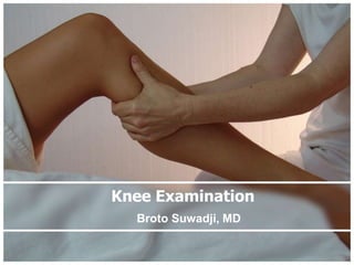
Knee examination
- 3. Anatomy Osseous Structures • Distal femur • Proksimal Tibia • Patella
- 4. • femoral condyles are two rounded prominences • Anteriorly flattened • medial condyle is longer • lateral condyle is wider
- 5. • two rather flat surfaces • separated in the midline by the intercondylar eminence • medial and lateral intercondylar tubercles • Anterior and posterior to the intercondylar eminence
- 6. • triangular sesamoid bone • wider at the proximal pole • patella is divided by a vertical ridge larger lateral articular facet • increase the power of extension
- 7. Extraarticular Tendinous Structures • Synovium • Capsule • collateral ligaments –medial n Lateral • Musculotendinous –gastrocnemius, the medial and lateral hamstring groups, the popliteus, and the iliotibial band)
- 8. Intraarticular Structures • medial and lateral menisci • the anterior and posterior cruciate ligaments
- 9. • Function – increase the contact area – Weight transmission • Prone to injury, particularly during unguarded movements • medial meniscus is especially vulnerable
- 10. • medial meniscus is especially vulnerable because –firmly attached at three widely separated points: the anterior horn, the posterior horn and to the medial collateral ligament –deep portion of the medial collateral ligament blends with the posteromedial capsule
- 11. Knee Examination
- 12. SYMPTOMS • Pain • Swelling • Stiffness’ • Locking • Deformity • Giving way • Limp
- 13. SIGNS Look – Feel – Move • SIGNS WITH THE PATIENT UPRIGHT • SIGNS WITH THE PATIENT SITTING • SIGNS WITH THE PATIENT LYING SUPINE • SIGNS WITH THE PATIENT LYING PRONE
- 14. SIGNS WITH THE PATIENT UPRIGHT • Deformity – (valgus or varus or hyperextension (Normally the knees and ankles can touch in the midline) • Quadriceps,patella and patellar ligament • Gait – stance phase • fixed flexion deformity or a hyperextension deformity • lateral or medial thrust – swing phase • knee moves freely or is held in one position
- 18. SIGNS WITH THE PATIENT SITTING • knees dangling at 90 degrees of flexion – patella alta – patella baja • ask the patient to straighten each knee in turn , observe how the patella moves upwards • Q-angle14 degrees in men and 17 degrees in women
- 23. SIGNS WITH THE PATIENT LYING SUPINELook • position of the knee – valgus or varus, – incompletely extended, – hyperextended – lumps or bumps • Wasting • Bruising • scars or sinuses • shape and position of the patella
- 25. Feel • skin temperature • soft tissues and bony outlines • Synovial thickening
- 26. soft tissues and bony outlines
- 28. 1, quadriceps tendon; 2, edge of patella; 3, medial collateral ligament; 4, joint line; 5, lateral collateral ligament; 6, patellar ligament
- 30. SIGNS WITH THE PATIENT LYING SUPINE Move • Passive extension • Active extension • Passive and active flexion – The ‘heel-to-buttock’ distance • Internal and external rotation • Crepitus • Movement with compartmental loading
- 31. • A : Normal Range : – 0-135⁰ • B : Hiper extension – 5⁰ hyperext - 140⁰ • C : Fixed flexion deformity – 10⁰ – 60⁰
- 33. Tests • Tests for intra-articular fluid • The patello-femoral joint • Tests for stability • Tests for meniscal injuries
- 34. Tests for intra-articular fluid • Juxtapatellar hollow • Patellar tap test • Bulge test • Cross fluctuation
- 37. Bulge test
- 39. The patello-femoral joint • size, shape and position of the patella are noted • the ‘friction test’ – Moving the patella up and down while pressing it lightly against the femur • Apprehension test – Pressing the patella laterally with the thumb while flexing the knee slowly may induce anxiety and sharp resistance
- 41. Tests for stability • Collateral ligaments – Varus – Valgus stree test – The test is performed at full extension and again at 30 degrees of flexion • Cruciate ligaments – both knees flexed 90 degrees – feet resting on the couch – upper tibia is inspected from the side ‘sag sign drawer test Lachman test
- 42. Varus Valgus stress test
- 43. Sag Sign
- 44. Drawer Test
- 45. Lachman Test • knee is flexed 20 deg • one hand grasping the lower thigh • Other upper part of the leg • Shifted backwards and forwards
- 46. Tests for meniscal injuries • McMurray’s test – knee is flexed as far as possible – hand steadies the joint – other rotates the leg medially and laterally – knee is slowly extended – repeated several times – knee stressed in valgus or varus – feeling and listening for the click A positive test is helpful but not pathognomonic; a negative test does not exclude a tear
- 48. • Thessaly test • based on a dynamic reproduction of load transmission • affected knee flexed to 20 degrees • foot placed flat on the ground • instructed to twist his or her body to one side
- 50. SIGNS WITH THE PATIENT LYING PRONE • Scars or lumps in the popliteal fossa • Swelling: midline (capsule), one side (bursa), Baker’s cyst • Apley’s test • Lachman’s test
- 51. Apley’s test • patient prone • knee is flexed to 90 degrees • rotated while a compression force applied • grinding test meniscus is torn • distraction test ligament damage