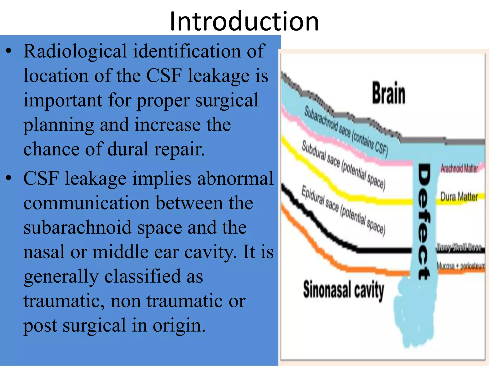
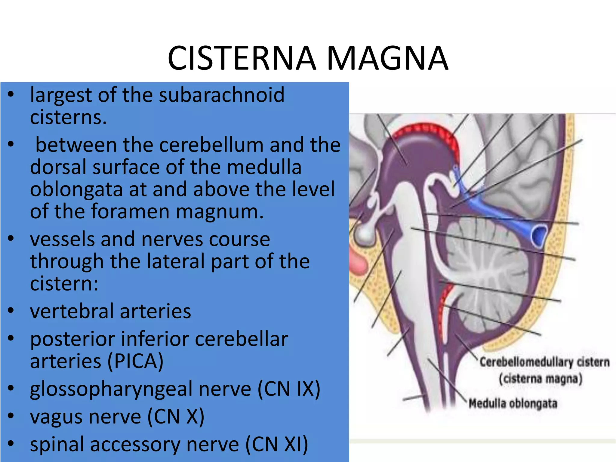
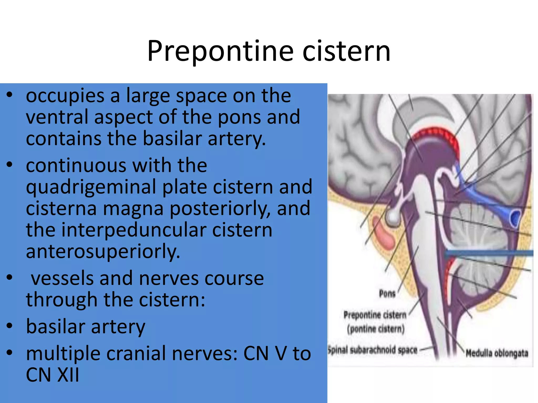
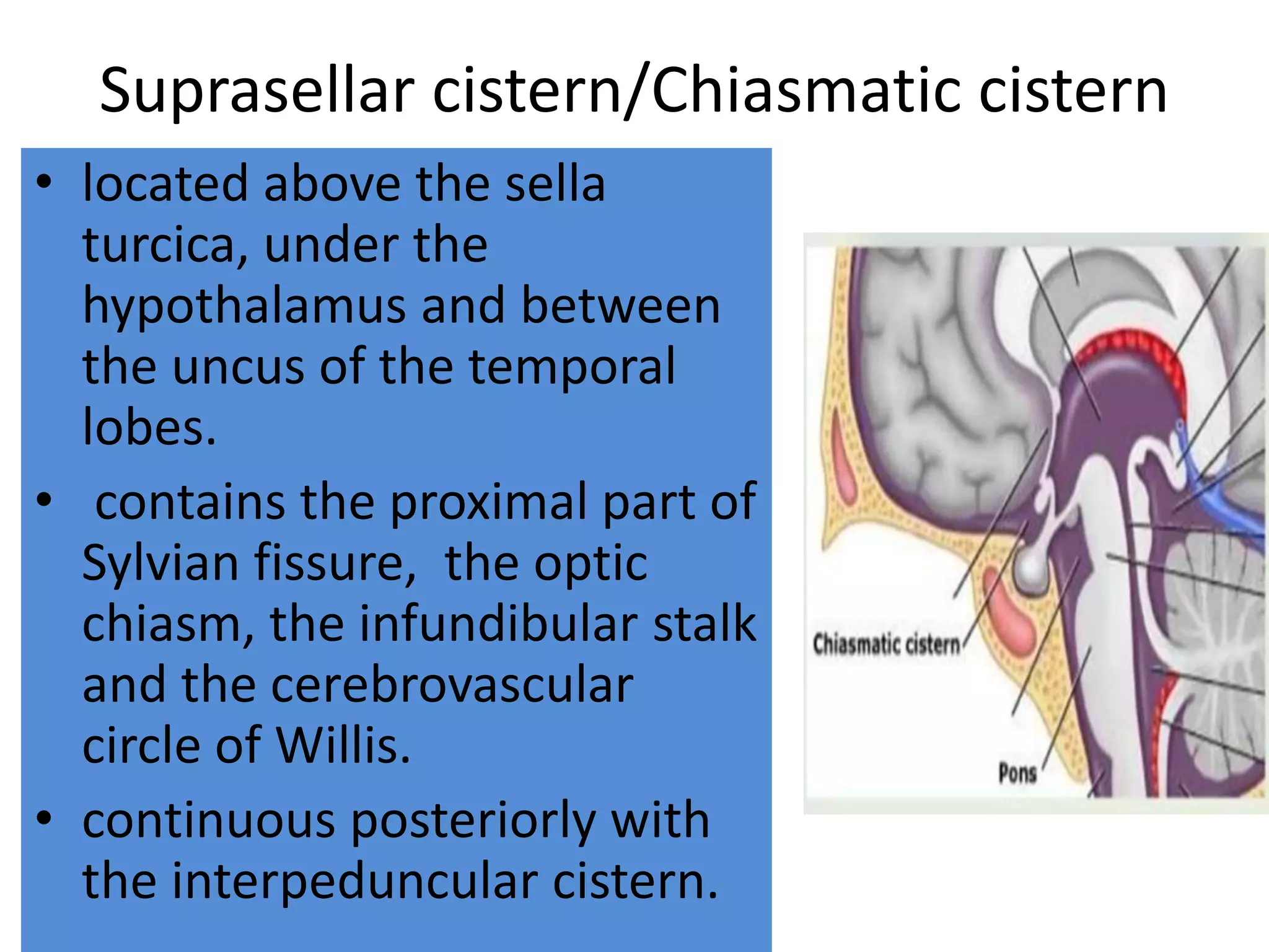
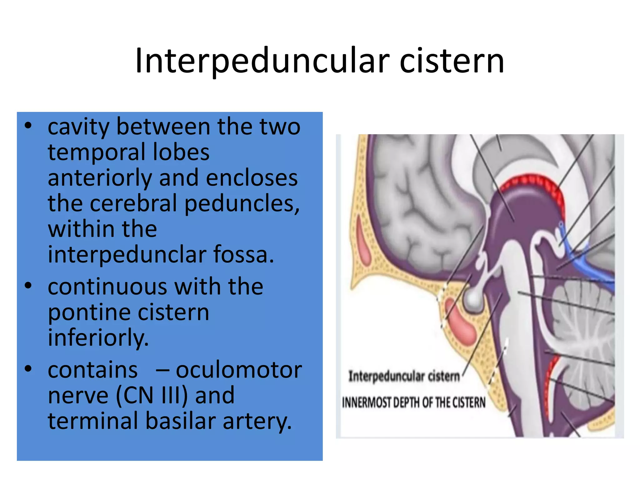
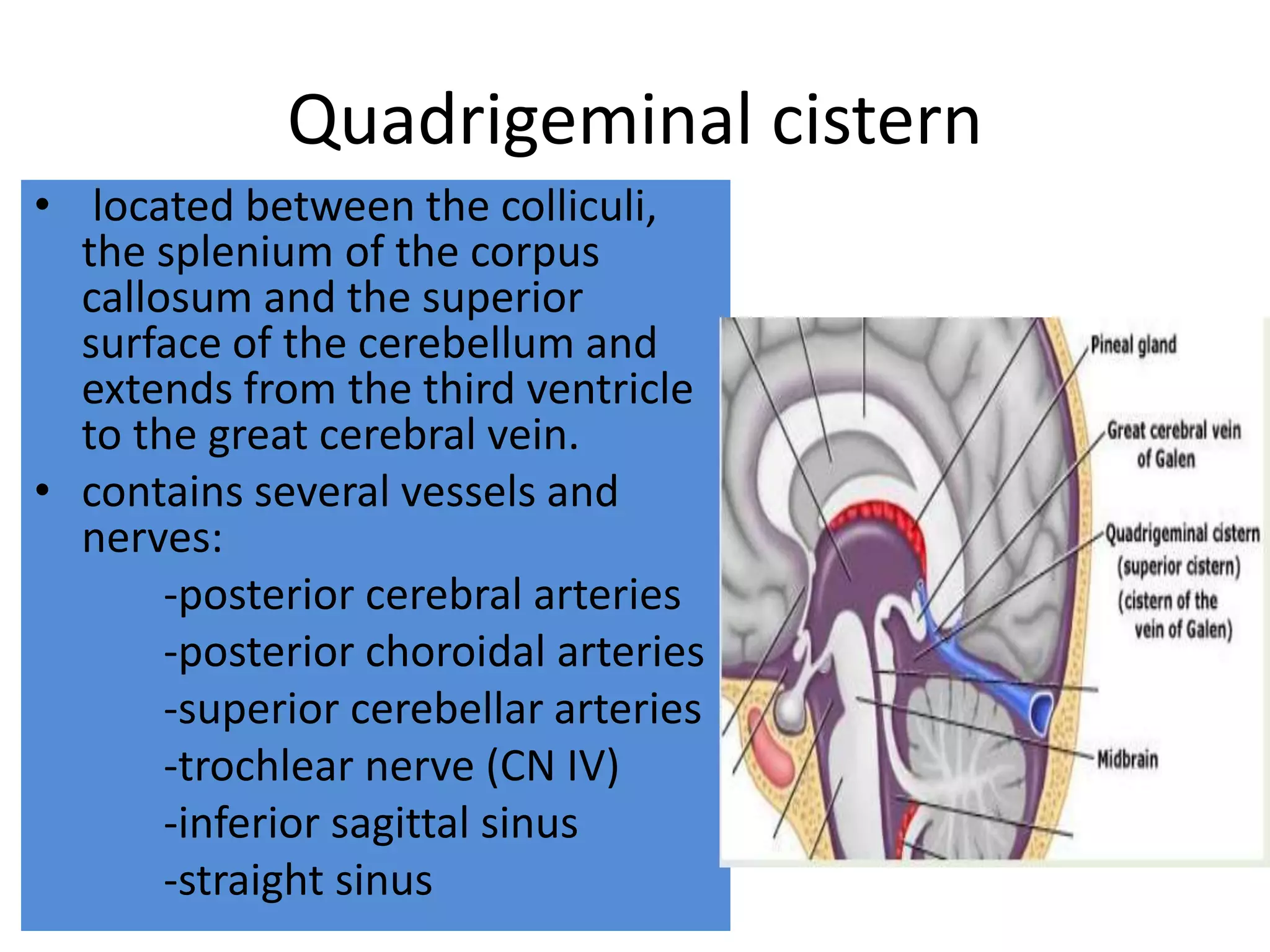
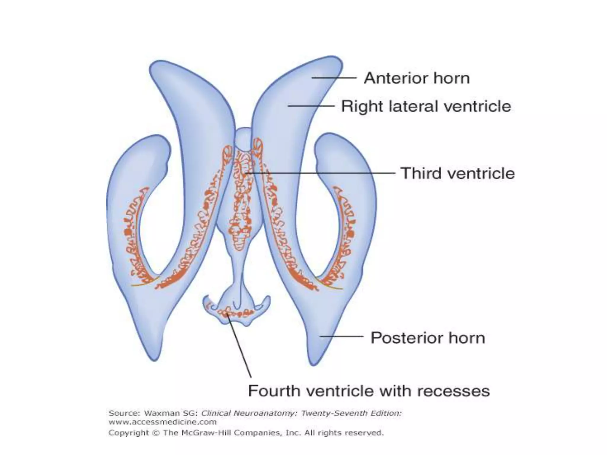
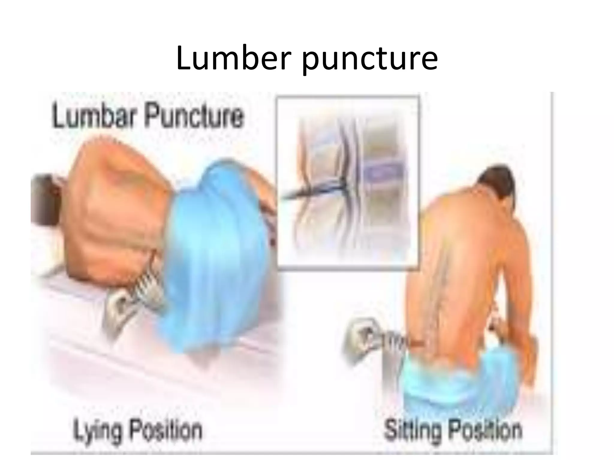
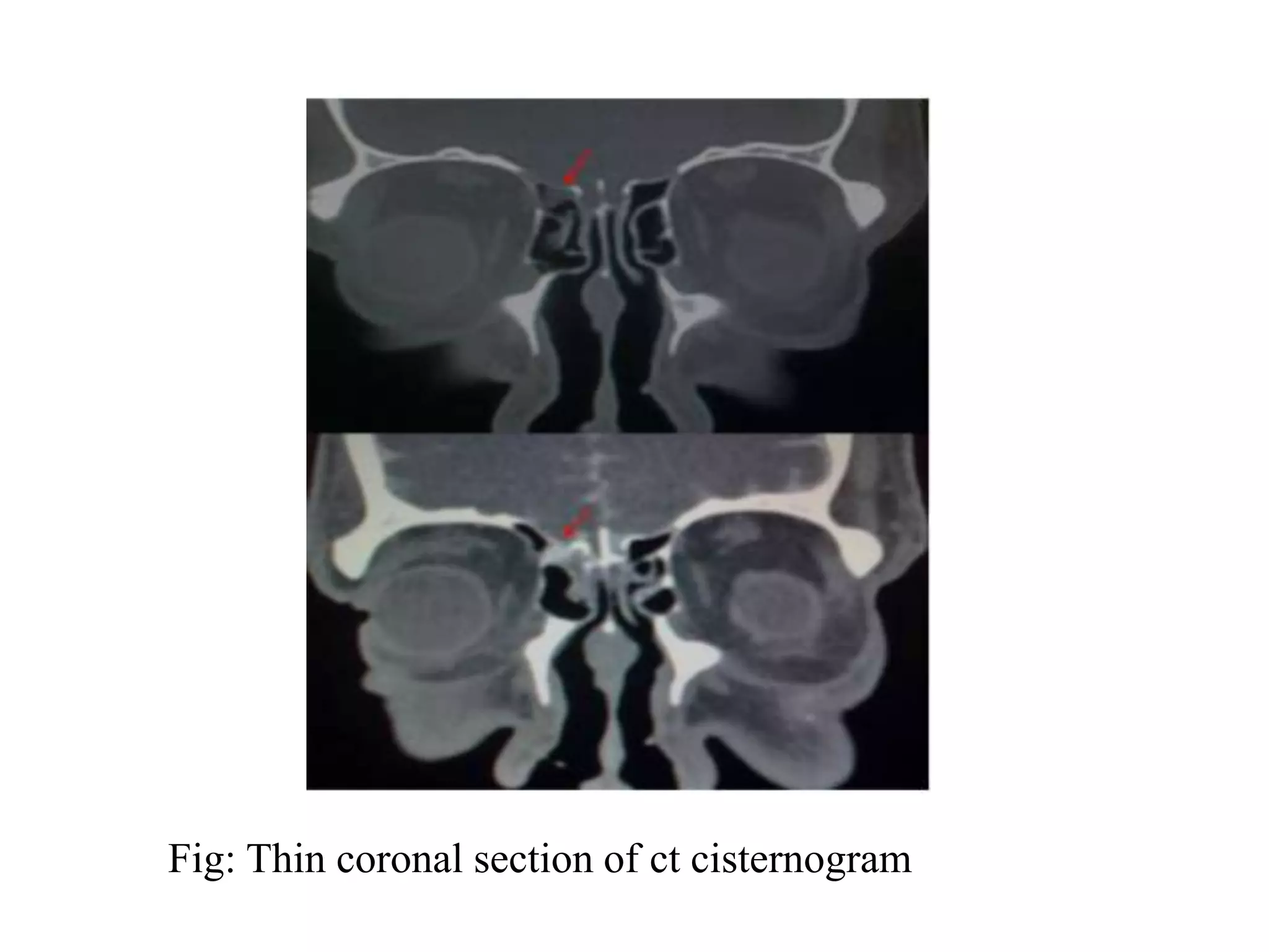
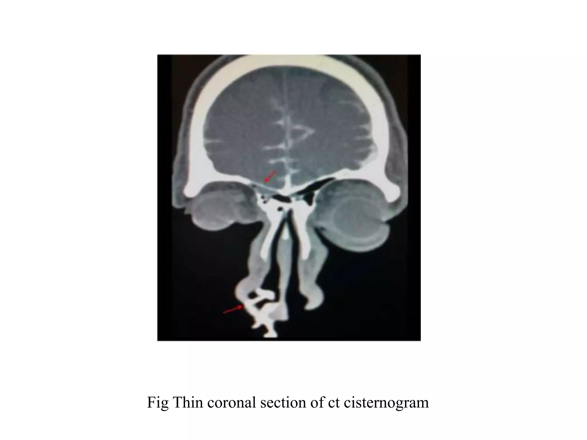
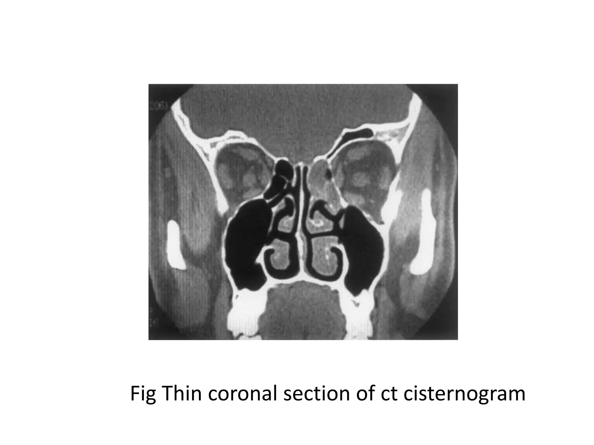
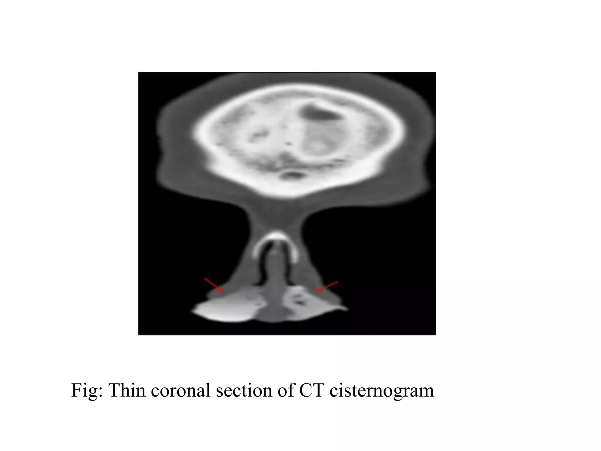
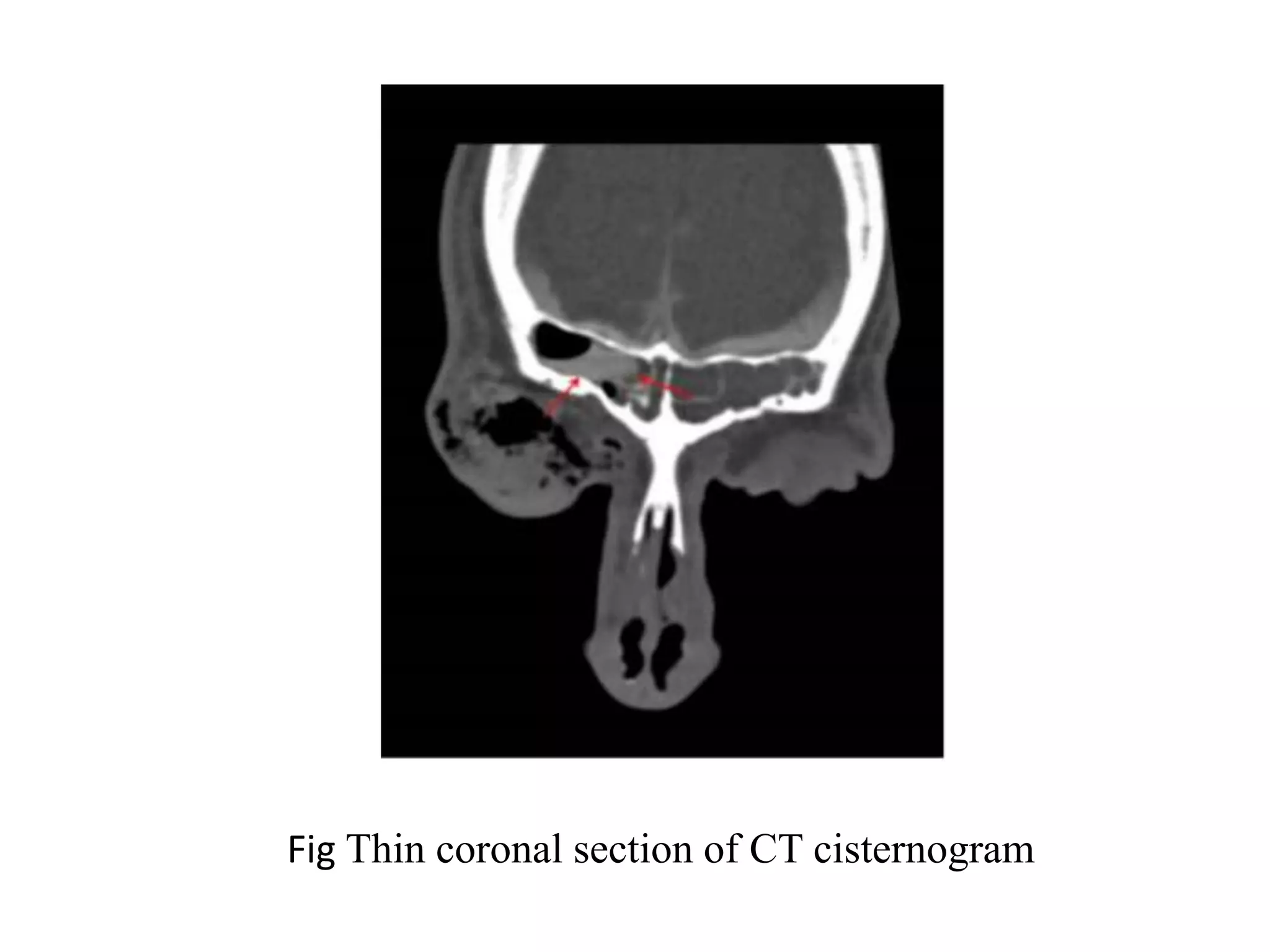
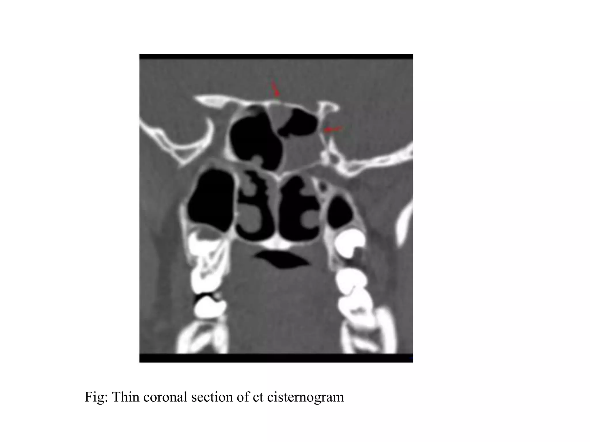
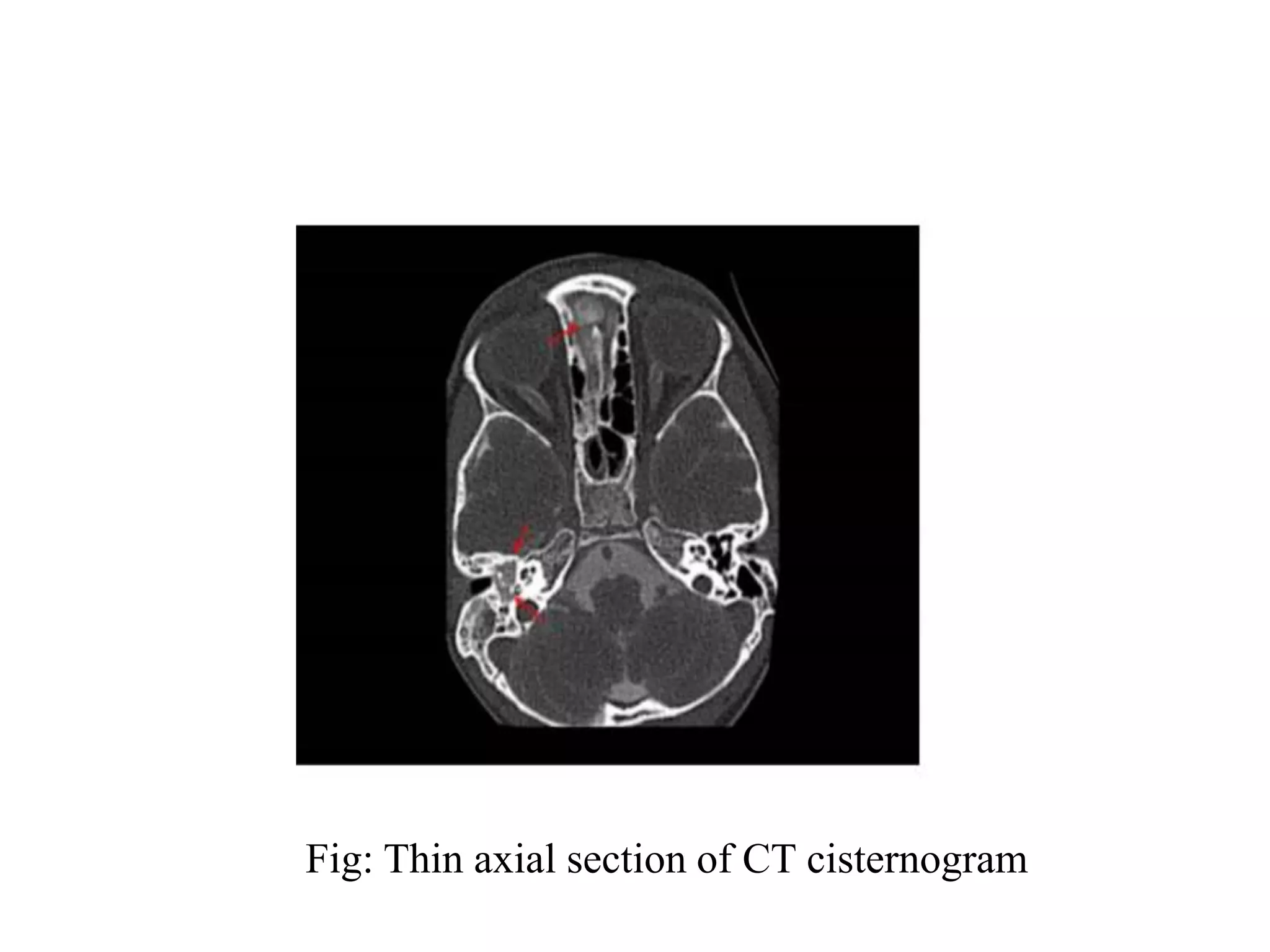
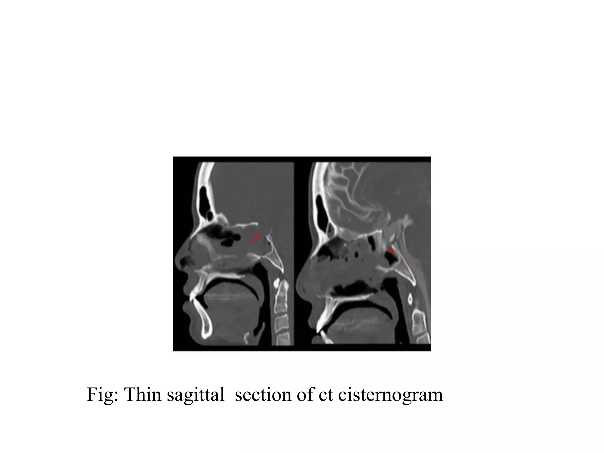
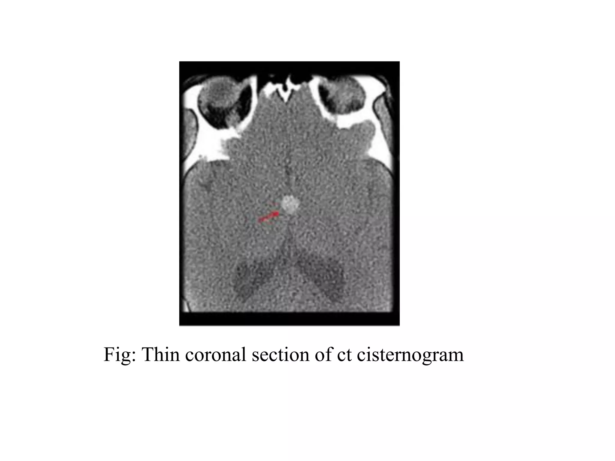
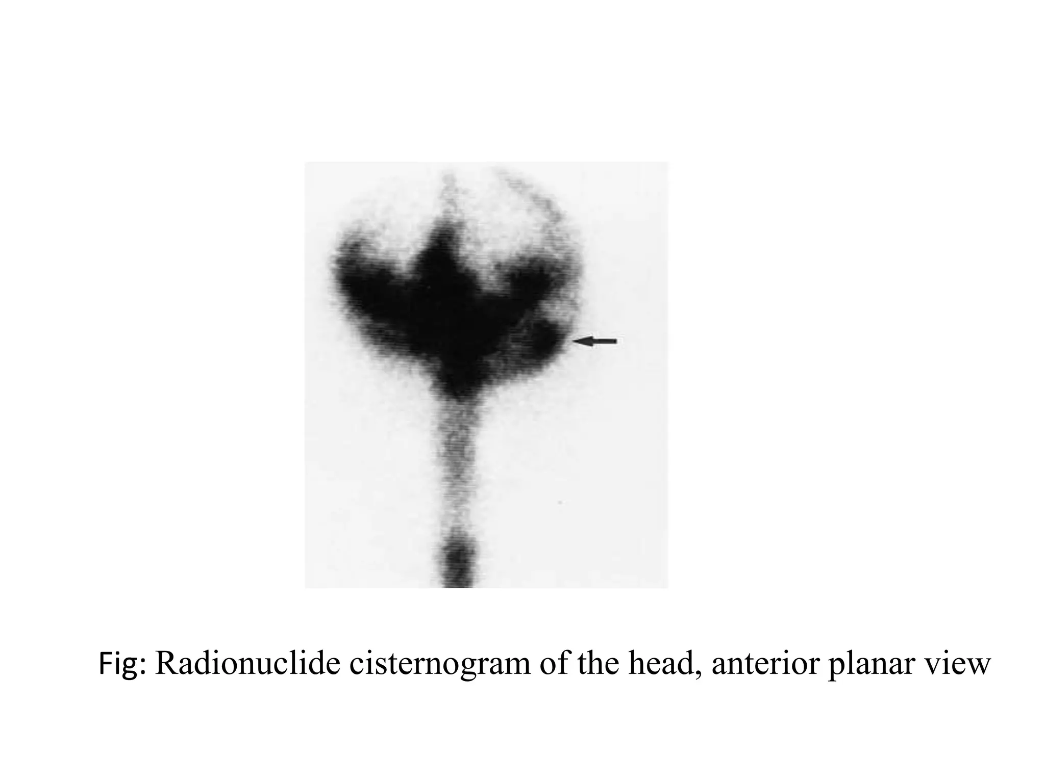
Cisternography
Presenter: Sujan Karki
B. Sc. MIT 2nd year
National Academy of Medical Sciences, Bir
Hospital
Overview
• Introduction
• Anatomy
• Indications and Contraindications
• Patient preparation, procedure, post care
• Technique for lumber puncture
• Procedure
• References
Introduction
• Radiological identification of
location of the CSF leakage is
important for proper surgical
planning and increase the
chance of dural repair.
• CSF leakage implies abnormal
communication between the
subarachnoid space and the
nasal or middle ear cavity. It is
generally classified as
traumatic, non traumatic or
post surgical in origin.
History
• CSF leak was first described by Galen in the
second century as a normal ,periodic release of
CSF into the nose via the sella turcica and
ethmoid regions.
• This process was accepted as a physiological
communication for many centuries until the late
1800s when St. Clair Thompson described as
termed it as rhinorrhea.
A CSF leak can be identified by various
modalities,they are as follows:
-MR Cisternography
-CT Cisternography
-Combination of MRI and HRCT temporal bone
-Nasal endoscopy
-Radionuclide Cisternography
Traumatic leakage
• Post traumatic CSF leak accounts for nearly
90% of all the cases, with approximately 80%
manifesting as rhinorrhea and 20% as otorrhea.
• Included in the category of traumatic are CSF
leaks are postoperative or iatrogenic defects.
• The most common sites of injury involves the
anterior cranial fossa, with fracture through the
frontal sinus or cribriform plates of ethmoid
bones.
Non- Traumatic leakage
• The non traumatic group is associated to brain
tumors (intracranial and extracranial tumors
,cholesteatoma, or tuberculoma which are
known to erode the bone directly.)
• Skull based congenital defects and
meningoceles or meningoencephocles.
SUBARACHNOID CISTERNS
• The subarachnoid cisterns are spaces within
the subarachnoid space where the pia mater
and arachnoid membrane are not in close
approximation.
• Subarachnoid space and cerebrospinal fluid
(CSF) gathers to form pools or cisterns (Latin:
“box”). Being within the subarachnoid space,
the cisterns may have vessels and/or cranial
nerves passing through them.
Major Cisterns of the brain
• cisterna magna: the largest of the subarachnoid
cisterns
• prepontine cistern: anterior to the pons
• suprasellar cistern: surrounding the infundibulum
• interpeduncular cistern: between the cerebral
crura
• quadrigeminal cistern: superior cistern or cistern of
the great cerebral vein
• ambient cistern: surrounds the back of the
midbrain
• sylvian cistern: superficial to the insular cortex
CISTERNA MAGNA
• largest of the subarachnoid
cisterns.
• between the cerebellum and the
dorsal surface of the medulla
oblongata at and above the level
of the foramen magnum.
• vessels and nerves course
through the lateral part of the
cistern:
• vertebral arteries
• posterior inferior cerebellar
arteries (PICA)
• glossopharyngeal nerve (CN IX)
• vagus nerve (CN X)
• spinal accessory nerve (CN XI)
Prepontine cistern
• occupies a large space on the
ventral aspect of the pons and
contains the basilar artery.
• continuous with the
quadrigeminal plate cistern and
cisterna magna posteriorly, and
the interpeduncular cistern
anterosuperiorly.
• vessels and nerves course
through the cistern:
• basilar artery
• multiple cranial nerves: CN V to
CN XII
Suprasellar cistern/Chiasmatic cistern
• located above the sella
turcica, under the
hypothalamus and between
the uncus of the temporal
lobes.
• contains the proximal part of
Sylvian fissure, the optic
chiasm, the infundibular stalk
and the cerebrovascular
circle of Willis.
• continuous posteriorly with
the interpeduncular cistern.
Interpeduncular cistern
• cavity between the two
temporal lobes
anteriorly and encloses
the cerebral peduncles,
within the
interpedunclar fossa.
• continuous with the
pontine cistern
inferiorly.
• contains – oculomotor
nerve (CN III) and
terminal basilar artery.
Quadrigeminal cistern
• located between the colliculi,
the splenium of the corpus
callosum and the superior
surface of the cerebellum and
extends from the third ventricle
to the great cerebral vein.
• contains several vessels and
nerves:
-posterior cerebral arteries
-posterior choroidal arteries
-superior cerebellar arteries
-trochlear nerve (CN IV)
-inferior sagittal sinus
-straight sinus
Cerebrospinal fluid
secreted by the choroid plexus - 500 ml per day, 0.35 ml
per minute, the usual volume is 150 ml.
Formation occurs as a result of a two-step process.
Fluid is first filtered through the core capillaries of the
choroid plexus into the extracellular space surrounding
the choroidal cells. This fluid is a plasma ultrafiltrate.
Then sodium is actively transported by sodium–
potassium activated adenosine triphosphatase (ATPase)
across the choroidal cells into the CSF; water follows
down an osmotic: gradient.
Cholinergic stimulation increases production and
adrenergic stimulation decreases production.
Some CSF is apparently produced in the ependyma of
the brain, in addition to the choroid plexus.
Function of CSF
• Nourishment of nervous tissue
• Removal of waste product associated with
neuronal activity
• Fluid cushion to protect brain and spinal cord
from injury
• Regulation of ICP
• protection
•
Formation
• Mostly from choroid plexuses of lateral
ventricles, 3rd ventricle and 4th ventricle.
• Small amount derived from interstitial spaces
of brain through ependymal epithelium and
from the blood capillaries of pia arachnoid.
Arachnoid villi(pacchionian bodies)
• Small granular bodies along venous sinuses
especially .
• Represent invasion of dura by arachnoid
membrane, covered by endothelium of
venous sinuses.
Sites of leakage
1. CSF from anterior cranial fossa reaches the nose
via
A. Cribriform plate
B. Roof of ethmoid
C. Frontal sinus
2. CSF from middle cranial fossa
A. Injuries to sphenoid sinus
B. In fracture of temporal bone ( middle ear-
Eustachian tube-nose)
FIG: Coronal image showing cribriform plate
FIG: Sagittal CT showing the sites of perisellar CSF leak
Laboratory
• Performance of Beta-2 transferrin test of the fluid
should be conducted prior to the procedure.
• Beta-2 transferrin is a carbohydrate-free isoform
of transferrin, which is almost exclusively found
in the CSF and blood or nasal secretion does not
disturb the test .
• Beta-2 transferrin is not present in blood, nasal
mucus, tears or mucosal discharge. This protein
was first described by Irjala et al in 1979 .
Major indications of cisternography
• Trauma
• Cerebrospinal leakage
• Orthostatic headache
• Spontaneous intracranial hypotension
• Diagnostic evaluation of spinal or basal
cisternal disease.
• Cerebrospinal blockages
• Cerebrospinal circulation problems
• Normal-pressure hydrocephalus
Contraindications
• Known significant intracranial process with
increased intracranial pressure
• Historical or laboratory evidence of bleeding
disorder or coagulopathy
• Recent myelography performed within 1
week
• Previous surgical procedure in anticipated
puncture site (can choose alternative puncture
site)
Cont….
• History of significant adverse reaction to
iodinated contrast media
• History of seizures (patient may be
premedicated)
• Localized infection at region of puncture site
• Pregnancy
Equipment and accessories
• High-quality radiographic/fluoroscopic imaging
equipment, film or digital records of the
examination, and a tilt table. The tilt table should
be capable of –30 degrees of tilt in the head
downward direction. A proper support device for
securing the patient on the tilt table should be
available.
• A CT scanner to perform CT cisternography
studies. Multiplanar reconstruction capability for
CT is highly desirable.
Materials required
• Betadine
• Alcohol
• Antiseptic tape
• 2% xylocaine
• Contrast
• 16G monoject needles(spinal needle)
Monoject needle
Patient preparation
• Patient must be NPO (no food) six hours prior
to the lumbar puncture.
• The patient should be adequately hydrated
• The patient should be observed following the
examination.
• The patient should be properly instructed
regarding limitations following the procedure
(e.g., driving)
• Remove all the dentures and nasal ornaments
that can produce artifacts
• Clean nose thoroughly prior to the post-thecal
injection of contrast so that even minute
leakage of contrast can be picked up in nasal
cavity
• Instructions regarding post procedural care,
including warning signs of adverse reactions
and the possibility of persistent headaches,
should be given to the patient. The instructions
should include a recommendation that the
patient should be in the company of a
responsible adult for 12 hours following the
procedure.
• A physician should be available to answer
questions and provide patient management
following the procedure.
Consent , procedure and patient care
• Written and verbal consent should be taken.
• A medical history should be obtained before the
procedure is begun.
• This should include questions intended to assess
the patient stability to tolerate the contrast
injection (eg: allergy history , cardiac /
pulmonary status )
• the patient also will be interviewed regarding
medication history and symptoms
Cont…
• Medication history is important because some
medications are anti-coagulants and will cause
excessive bleeding during and after the
procedure.
• Knowing the medication history is also important
when one is selecting the premedication.
• Previous laboratory reports and other data are
reviewed as well .
Cont…
• A detailed explanation of the procedure will be
given to the patient , which is important to
ensure full understanding and cooperation .
• The explanation will include possible risks and
complications of the procedure , so that the
patient is fully informed before signing the
consent .
• Vital signs are obtained and recorded .
• Continual communication and monitoring of
the patient by the technologist should be done.
• Then the contrast can be injected in the lumber
arachnoids space and the patient is asked to lie
down in trendelenburg’s position(head low,
leg up) for 20 to 30 minutes
•
Technique For lumber puncture
• Position of patient
• Usually placed on their side(left more
common then right)
• Patient bends the neck so the chin is closed
to the chest, hunches the back, and brings
knee towards the chest(same as fetal
position)
• Also can be done on sitting position on a
stool and bend their head and shoulder
forward.
• Area around the lower back is prepared using
aseptic technique
• Appropriate location is palpated and local
anesthesia is infiltrated under the skin
• Spinal needle is inserted between lumber
vertebrae(L3/L4)
• The spinal needle is inserted upto the sub-
arachnoid space
• The stylet from the spinal needle is then
withdrawn and drops of CSF is collected
• Normal CSF pressure measured by manometer
is 50-150 mm H2O.(8-10 mmHg)
Lumber puncture
Contrast Media
• Non ionic contrast media is used
• Non ionic dimer is more viscous so LOCM
non ionic monomer is the choice(metrizimide)
• Concentration: 300mgI/ml
• Volume :10-15 ml
Patient positioning for CT
cisternography
• Patient is kept prone on the couch with head
first
• Arms are kept beside the trunk
• Neck is hyper extended
• Scan delay- 20-30min
• The use of head rest and chin rest is
recommended for positioning
• Baseline non contrast scans should be taken in
helical mode and reconstructed with the same
slice thickness and interval
• Ensure that the patient does not move in between
the noncontrast and contrast enhanced scans
Technical considerations
• Topogram/landmark: lateral 2-3 cm anterior
to the tip of the nose
• Mode of scanning: helical
• Scan orientation: posterior to anterior
starting location- level of clivus
end location- anterior part of the nasal
cavity anterior to the frontal sinus
• Gantry tilt: required degree to make scanning
parallel to face or perpendicular to the hard
palate
• FOV- include the region of interest only.
• Contrast administration- intrathecal or
cisternal
• In the case of coexistent spinal block or other
pathology that is responsible for poor contrast in
cisterns, cisternal puncture can be done for
injection of the contrast.
• Volume of contrast- 10-15 ml
• Slice thickness in reconstruction- 1.0 to
1.5mm
• Slice interval in reconstruction- 0.5 to
0.75mm
• Reconstruction algorithm/kernel- medium
smooth for the soft tissues and sharp for the
bones
• 3D reconstructions- MPR, MIP
Criteria of image quality
• Symmetric position with the orbital plates
overlapping with each other.
• Absence of the motion artifacts.
• Absence of beam hardening.
• Optimal opacification of the cisterns and the
sulcal spaces.
Fig: Thin coronal section of ct cisternogram
Fig Thin coronal section of ct cisternogram
Fig: CT Coronal view
Fig: Thin coronal section of ct cisternogram
Fig Thin coronal section of ct cisternogram
Fig: Thin coronal section of CT cisternogram
Fig Thin coronal section of CT cisternogram
Fig: Thin coronal section of ct cisternogram
Fig: Thin axial section of CT cisternogram
Fig: Thin sagittal section of ct cisternogram
Fig: Thin coronal section of ct cisternogram
Fig: Radionuclide cisternogram of the head, anterior planar view
• Gd-enhanced MR cisternography (Gd-MRC) to confirm and
localize CSF leaks in all patients.
• All lumbar puncture procedures were performed by using a
22-gauge needle at the L4-L5 level under sterile conditions
with the patient sitting in an upright position.
• Saline (4 mL) was mixed with 0.5 mL of gadopentetate
dimeglumine (Gd-DTPA or Magnevist to produce a solution
of 469.01 mg/mL.
• This solution was injected into the subarachnoid space, and
the needle was removed.
• The patients remained in the knee-to-elbow position for 15
minutes after injection to maximize accumulation of the
contrast medium in the basal cisterns and to facilitate its
passage into the CSF fistula.
MR Cisternography Procedure
Contd.
• One hour after injection, the patient was moved
into a prone position and fat-saturated T1-
weighted images were obtained in 3 orthogonal
planes.
• Coronal and sagittal T1-weighted images (TR/TE,
500/17 ms; 2 signals acquired) and axial T1-
weighted images (TR/TE, 600/17 ms; 2 signals
acquired) were obtained by using a 1.5T MR
imaging unit
• In cases in which the CSF leak could not be
accurately visualized on MR images, additional
scans were obtained in the third and fifth hours.
Sequences used for”MRI
cisternography protocols
• Patient position prone on a head coil
• Following sequences are taken
• T2 axial/coronal
• Heavily weighted T2 coronal to localize the site
of leakage(BASG,DRIVE,CISS)
• T2 fat sat
T1 weighted sequence (axial or coronal to
characterize any leasons)
Fig:Coronal T1-weighted fat-saturated MR
FIG:Sagittal T1-weighted fat-suppressed MR image
• The main disadvantage of MRI is its poor
spatial resolution and leaks of bony details.
Thus CT and MRI seem to be complementary
in the diagnosis of CSF leaks.
• For CT sometimes difficult to differentiate the
CSF and sinus opacification and is not an
active leak or intermittent CSF leaks means
sometimes difficult to determine only in CT,
intracranial hypotension patients also MRI
more usefull.
References
1)Intrathecal Gadolinium-Enhanced MR Cisternography
in the Evaluation of CSF Leakage H. Selcuk, S. Albayram,
H. Ozer, S
2)Combined HRCT and MRI in the Detection of CSF
RhinorrheaBadr Eldin Mostafa, M.D.1 and Ahmed
Khafagi, M.D
3)Current Approach to Cerebrospinal Fluid Rhinorrhea
Diagnosis and Management Ibrahim Sumaily,
Otorhinolaryngologist, Asir central Hospital, Abha, KSA,
Saudi Journal of Otolaryngology-ENT Research
4)www.myESR.com
5)Radiopedia.org
Questions
• list the Major indication for cisternogram?
• List the major cisterns of the brain?
• Which are the most common site of csf leakage?
• Which test is gold standerd to confirm that the
nasal secretion is csf and why?
• Why is patient kept in trendelenburg’s position
after the intrathecal administration contrast media?

Cisternography sujan

  • 1.
    Cisternography Presenter: Sujan Karki B.Sc. MIT 2nd year National Academy of Medical Sciences, Bir Hospital
  • 2.
    Overview • Introduction • Anatomy •Indications and Contraindications • Patient preparation, procedure, post care • Technique for lumber puncture • Procedure • References
  • 3.
    Introduction • Radiological identificationof location of the CSF leakage is important for proper surgical planning and increase the chance of dural repair. • CSF leakage implies abnormal communication between the subarachnoid space and the nasal or middle ear cavity. It is generally classified as traumatic, non traumatic or post surgical in origin.
  • 4.
    History • CSF leakwas first described by Galen in the second century as a normal ,periodic release of CSF into the nose via the sella turcica and ethmoid regions. • This process was accepted as a physiological communication for many centuries until the late 1800s when St. Clair Thompson described as termed it as rhinorrhea.
  • 5.
    A CSF leakcan be identified by various modalities,they are as follows: -MR Cisternography -CT Cisternography -Combination of MRI and HRCT temporal bone -Nasal endoscopy -Radionuclide Cisternography
  • 6.
    Traumatic leakage • Posttraumatic CSF leak accounts for nearly 90% of all the cases, with approximately 80% manifesting as rhinorrhea and 20% as otorrhea. • Included in the category of traumatic are CSF leaks are postoperative or iatrogenic defects. • The most common sites of injury involves the anterior cranial fossa, with fracture through the frontal sinus or cribriform plates of ethmoid bones.
  • 7.
    Non- Traumatic leakage •The non traumatic group is associated to brain tumors (intracranial and extracranial tumors ,cholesteatoma, or tuberculoma which are known to erode the bone directly.) • Skull based congenital defects and meningoceles or meningoencephocles.
  • 8.
    SUBARACHNOID CISTERNS • Thesubarachnoid cisterns are spaces within the subarachnoid space where the pia mater and arachnoid membrane are not in close approximation. • Subarachnoid space and cerebrospinal fluid (CSF) gathers to form pools or cisterns (Latin: “box”). Being within the subarachnoid space, the cisterns may have vessels and/or cranial nerves passing through them.
  • 9.
    Major Cisterns ofthe brain • cisterna magna: the largest of the subarachnoid cisterns • prepontine cistern: anterior to the pons • suprasellar cistern: surrounding the infundibulum • interpeduncular cistern: between the cerebral crura • quadrigeminal cistern: superior cistern or cistern of the great cerebral vein • ambient cistern: surrounds the back of the midbrain • sylvian cistern: superficial to the insular cortex
  • 10.
    CISTERNA MAGNA • largestof the subarachnoid cisterns. • between the cerebellum and the dorsal surface of the medulla oblongata at and above the level of the foramen magnum. • vessels and nerves course through the lateral part of the cistern: • vertebral arteries • posterior inferior cerebellar arteries (PICA) • glossopharyngeal nerve (CN IX) • vagus nerve (CN X) • spinal accessory nerve (CN XI)
  • 11.
    Prepontine cistern • occupiesa large space on the ventral aspect of the pons and contains the basilar artery. • continuous with the quadrigeminal plate cistern and cisterna magna posteriorly, and the interpeduncular cistern anterosuperiorly. • vessels and nerves course through the cistern: • basilar artery • multiple cranial nerves: CN V to CN XII
  • 12.
    Suprasellar cistern/Chiasmatic cistern •located above the sella turcica, under the hypothalamus and between the uncus of the temporal lobes. • contains the proximal part of Sylvian fissure, the optic chiasm, the infundibular stalk and the cerebrovascular circle of Willis. • continuous posteriorly with the interpeduncular cistern.
  • 13.
    Interpeduncular cistern • cavitybetween the two temporal lobes anteriorly and encloses the cerebral peduncles, within the interpedunclar fossa. • continuous with the pontine cistern inferiorly. • contains – oculomotor nerve (CN III) and terminal basilar artery.
  • 14.
    Quadrigeminal cistern • locatedbetween the colliculi, the splenium of the corpus callosum and the superior surface of the cerebellum and extends from the third ventricle to the great cerebral vein. • contains several vessels and nerves: -posterior cerebral arteries -posterior choroidal arteries -superior cerebellar arteries -trochlear nerve (CN IV) -inferior sagittal sinus -straight sinus
  • 15.
    Cerebrospinal fluid secreted bythe choroid plexus - 500 ml per day, 0.35 ml per minute, the usual volume is 150 ml. Formation occurs as a result of a two-step process. Fluid is first filtered through the core capillaries of the choroid plexus into the extracellular space surrounding the choroidal cells. This fluid is a plasma ultrafiltrate. Then sodium is actively transported by sodium– potassium activated adenosine triphosphatase (ATPase) across the choroidal cells into the CSF; water follows down an osmotic: gradient. Cholinergic stimulation increases production and adrenergic stimulation decreases production. Some CSF is apparently produced in the ependyma of the brain, in addition to the choroid plexus.
  • 16.
    Function of CSF •Nourishment of nervous tissue • Removal of waste product associated with neuronal activity • Fluid cushion to protect brain and spinal cord from injury • Regulation of ICP • protection •
  • 17.
    Formation • Mostly fromchoroid plexuses of lateral ventricles, 3rd ventricle and 4th ventricle. • Small amount derived from interstitial spaces of brain through ependymal epithelium and from the blood capillaries of pia arachnoid.
  • 19.
    Arachnoid villi(pacchionian bodies) •Small granular bodies along venous sinuses especially . • Represent invasion of dura by arachnoid membrane, covered by endothelium of venous sinuses.
  • 22.
    Sites of leakage 1.CSF from anterior cranial fossa reaches the nose via A. Cribriform plate B. Roof of ethmoid C. Frontal sinus 2. CSF from middle cranial fossa A. Injuries to sphenoid sinus B. In fracture of temporal bone ( middle ear- Eustachian tube-nose)
  • 23.
    FIG: Coronal imageshowing cribriform plate
  • 24.
    FIG: Sagittal CTshowing the sites of perisellar CSF leak
  • 27.
    Laboratory • Performance ofBeta-2 transferrin test of the fluid should be conducted prior to the procedure. • Beta-2 transferrin is a carbohydrate-free isoform of transferrin, which is almost exclusively found in the CSF and blood or nasal secretion does not disturb the test . • Beta-2 transferrin is not present in blood, nasal mucus, tears or mucosal discharge. This protein was first described by Irjala et al in 1979 .
  • 28.
    Major indications ofcisternography • Trauma • Cerebrospinal leakage • Orthostatic headache • Spontaneous intracranial hypotension • Diagnostic evaluation of spinal or basal cisternal disease. • Cerebrospinal blockages • Cerebrospinal circulation problems • Normal-pressure hydrocephalus
  • 29.
    Contraindications • Known significantintracranial process with increased intracranial pressure • Historical or laboratory evidence of bleeding disorder or coagulopathy • Recent myelography performed within 1 week • Previous surgical procedure in anticipated puncture site (can choose alternative puncture site)
  • 30.
    Cont…. • History ofsignificant adverse reaction to iodinated contrast media • History of seizures (patient may be premedicated) • Localized infection at region of puncture site • Pregnancy
  • 31.
    Equipment and accessories •High-quality radiographic/fluoroscopic imaging equipment, film or digital records of the examination, and a tilt table. The tilt table should be capable of –30 degrees of tilt in the head downward direction. A proper support device for securing the patient on the tilt table should be available. • A CT scanner to perform CT cisternography studies. Multiplanar reconstruction capability for CT is highly desirable.
  • 32.
    Materials required • Betadine •Alcohol • Antiseptic tape • 2% xylocaine • Contrast • 16G monoject needles(spinal needle)
  • 33.
  • 34.
    Patient preparation • Patientmust be NPO (no food) six hours prior to the lumbar puncture. • The patient should be adequately hydrated • The patient should be observed following the examination. • The patient should be properly instructed regarding limitations following the procedure (e.g., driving)
  • 35.
    • Remove allthe dentures and nasal ornaments that can produce artifacts • Clean nose thoroughly prior to the post-thecal injection of contrast so that even minute leakage of contrast can be picked up in nasal cavity
  • 36.
    • Instructions regardingpost procedural care, including warning signs of adverse reactions and the possibility of persistent headaches, should be given to the patient. The instructions should include a recommendation that the patient should be in the company of a responsible adult for 12 hours following the procedure. • A physician should be available to answer questions and provide patient management following the procedure.
  • 37.
    Consent , procedureand patient care • Written and verbal consent should be taken. • A medical history should be obtained before the procedure is begun. • This should include questions intended to assess the patient stability to tolerate the contrast injection (eg: allergy history , cardiac / pulmonary status ) • the patient also will be interviewed regarding medication history and symptoms
  • 38.
    Cont… • Medication historyis important because some medications are anti-coagulants and will cause excessive bleeding during and after the procedure. • Knowing the medication history is also important when one is selecting the premedication. • Previous laboratory reports and other data are reviewed as well .
  • 39.
    Cont… • A detailedexplanation of the procedure will be given to the patient , which is important to ensure full understanding and cooperation . • The explanation will include possible risks and complications of the procedure , so that the patient is fully informed before signing the consent .
  • 40.
    • Vital signsare obtained and recorded . • Continual communication and monitoring of the patient by the technologist should be done. • Then the contrast can be injected in the lumber arachnoids space and the patient is asked to lie down in trendelenburg’s position(head low, leg up) for 20 to 30 minutes •
  • 41.
    Technique For lumberpuncture • Position of patient • Usually placed on their side(left more common then right) • Patient bends the neck so the chin is closed to the chest, hunches the back, and brings knee towards the chest(same as fetal position) • Also can be done on sitting position on a stool and bend their head and shoulder forward.
  • 42.
    • Area aroundthe lower back is prepared using aseptic technique • Appropriate location is palpated and local anesthesia is infiltrated under the skin • Spinal needle is inserted between lumber vertebrae(L3/L4) • The spinal needle is inserted upto the sub- arachnoid space
  • 43.
    • The styletfrom the spinal needle is then withdrawn and drops of CSF is collected • Normal CSF pressure measured by manometer is 50-150 mm H2O.(8-10 mmHg)
  • 44.
  • 45.
    Contrast Media • Nonionic contrast media is used • Non ionic dimer is more viscous so LOCM non ionic monomer is the choice(metrizimide) • Concentration: 300mgI/ml • Volume :10-15 ml
  • 46.
    Patient positioning forCT cisternography • Patient is kept prone on the couch with head first • Arms are kept beside the trunk • Neck is hyper extended • Scan delay- 20-30min
  • 47.
    • The useof head rest and chin rest is recommended for positioning • Baseline non contrast scans should be taken in helical mode and reconstructed with the same slice thickness and interval • Ensure that the patient does not move in between the noncontrast and contrast enhanced scans
  • 48.
    Technical considerations • Topogram/landmark:lateral 2-3 cm anterior to the tip of the nose • Mode of scanning: helical • Scan orientation: posterior to anterior starting location- level of clivus end location- anterior part of the nasal cavity anterior to the frontal sinus • Gantry tilt: required degree to make scanning parallel to face or perpendicular to the hard palate
  • 49.
    • FOV- includethe region of interest only. • Contrast administration- intrathecal or cisternal • In the case of coexistent spinal block or other pathology that is responsible for poor contrast in cisterns, cisternal puncture can be done for injection of the contrast.
  • 50.
    • Volume ofcontrast- 10-15 ml • Slice thickness in reconstruction- 1.0 to 1.5mm • Slice interval in reconstruction- 0.5 to 0.75mm • Reconstruction algorithm/kernel- medium smooth for the soft tissues and sharp for the bones • 3D reconstructions- MPR, MIP
  • 51.
    Criteria of imagequality • Symmetric position with the orbital plates overlapping with each other. • Absence of the motion artifacts. • Absence of beam hardening. • Optimal opacification of the cisterns and the sulcal spaces.
  • 52.
    Fig: Thin coronalsection of ct cisternogram
  • 53.
    Fig Thin coronalsection of ct cisternogram
  • 54.
  • 55.
    Fig: Thin coronalsection of ct cisternogram
  • 56.
    Fig Thin coronalsection of ct cisternogram
  • 57.
    Fig Thin coronalsection of ct cisternogram
  • 58.
    Fig: Thin coronalsection of CT cisternogram
  • 59.
    Fig: Thin coronalsection of CT cisternogram
  • 60.
    Fig Thin coronalsection of CT cisternogram
  • 61.
    Fig: Thin coronalsection of ct cisternogram
  • 62.
    Fig: Thin axialsection of CT cisternogram
  • 63.
    Fig: Thin sagittalsection of ct cisternogram
  • 64.
    Fig: Thin coronalsection of ct cisternogram
  • 65.
    Fig: Radionuclide cisternogramof the head, anterior planar view
  • 66.
    • Gd-enhanced MRcisternography (Gd-MRC) to confirm and localize CSF leaks in all patients. • All lumbar puncture procedures were performed by using a 22-gauge needle at the L4-L5 level under sterile conditions with the patient sitting in an upright position. • Saline (4 mL) was mixed with 0.5 mL of gadopentetate dimeglumine (Gd-DTPA or Magnevist to produce a solution of 469.01 mg/mL. • This solution was injected into the subarachnoid space, and the needle was removed. • The patients remained in the knee-to-elbow position for 15 minutes after injection to maximize accumulation of the contrast medium in the basal cisterns and to facilitate its passage into the CSF fistula. MR Cisternography Procedure
  • 67.
    Contd. • One hourafter injection, the patient was moved into a prone position and fat-saturated T1- weighted images were obtained in 3 orthogonal planes. • Coronal and sagittal T1-weighted images (TR/TE, 500/17 ms; 2 signals acquired) and axial T1- weighted images (TR/TE, 600/17 ms; 2 signals acquired) were obtained by using a 1.5T MR imaging unit • In cases in which the CSF leak could not be accurately visualized on MR images, additional scans were obtained in the third and fifth hours.
  • 68.
    Sequences used for”MRI cisternographyprotocols • Patient position prone on a head coil • Following sequences are taken • T2 axial/coronal • Heavily weighted T2 coronal to localize the site of leakage(BASG,DRIVE,CISS) • T2 fat sat T1 weighted sequence (axial or coronal to characterize any leasons)
  • 69.
  • 70.
  • 71.
    • The maindisadvantage of MRI is its poor spatial resolution and leaks of bony details. Thus CT and MRI seem to be complementary in the diagnosis of CSF leaks. • For CT sometimes difficult to differentiate the CSF and sinus opacification and is not an active leak or intermittent CSF leaks means sometimes difficult to determine only in CT, intracranial hypotension patients also MRI more usefull.
  • 72.
    References 1)Intrathecal Gadolinium-Enhanced MRCisternography in the Evaluation of CSF Leakage H. Selcuk, S. Albayram, H. Ozer, S 2)Combined HRCT and MRI in the Detection of CSF RhinorrheaBadr Eldin Mostafa, M.D.1 and Ahmed Khafagi, M.D 3)Current Approach to Cerebrospinal Fluid Rhinorrhea Diagnosis and Management Ibrahim Sumaily, Otorhinolaryngologist, Asir central Hospital, Abha, KSA, Saudi Journal of Otolaryngology-ENT Research 4)www.myESR.com 5)Radiopedia.org
  • 73.
    Questions • list theMajor indication for cisternogram? • List the major cisterns of the brain? • Which are the most common site of csf leakage? • Which test is gold standerd to confirm that the nasal secretion is csf and why? • Why is patient kept in trendelenburg’s position after the intrathecal administration contrast media?

Editor's Notes

  • #16  CSF is derived from blood plasma and is largely similar to it, except that CSF is nearly protein-free compared with plasma 
  • #24  Spontaneous CSF fistulas may occur anywhere in the skull base, but they are more common at the cribriform plate, tegmen tympani or sphenoid sinus. Redline :lamella lateralis(a thin vertical strut joining the medial aspect of ethmoidal roof with cribriform plate.)
  • #25  Thin green :tuberculum sellae Thick red arrow:sellar floor Thick orange:spheno-occipital synchondrosis