Downloaded 825 times

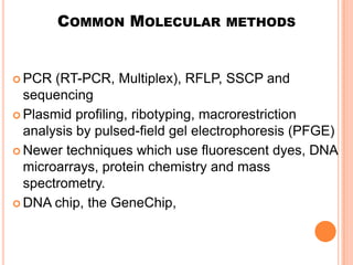
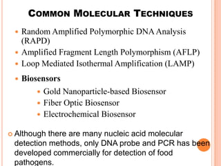


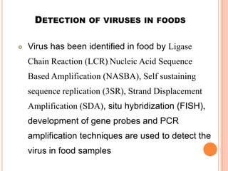


This document provides an overview of molecular detection techniques used in food quality control. It discusses how chemistry alone cannot solve all detection problems and that molecular biology methods like PCR, RFLP, and sequencing are better alternatives as they are more accurate, rapid and cost-effective. It describes several common molecular detection methods and their applications in detecting food pathogens, adulterants, allergens and GM ingredients. The document emphasizes that molecular methods can identify microbes at the strain level and detect viable cells, but may not be able to find non-authorized GMOs due to lack of molecular information.


