SlideShare a Scribd company logo
1 of 10
Download to read offline
FORMULATION AND EVALUATION OF MICROSPHERES 
INTRODUCTION: 
• A well designed controlled drug delivery system can overcome some of the problems 
of conventional therapy and enhance the therapeutic efficacy of a given drug. 
• To achieve maximum therapeutic efficacy, it becomes necessary to deliver the agent 
to the target tissue in the optimal amount in the right period of time there by causing 
little toxicity and minimal side effects. 
• There are various approaches in delivering a therapeutic substance to the target site in 
a sustained controlled release fashion. One such approach is using microspheres as 
carriers for drugs. 
• Microspheres are characteristically free flowing powders consisting of proteins or 
synthetic polymers which are biodegradable in nature. 
• Microspheres are defined as “Monolithic sphere or therapeutic agent distributed 
throughout the matrix either as a molecular dispersion of particles” or can be defined 
as structure made up of continuous phase of one or more miscible polymers in which 
drug particles are dispersed at the molecular level or macroscopic level. 
• It has particle size of 1-1000 nm. 
• Due to smaller particle size it spreads to a large area in gastrointestinal tract which 
improves drug absorption and reduces side effects due to localized buildup of 
irritating drugs against the gastrointestinal mucosa. 
TYPES OF MICROSPHERES: 
• Bio-adhesive microspheres: 
Adhesion can be defined as sticking of drug to the membrane by using the sticking 
property of the water soluble polymers. Adhesion of drug delivery device to the 
mucosal membrane such as buccal, ocular, rectal, nasal etc can be termed as bio-adhesion. 
These kinds of microspheres exhibit a prolonged residence time at the site 
of application and causes intimate contact with the absorption site and produces better 
therapeutic action. 
• Magnetic microspheres: 
This kind of delivery system is very much important which localises the drug to the 
disease site. In this larger amount of freely circulating drug can be replaced by smaller 
amount of magnetically targeted drug. Magnetic carriers receive magnetic responses 
to a magnetic field from incorporated materials that are used for magnetic 
microspheres. 
• Floating microspheres: 
In floating types the bulk density is less than the gastric fluid and so remains buoyant 
in stomach without affecting gastric emptying rate. The drug is released slowly at the
desired rate and increases gastric residence and fluctuation in plasma concentration. It 
also reduces chances of sticking and dose dumping. 
• Radioactive microspheres: 
In Radio emobilisation therapy microspheres sized of 10-30 nm are larger than 
capillaries and gets tapped in first capillary bed when they come across. They are 
injected to the arteries that lead to tumour of interest. In all these conditions 
radioactive microspheres deliver high radiation dose to the targeted areas without 
damaging the normal surrounding tissues. 
• Polymeric microspheres: 
The different types of polymeric microspheres can be classified as follows: 
a) Biodegradable polymeric microspheres: 
Natural polymers such as starch are used with the concept that they are biodegradable, 
biocompatible, and also bio adhesive in nature. Biodegradable polymers prolongs the 
residence time when contact with mucous membrane due to its high degree of 
swelling property with aqueous medium, results gel formation. 
b) Synthetic polymeric microspheres: 
The interest of synthetic polymeric microspheres are widely used in clinical 
application, moreover that also used as bulking agent, fillers, embolic particles, drug 
delivery vehicles etc and proved to be safe and biocompatible. But the main 
disadvantage of these kinds of microspheres, are tend to migrate away from injection 
site and lead to potential risk, embolism and further organ damage. 
METHODS OF PREPARATIONS 
Different methods used for various microspheres preparation depends on particle size, 
route of administration, duration of drug release and these above characters related to 
rpm, method of cross linking, drug of cross linking, evaporation time, co-precipitation 
etc. The various methods of preparations are 
1. Emulsion solvent evaporation technique 
In this technique the drug is dissolved in polymer which was previously dissolved 
in chloroform and the resulting solution is added to aqueous phase containing 
0.2% sodium of PVP as Emulsifying agent. The above mixture was agitated at 
500 rpm then the drug and polymer was transformed into fine droplet which 
solidified into rigid microspheres by solvent evaporation and then collected by 
filtration and washed with demineralised water and desiccated at room 
temperature for 24 hours. Diclofenac microspheres are prepared by this method. 
2. Emulsion cross linking method 
In this method drug was dissolved in aqueous gelatine solution which was 
previously heated for 1 hour at 40oC. The solution was added drop wise to liquid
paraffin while stirring the mixture at 1500 rpm for 10 min at 35oC, results in w/o 
emulsion then further stirring is done for 10 min at 15oC. Thus the produced 
microspheres were washed respectively three times with acetone and isopropyl 
alcohol which then air dried and dispersed in 5 mL of aqueous glutaraldehyde 
saturated toluene solution at room temperature for 3 hours for cross linking and 
then was treated with 100 mL of 10 mm glycerine solution containing 0.1% w/v 
of tween 80 at 37oC for 10 min to block unreacted glutaraldehyde. Examples for 
this technique is Gelatin A microspheres. 
3. Co-acervation method 
a) Co-acervation thermal change: 
Performed by weighed amount of ethyl cellulose was dissolved in cyclohexane 
with vigorous stirring on the above solution and phase separation was done by 
reducing temperature and using ice bath. Then above product is washed with 
cyclohexane and air dried then passed through sieve (sieve no. 40) to obtain 
individual microcapsule. 
b) Co-acervation non solvent addition: 
Developed by weighed amount of ethyl cellulose was dissolved in toluene 
containing propyl isobutylene in closed beaker with magnetic stirring for 6 hours 
at 500 rpm and the drug is dispersed in it and stirring is continued for 15 mins. 
Then phase separation is done by petroleum benzoin with continuous stirring. 
After that the microcapsules were washed with n-hexane and air dried for 2 hours 
and then in oven at 50oC for 4 hours. 
4. Spray drying technique 
It involves dispersing the core material into liquefied coating material and then 
spraying the mixture in the environment for solidification of coating followed by 
rapid evaporation of solvent. Organic solution of poly (epsilon-caprolactone) 
(PCL) and cellulose acetate butyrate (CAB), in different weight ratios and 
ketoprofen were prepared and sprayed in different experimental condition 
achieving drug loaded microspheres. 
5. Emulsion-solvent diffusion technique 
The drug polymer mixture was dissolved in a mixture of ethanol and 
dichloromethane (1:1) and then the mixture was added drop wise to sodium lauryl 
sulphate (SLS) solution. The solution was stirred with propeller type agitator at 
room temperature at 150 rpm for 1hour. Thus the formed floating microspheres 
were washed and dried in a desiccator at room temperature. The following micro 
particles were sieved and collected. 
6. Multiple emulsion method 
In the beginning powder drug was dispersed in solution (methyl cellulose) 
followed by emulsification in ethyl cellulose solution in ethyl acetate. The primary
emulsion was then re emulsified in aqueous medium. Under optimised condition 
discrete microspheres were formed during this phase. 
7. Ionic gelation 
Alginate/chitosan particulate system for Diclofenac release was prepared using 
this technique. 25% (w/v) of Diclofenac was added to 1.2 % (w/v) aqueous 
solution of sodium alginate. In order to get the complete solution stirring is 
continued and after that it was added drop wise to a solution containing Ca2+/Al3+ 
and chitosan solution in acetic acid. Microspheres which were formed were kept 
in original solution for 24 hours for internal gellification followed by filtration for 
separation. 
ADVANTAGES: 
• Controlled release delivery there by reducing side effects and eliminating the 
inconvenience of repeated injections. 
• Protein/peptide stability – microspheres helps to protect proteins because they are not 
allowed to react with anything until the polymer is degraded, thus minimizing the 
contact with solutions that could cause the proteins to react. 
Ex: albumin prototype and lyzozymes. 
• Drug targeting – it is the greatest advantage. Most drugs are targeted in the body to 
give desired results either in specific tissues or organs. 
Ex: It could be employed in targeting cancer cells in chemotherapy, as drugs and 
chemical agents attack cancer cells but have a toxic effect on healthy ones which 
could easily cause them to die. 
• Gene delivery – Encapsulation of therapeutic agents such as DNA in microspheres 
protects the agent from enzymatic degradation, enhances tissue specificity due to 
localized delivery, eliminates the need for multiple administrations and allows for 
sustained and controlled delivery. 
• Microspheres are used with Gamma emitters such as Tc99 and 1131 for diagnostic 
purposes. 
APPLICATIONS: 
• Microspheres in vaccine delivery: The prerequisite of a vaccine is protection against 
the microorganism or its toxic product. An ideal vaccine must fulfil the requirement 
of efficacy, safety, convenience in application and cost. Biodegradable delivery 
systems for vaccines that are given by parenteral route may overcome the 
shortcoming of the conventional vaccines. 
• Targeting using micro particulate carriers: The therapeutic efficacy of the drug 
relies on its access and specific interaction with its candidate receptors. Placement of 
the particles in discrete anatomical compartment leads to their retention either because 
of the physical properties of the environment or biophysical interaction of the 
particles with the cellular content of the target tissue.
• Monoclonal antibodies mediated microspheres targeting: Monoclonal antibodies 
targeting microspheres are immune-microspheres. This targeting is a method used to 
achieve selective targeting to the specific sites. Monoclonal antibodies are extremely 
specific molecules. This extreme specificity of monoclonal antibodies (Mabs) can be 
utilized to target microspheres loaded bioactive molecules to selected sites. The Mabs 
can be attached to microspheres by any of the following methods 
1. Non specific adsorption 
2. Specific adsorption 
3. Direct coupling 
4. Coupling via reagents 
• Chemoembolisation: Chemoembolisation is an endovascular therapy, which involves 
the selective arterial embolisation of a tumor together with simultaneous or 
subsequent local delivery to chemotherapeutic agent. The theoretical advantage is that 
such embolisations will not only provide vascular occlusion but will bring about 
sustained therapeutic levels of chemotherapeutics in the areas of tumor. 
• Imaging: The microspheres have been extensively studied and used for the targeting 
purposes. Various cells, cell lines, tissues and organs can be imaged using radio 
labelled microspheres. The particle size range of microspheres is an important factor 
in determining the imaging of particular sites. 
• Topical porous microspheres: Micro sponges are porous microspheres having 
myriad of interconnected voids of particle size range 5-300 μm. These micro sponges 
having capacity to entrap wide range of active ingredients such as emollients, 
fragrances, essential oils etc., are used as the topical carrier system further, these 
porous microspheres with active ingredients can be incorporated into formulations 
such as creams, lotions and powders. 
• Surface modified microspheres: Different approaches have been utilized to change 
the surface properties of carriers to protect them against phagocytic clearance and to 
alter their body distribution patterns. The adsorption of the poloxamer on the surface 
of the polystyrene, polyester or poly methacrylate microspheres renders them more 
hydrophilic and hence decreases their MPS uptake.
AIM: 
To formulate Diclofenac micro-bead using two different polymeric systems: 
(1) Sodium alginate microspheres using ionotropic gelation technique 
(2) Ethyl cellulose microcapsules using solvent evaporation technique 
REQUIREMENTS: 
Chemicals required: 
1) Polymers: Sodium alginate, Ethyl cellulose 
2) Drug: Diclofenac 
3) Solvent system: Isopropyl alcohol, Dichloro-methane, Distilled water 
4) Surfactant: Tween 80 
Equipments required: 
Magnetic stirrer, 18-gauge hypodermic needle, 10 ml glass syringe, filters, hot air oven, 
desiccators, beakers, glass rods etc. 
FORMULATION: 
Table 1: Formulation 1 (Sodium Alginate microbeads) 
MATERIALS QUANTITY 
Diclofenac (g) 0.1 
Sodium alginate (g) 0.8 
Water (ml) Q.S 
Calcium chloride Q.S to make 4% w/w solution 
Table 2: Formulation 2 (Ethyl cellulose microspheres) 
MATERIALS F1 F2 F3 
Diclofenac (g) 0.1 0.2 0.4 
Ethyl cellulose (g) 0.8 0.7 0.5 
PVP (g) 0.1 0.1 0.1 
Dichloromethane & Isopropanol (1:1) 30 ml 30 ml 30 ml 
Water 250 ml 250 ml 250 ml 
Tween 80 1 ml 1 ml 1 ml 
PROCEDURE: 
1) Preparation of Diclofenac loaded Sodium Alginate micro-beads: 
• The micro beads were prepared by ionotropic external gelation technique.
• Sodium alginate was dissolved in water using gentle heat and magnetic 
stirring. 
• On complete solution, an accurately weighed quantity of Diclofenac sodium 
added and dispersed uniformly. 
• The dispersion was sonicated for 30 min to remove any air bubbles formed 
during the stirring process. 
• The bubble free sodium alginate-drug dispersion (50ml) were added drop wise 
via hypodermic needle into a mixture of 4% solution of Calcium chloride in 
water & stirred at 500-1000 rpm for 30 min. 
• The droplets from the dispersion gelled into discrete matrices upon contact 
with the solution of gelling agent. 
• The formed drug loaded micro-beads were stirred in solution of gelling agent 
for an additional 1 hour. 
• After specified time & stirring speed the gelled beads separated by filtration, 
washed with H2O, dried at 60oC for 2 hours in hot air oven. 
2) Preparation of Diclofenac loaded Ethyl cellulose microspheres: 
• The micro-beads were prepared by double emulsion solvent evaporation 
technique. 
• Diclofenac was weighed, blended with Ethyl cellulose represented as F1, F2 
and F3. 
• The blend is added to the mixture of Isopropyl alcohol and dichloromethane 
taken in the ratio 1:1. 
• The dispersion was sonicated for 30 min to remove any air bubbles that may 
formed during the stirring process. 
• The above solution is mixed, added drop wise through hypodermic needle into 
250 ml beaker of water (40oC) containing 1% tween and stirred at 500-1000 
rpm for 30 min. 
• The microspheres formed were stirred further for 0.5 – 3 hours. 
• The entire solvent is allowed to evaporate & microspheres formed are 
collected and stored. 
EVALUATION OF MICROSPHERS 
1. Assay: 
• Diclofenac equivalent to 10 mg was weighed, transferred into a glass 
mortar and crushed. 
• To this 10-15 ml of methanol was added and transferred into the 100 ml 
volumetric flask. 
• Mortar is rinsed and the absorbance was checked at 274 nm on UV visible 
spectrophotometer. 
• The % assay was calculated from the absorbance of a standard drug 
solution similarly prepared and diluted.
2. In-vitro dissolution studies: 
The in-vitro release of Diclofenac from microspheres was measured in phosphate 
buffer medium (pH 7.4) by using UV spectrometer. Microspheres equivalent to 
100 mg of the drug was taken into basket and the dissolution was performed for 2 
hours. 5 ml of the sample was withdrawn for every 15 min, filtered and replaced 
with fresh medium in order to maintain the sink condition. Suitable dilutions were 
made and the absorbance was measured at 274 nm.
Table 3: Calibration curve of Diclofenac in 7.5 pH buffer at 275 nm 
Concentation 
(μg/ml) 
Absorbance 
2 0.057 
4 0.106 
6 0.151 
8 0.185 
10 0.218 
Figure 1: Calibration curve of Diclofenac in 7.5 pH buffer at 275 nm 
y = 0.020x + 0.023 
0.25 
0.2 
0.15 
0.1 
0.05 
Evaluation of microbeads (Sodium Alginate beads) 
Table 4: Assay 
Evaluation 
parameter 
Sodium 
alginate beads 
Diclofenac loaded Ethyl cellulose microsphere 
F1 F2 F3 
Assay (%w/w) 92.5 
R² = 0.991 
0 
0 2 4 6 8 10 12 
Series1 
Linear (Series1)
Table 5: Dissolution profile of Diclofenac sodium alginate microbeads 
Time 
(hrs) 
Absorbance Conc. 
Conc. 
* DF 
(a) 
a*5 
ml 
Cum. in 
5ml 
a*900 
ml 
Cum. in 900 
ml 
Amount 
(mg) 
Cum. 
% Drug 
Release 
0.5 0.006 -0.85 -0.85 -4.25 -4.25 -765 -765 -0.76 -7.6 
1 0.067 2.2 2.2 11 11 1980 1980 1.98 19.8 
2 0.098 3.75 3.75 18.75 29.75 3375 3386 3.386 33.86 
3 0.118 4.75 4.75 23.75 53.5 4275 4304.75 4.30475 43.0475 
4 0.156 6.65 6.65 33.25 86.75 5985 6038.5 6.0385 60.385 
5 0.181 7.9 7.9 39.5 126.25 7110 7196.75 7.19675 71.9675 
6 0.198 8.75 8.75 43.75 170 7875 8001.25 8.00125 80.0125 
Figure 2: Dissolution profile of Diclofenac sodium alginate microbeads 
90 
80 
70 
60 
50 
40 
30 
20 
10 
0 
0 1 2 3 4 5 6 7 
Series1

More Related Content

What's hot

Rate limiting steps in drug absorption [autosaved]
Rate limiting steps in drug absorption [autosaved]Rate limiting steps in drug absorption [autosaved]
Rate limiting steps in drug absorption [autosaved]Nagaraju Ravouru
 
Dissolution profile comparison
Dissolution profile comparisonDissolution profile comparison
Dissolution profile comparisonMohammad Imran
 
Drug excipient interaction different method
Drug excipient interaction different methodDrug excipient interaction different method
Drug excipient interaction different methodROHIT
 
Statistical modeling in pharmaceutical research and development
Statistical modeling in pharmaceutical research and developmentStatistical modeling in pharmaceutical research and development
Statistical modeling in pharmaceutical research and developmentPV. Viji
 
Self Micro Emulsifying Drug Delivery System
Self Micro Emulsifying Drug Delivery SystemSelf Micro Emulsifying Drug Delivery System
Self Micro Emulsifying Drug Delivery SystemSagar Savale
 
Mucoadhesive drug delivery system
Mucoadhesive drug delivery systemMucoadhesive drug delivery system
Mucoadhesive drug delivery systemAnita Duduskar
 
Self Emulsifying Drug Delivery System (SEDDS)
Self Emulsifying Drug Delivery System (SEDDS)Self Emulsifying Drug Delivery System (SEDDS)
Self Emulsifying Drug Delivery System (SEDDS)Ashutosh Panke
 
ICH Q8 GUIDELINES OF QUALITY BY DESIGN(PRODUCT DEVELOPEMENT)
ICH Q8 GUIDELINES OF QUALITY BY DESIGN(PRODUCT DEVELOPEMENT)ICH Q8 GUIDELINES OF QUALITY BY DESIGN(PRODUCT DEVELOPEMENT)
ICH Q8 GUIDELINES OF QUALITY BY DESIGN(PRODUCT DEVELOPEMENT)ROHIT
 
Evaluation methods for drug excipients and container interaction
Evaluation methods for drug excipients and container interactionEvaluation methods for drug excipients and container interaction
Evaluation methods for drug excipients and container interactionSagar Savale
 
Microsphere & microcapsules
Microsphere & microcapsulesMicrosphere & microcapsules
Microsphere & microcapsulesPravin Chinchole
 
Rate-Controlled Drug Delivery System
Rate-Controlled Drug Delivery SystemRate-Controlled Drug Delivery System
Rate-Controlled Drug Delivery SystemKailas Mali
 
NIOSOME, ITS PREPARATION AND EVALUATION
NIOSOME, ITS PREPARATION AND EVALUATIONNIOSOME, ITS PREPARATION AND EVALUATION
NIOSOME, ITS PREPARATION AND EVALUATIONMUSTAFIZUR RAHMAN
 
MEETING DISSOLUTION REQUIREMENTS PROBLEMS OF VARIABLE CONTROL IN DISSOLUTION ...
MEETING DISSOLUTION REQUIREMENTS PROBLEMS OF VARIABLE CONTROL IN DISSOLUTION ...MEETING DISSOLUTION REQUIREMENTS PROBLEMS OF VARIABLE CONTROL IN DISSOLUTION ...
MEETING DISSOLUTION REQUIREMENTS PROBLEMS OF VARIABLE CONTROL IN DISSOLUTION ...MukeshKumarBhagat
 

What's hot (20)

Niosomes
NiosomesNiosomes
Niosomes
 
Rate limiting steps in drug absorption [autosaved]
Rate limiting steps in drug absorption [autosaved]Rate limiting steps in drug absorption [autosaved]
Rate limiting steps in drug absorption [autosaved]
 
Dissolution profile comparison
Dissolution profile comparisonDissolution profile comparison
Dissolution profile comparison
 
Drug excipient interaction different method
Drug excipient interaction different methodDrug excipient interaction different method
Drug excipient interaction different method
 
Statistical modeling in pharmaceutical research and development
Statistical modeling in pharmaceutical research and developmentStatistical modeling in pharmaceutical research and development
Statistical modeling in pharmaceutical research and development
 
Self Micro Emulsifying Drug Delivery System
Self Micro Emulsifying Drug Delivery SystemSelf Micro Emulsifying Drug Delivery System
Self Micro Emulsifying Drug Delivery System
 
Mucoadhesive drug delivery system
Mucoadhesive drug delivery systemMucoadhesive drug delivery system
Mucoadhesive drug delivery system
 
Self Emulsifying Drug Delivery System (SEDDS)
Self Emulsifying Drug Delivery System (SEDDS)Self Emulsifying Drug Delivery System (SEDDS)
Self Emulsifying Drug Delivery System (SEDDS)
 
Controlled Release Oral Drug Delivery System
Controlled Release Oral Drug Delivery SystemControlled Release Oral Drug Delivery System
Controlled Release Oral Drug Delivery System
 
ICH Q8 GUIDELINES OF QUALITY BY DESIGN(PRODUCT DEVELOPEMENT)
ICH Q8 GUIDELINES OF QUALITY BY DESIGN(PRODUCT DEVELOPEMENT)ICH Q8 GUIDELINES OF QUALITY BY DESIGN(PRODUCT DEVELOPEMENT)
ICH Q8 GUIDELINES OF QUALITY BY DESIGN(PRODUCT DEVELOPEMENT)
 
Microspheres
MicrospheresMicrospheres
Microspheres
 
Liposome preparation and evaluation
Liposome preparation and evaluationLiposome preparation and evaluation
Liposome preparation and evaluation
 
Evaluation methods for drug excipients and container interaction
Evaluation methods for drug excipients and container interactionEvaluation methods for drug excipients and container interaction
Evaluation methods for drug excipients and container interaction
 
Niosome
Niosome Niosome
Niosome
 
Microsphere & microcapsules
Microsphere & microcapsulesMicrosphere & microcapsules
Microsphere & microcapsules
 
Rate-Controlled Drug Delivery System
Rate-Controlled Drug Delivery SystemRate-Controlled Drug Delivery System
Rate-Controlled Drug Delivery System
 
Microcapsules and microspheres
Microcapsules and microspheresMicrocapsules and microspheres
Microcapsules and microspheres
 
NIOSOME, ITS PREPARATION AND EVALUATION
NIOSOME, ITS PREPARATION AND EVALUATIONNIOSOME, ITS PREPARATION AND EVALUATION
NIOSOME, ITS PREPARATION AND EVALUATION
 
MEETING DISSOLUTION REQUIREMENTS PROBLEMS OF VARIABLE CONTROL IN DISSOLUTION ...
MEETING DISSOLUTION REQUIREMENTS PROBLEMS OF VARIABLE CONTROL IN DISSOLUTION ...MEETING DISSOLUTION REQUIREMENTS PROBLEMS OF VARIABLE CONTROL IN DISSOLUTION ...
MEETING DISSOLUTION REQUIREMENTS PROBLEMS OF VARIABLE CONTROL IN DISSOLUTION ...
 
aquasomes
aquasomesaquasomes
aquasomes
 

Viewers also liked

Microspheres as drug delivery system
Microspheres as drug delivery systemMicrospheres as drug delivery system
Microspheres as drug delivery systemGaJendra Gupta
 
Formulation and evaluation_of_microspheres[1]
Formulation and evaluation_of_microspheres[1]Formulation and evaluation_of_microspheres[1]
Formulation and evaluation_of_microspheres[1]Dadhichi Thakkar
 
Factors affecting design of Controlled Release Drug Delivery Systems (write-up)
Factors affecting design of Controlled Release Drug Delivery Systems (write-up)Factors affecting design of Controlled Release Drug Delivery Systems (write-up)
Factors affecting design of Controlled Release Drug Delivery Systems (write-up)Suraj Choudhary
 
Floating drug delivery system review
Floating drug delivery system reviewFloating drug delivery system review
Floating drug delivery system reviewakshaymundhe
 
Buccal drug delivery system
Buccal drug delivery systemBuccal drug delivery system
Buccal drug delivery systemShivaram
 
Floating drug delivery system ppt
Floating drug delivery system ppt Floating drug delivery system ppt
Floating drug delivery system ppt Shireen Zeba
 
Formulation and evaluation of sustained release microspheres of
Formulation and evaluation of sustained release microspheres ofFormulation and evaluation of sustained release microspheres of
Formulation and evaluation of sustained release microspheres ofReshma Fathima .K
 
floating drug delivery system
floating drug delivery systemfloating drug delivery system
floating drug delivery systemfirdous asma
 
Jeff Leach Master's thesis- Magnetic Targeted Drug Delivery
Jeff Leach Master's thesis- Magnetic Targeted Drug DeliveryJeff Leach Master's thesis- Magnetic Targeted Drug Delivery
Jeff Leach Master's thesis- Magnetic Targeted Drug DeliveryJeff Leach
 
Magnetic microspheres
Magnetic microspheresMagnetic microspheres
Magnetic microspheresSonam Gandhi
 
MAGNETIC MICROSPHERE
MAGNETIC MICROSPHEREMAGNETIC MICROSPHERE
MAGNETIC MICROSPHEREBiocon ltd
 
Stability of packaging in pharmacy
Stability of packaging in pharmacyStability of packaging in pharmacy
Stability of packaging in pharmacyRahul Pandit
 
Resealed erythrocytes
Resealed erythrocytesResealed erythrocytes
Resealed erythrocytesBINDIYA PATEL
 
buccal drug delivery system
buccal drug delivery systembuccal drug delivery system
buccal drug delivery systemDanish Kurien
 

Viewers also liked (20)

Microspheres as drug delivery system
Microspheres as drug delivery systemMicrospheres as drug delivery system
Microspheres as drug delivery system
 
Formulation and evaluation_of_microspheres[1]
Formulation and evaluation_of_microspheres[1]Formulation and evaluation_of_microspheres[1]
Formulation and evaluation_of_microspheres[1]
 
Microspheres
MicrospheresMicrospheres
Microspheres
 
Factors affecting design of Controlled Release Drug Delivery Systems (write-up)
Factors affecting design of Controlled Release Drug Delivery Systems (write-up)Factors affecting design of Controlled Release Drug Delivery Systems (write-up)
Factors affecting design of Controlled Release Drug Delivery Systems (write-up)
 
Floating drug delivery system review
Floating drug delivery system reviewFloating drug delivery system review
Floating drug delivery system review
 
Buccal drug delivery system
Buccal drug delivery systemBuccal drug delivery system
Buccal drug delivery system
 
Floating drug delivery system ppt
Floating drug delivery system ppt Floating drug delivery system ppt
Floating drug delivery system ppt
 
Formulation and evaluation of sustained release microspheres of
Formulation and evaluation of sustained release microspheres ofFormulation and evaluation of sustained release microspheres of
Formulation and evaluation of sustained release microspheres of
 
floating drug delivery system
floating drug delivery systemfloating drug delivery system
floating drug delivery system
 
Metformin & glipizide microspheres
Metformin & glipizide microspheresMetformin & glipizide microspheres
Metformin & glipizide microspheres
 
Jeff Leach Master's thesis- Magnetic Targeted Drug Delivery
Jeff Leach Master's thesis- Magnetic Targeted Drug DeliveryJeff Leach Master's thesis- Magnetic Targeted Drug Delivery
Jeff Leach Master's thesis- Magnetic Targeted Drug Delivery
 
Magnetic microspheres
Magnetic microspheresMagnetic microspheres
Magnetic microspheres
 
Naresh ppt
Naresh pptNaresh ppt
Naresh ppt
 
floating dds
floating dds floating dds
floating dds
 
MAGNETIC MICROSPHERE
MAGNETIC MICROSPHEREMAGNETIC MICROSPHERE
MAGNETIC MICROSPHERE
 
FLOATING DRUG DELIVERY SYSTEM
FLOATING DRUG DELIVERY SYSTEMFLOATING DRUG DELIVERY SYSTEM
FLOATING DRUG DELIVERY SYSTEM
 
Floating drug mm
Floating drug  mmFloating drug  mm
Floating drug mm
 
Stability of packaging in pharmacy
Stability of packaging in pharmacyStability of packaging in pharmacy
Stability of packaging in pharmacy
 
Resealed erythrocytes
Resealed erythrocytesResealed erythrocytes
Resealed erythrocytes
 
buccal drug delivery system
buccal drug delivery systembuccal drug delivery system
buccal drug delivery system
 

Similar to Formulation and evaluation of microspheres

Microencapsulation ndds roll no.01
Microencapsulation  ndds roll no.01Microencapsulation  ndds roll no.01
Microencapsulation ndds roll no.01Hetal Hinglajia
 
Microspheres and microcapsules
Microspheres and microcapsulesMicrospheres and microcapsules
Microspheres and microcapsulesJayBhavsar41
 
microsphere and microencapsulation
microsphere and microencapsulationmicrosphere and microencapsulation
microsphere and microencapsulationDebash8
 
Metformine power point presentation .. .slide share
Metformine power point presentation .. .slide shareMetformine power point presentation .. .slide share
Metformine power point presentation .. .slide sharesriram kota
 
Microspheres drug delivery system
Microspheres  drug delivery  systemMicrospheres  drug delivery  system
Microspheres drug delivery systemPriyaManeDeshmukh
 
Applications of microsponge1
Applications of microsponge1Applications of microsponge1
Applications of microsponge1Madhavi Kuchekar
 
Magnetic Microspheres.pptx
Magnetic  Microspheres.pptxMagnetic  Microspheres.pptx
Magnetic Microspheres.pptxteekamchand3
 
Targeted drug delivery system
Targeted drug delivery systemTargeted drug delivery system
Targeted drug delivery systemSaiLakshmi110
 
Microspheres USED AS DRUG DELIVERY SYSTEM
Microspheres USED AS DRUG DELIVERY SYSTEMMicrospheres USED AS DRUG DELIVERY SYSTEM
Microspheres USED AS DRUG DELIVERY SYSTEMROHIT
 
Microspheresasdrugdeliverysystem 130313050012-phpapp01
Microspheresasdrugdeliverysystem 130313050012-phpapp01Microspheresasdrugdeliverysystem 130313050012-phpapp01
Microspheresasdrugdeliverysystem 130313050012-phpapp01Deepak chandra sharma
 
Nanosponges:Targeted drug delivery system
Nanosponges:Targeted drug delivery system Nanosponges:Targeted drug delivery system
Nanosponges:Targeted drug delivery system rasika walunj
 
Microencapsulation Unit 2 Novel Drug Delivery System
Microencapsulation Unit 2 Novel Drug Delivery SystemMicroencapsulation Unit 2 Novel Drug Delivery System
Microencapsulation Unit 2 Novel Drug Delivery SystemShubhangiKhade7
 
Nanoparticles ishita slideshare
Nanoparticles ishita slideshareNanoparticles ishita slideshare
Nanoparticles ishita slideshareIshita Bajpai
 

Similar to Formulation and evaluation of microspheres (20)

Microencapsulation ndds roll no.01
Microencapsulation  ndds roll no.01Microencapsulation  ndds roll no.01
Microencapsulation ndds roll no.01
 
Microspheres
Microspheres Microspheres
Microspheres
 
Microspheres ppt
Microspheres pptMicrospheres ppt
Microspheres ppt
 
Microspheres and microcapsules
Microspheres and microcapsulesMicrospheres and microcapsules
Microspheres and microcapsules
 
microsphere and microencapsulation
microsphere and microencapsulationmicrosphere and microencapsulation
microsphere and microencapsulation
 
Metformine power point presentation .. .slide share
Metformine power point presentation .. .slide shareMetformine power point presentation .. .slide share
Metformine power point presentation .. .slide share
 
Microspheres drug delivery system
Microspheres  drug delivery  systemMicrospheres  drug delivery  system
Microspheres drug delivery system
 
Applications of microsponge1
Applications of microsponge1Applications of microsponge1
Applications of microsponge1
 
Magnetic Microspheres.pptx
Magnetic  Microspheres.pptxMagnetic  Microspheres.pptx
Magnetic Microspheres.pptx
 
Nanosponge.pptx
Nanosponge.pptxNanosponge.pptx
Nanosponge.pptx
 
Targeted drug delivery system
Targeted drug delivery systemTargeted drug delivery system
Targeted drug delivery system
 
Seminar ppt
Seminar pptSeminar ppt
Seminar ppt
 
Microspheres
MicrospheresMicrospheres
Microspheres
 
Microspheres USED AS DRUG DELIVERY SYSTEM
Microspheres USED AS DRUG DELIVERY SYSTEMMicrospheres USED AS DRUG DELIVERY SYSTEM
Microspheres USED AS DRUG DELIVERY SYSTEM
 
Protein
ProteinProtein
Protein
 
Microspheresasdrugdeliverysystem 130313050012-phpapp01
Microspheresasdrugdeliverysystem 130313050012-phpapp01Microspheresasdrugdeliverysystem 130313050012-phpapp01
Microspheresasdrugdeliverysystem 130313050012-phpapp01
 
Nanosponges:Targeted drug delivery system
Nanosponges:Targeted drug delivery system Nanosponges:Targeted drug delivery system
Nanosponges:Targeted drug delivery system
 
Microencapsulation Unit 2 Novel Drug Delivery System
Microencapsulation Unit 2 Novel Drug Delivery SystemMicroencapsulation Unit 2 Novel Drug Delivery System
Microencapsulation Unit 2 Novel Drug Delivery System
 
Implants
ImplantsImplants
Implants
 
Nanoparticles ishita slideshare
Nanoparticles ishita slideshareNanoparticles ishita slideshare
Nanoparticles ishita slideshare
 

More from Malla Reddy College of Pharmacy (20)

Rna secondary structure prediction
Rna secondary structure predictionRna secondary structure prediction
Rna secondary structure prediction
 
Proteomics
ProteomicsProteomics
Proteomics
 
Proteins basics
Proteins basicsProteins basics
Proteins basics
 
Protein structure classification
Protein structure classificationProtein structure classification
Protein structure classification
 
Protein identication characterization
Protein identication characterizationProtein identication characterization
Protein identication characterization
 
Protein modeling
Protein modelingProtein modeling
Protein modeling
 
Primerdesign
PrimerdesignPrimerdesign
Primerdesign
 
Phylogenetic studies
Phylogenetic studiesPhylogenetic studies
Phylogenetic studies
 
Multiple sequence alignment
Multiple sequence alignmentMultiple sequence alignment
Multiple sequence alignment
 
Homology modeling tools
Homology modeling toolsHomology modeling tools
Homology modeling tools
 
Homology modeling
Homology modelingHomology modeling
Homology modeling
 
Genome assembly
Genome assemblyGenome assembly
Genome assembly
 
Genome analysis2
Genome analysis2Genome analysis2
Genome analysis2
 
Genome analysis
Genome analysisGenome analysis
Genome analysis
 
Fasta
FastaFasta
Fasta
 
Drug design intro
Drug design introDrug design intro
Drug design intro
 
Drug design
Drug designDrug design
Drug design
 
Data retrieval
Data retrievalData retrieval
Data retrieval
 
Blast
BlastBlast
Blast
 
Biological databases
Biological databasesBiological databases
Biological databases
 

Formulation and evaluation of microspheres

  • 1. FORMULATION AND EVALUATION OF MICROSPHERES INTRODUCTION: • A well designed controlled drug delivery system can overcome some of the problems of conventional therapy and enhance the therapeutic efficacy of a given drug. • To achieve maximum therapeutic efficacy, it becomes necessary to deliver the agent to the target tissue in the optimal amount in the right period of time there by causing little toxicity and minimal side effects. • There are various approaches in delivering a therapeutic substance to the target site in a sustained controlled release fashion. One such approach is using microspheres as carriers for drugs. • Microspheres are characteristically free flowing powders consisting of proteins or synthetic polymers which are biodegradable in nature. • Microspheres are defined as “Monolithic sphere or therapeutic agent distributed throughout the matrix either as a molecular dispersion of particles” or can be defined as structure made up of continuous phase of one or more miscible polymers in which drug particles are dispersed at the molecular level or macroscopic level. • It has particle size of 1-1000 nm. • Due to smaller particle size it spreads to a large area in gastrointestinal tract which improves drug absorption and reduces side effects due to localized buildup of irritating drugs against the gastrointestinal mucosa. TYPES OF MICROSPHERES: • Bio-adhesive microspheres: Adhesion can be defined as sticking of drug to the membrane by using the sticking property of the water soluble polymers. Adhesion of drug delivery device to the mucosal membrane such as buccal, ocular, rectal, nasal etc can be termed as bio-adhesion. These kinds of microspheres exhibit a prolonged residence time at the site of application and causes intimate contact with the absorption site and produces better therapeutic action. • Magnetic microspheres: This kind of delivery system is very much important which localises the drug to the disease site. In this larger amount of freely circulating drug can be replaced by smaller amount of magnetically targeted drug. Magnetic carriers receive magnetic responses to a magnetic field from incorporated materials that are used for magnetic microspheres. • Floating microspheres: In floating types the bulk density is less than the gastric fluid and so remains buoyant in stomach without affecting gastric emptying rate. The drug is released slowly at the
  • 2. desired rate and increases gastric residence and fluctuation in plasma concentration. It also reduces chances of sticking and dose dumping. • Radioactive microspheres: In Radio emobilisation therapy microspheres sized of 10-30 nm are larger than capillaries and gets tapped in first capillary bed when they come across. They are injected to the arteries that lead to tumour of interest. In all these conditions radioactive microspheres deliver high radiation dose to the targeted areas without damaging the normal surrounding tissues. • Polymeric microspheres: The different types of polymeric microspheres can be classified as follows: a) Biodegradable polymeric microspheres: Natural polymers such as starch are used with the concept that they are biodegradable, biocompatible, and also bio adhesive in nature. Biodegradable polymers prolongs the residence time when contact with mucous membrane due to its high degree of swelling property with aqueous medium, results gel formation. b) Synthetic polymeric microspheres: The interest of synthetic polymeric microspheres are widely used in clinical application, moreover that also used as bulking agent, fillers, embolic particles, drug delivery vehicles etc and proved to be safe and biocompatible. But the main disadvantage of these kinds of microspheres, are tend to migrate away from injection site and lead to potential risk, embolism and further organ damage. METHODS OF PREPARATIONS Different methods used for various microspheres preparation depends on particle size, route of administration, duration of drug release and these above characters related to rpm, method of cross linking, drug of cross linking, evaporation time, co-precipitation etc. The various methods of preparations are 1. Emulsion solvent evaporation technique In this technique the drug is dissolved in polymer which was previously dissolved in chloroform and the resulting solution is added to aqueous phase containing 0.2% sodium of PVP as Emulsifying agent. The above mixture was agitated at 500 rpm then the drug and polymer was transformed into fine droplet which solidified into rigid microspheres by solvent evaporation and then collected by filtration and washed with demineralised water and desiccated at room temperature for 24 hours. Diclofenac microspheres are prepared by this method. 2. Emulsion cross linking method In this method drug was dissolved in aqueous gelatine solution which was previously heated for 1 hour at 40oC. The solution was added drop wise to liquid
  • 3. paraffin while stirring the mixture at 1500 rpm for 10 min at 35oC, results in w/o emulsion then further stirring is done for 10 min at 15oC. Thus the produced microspheres were washed respectively three times with acetone and isopropyl alcohol which then air dried and dispersed in 5 mL of aqueous glutaraldehyde saturated toluene solution at room temperature for 3 hours for cross linking and then was treated with 100 mL of 10 mm glycerine solution containing 0.1% w/v of tween 80 at 37oC for 10 min to block unreacted glutaraldehyde. Examples for this technique is Gelatin A microspheres. 3. Co-acervation method a) Co-acervation thermal change: Performed by weighed amount of ethyl cellulose was dissolved in cyclohexane with vigorous stirring on the above solution and phase separation was done by reducing temperature and using ice bath. Then above product is washed with cyclohexane and air dried then passed through sieve (sieve no. 40) to obtain individual microcapsule. b) Co-acervation non solvent addition: Developed by weighed amount of ethyl cellulose was dissolved in toluene containing propyl isobutylene in closed beaker with magnetic stirring for 6 hours at 500 rpm and the drug is dispersed in it and stirring is continued for 15 mins. Then phase separation is done by petroleum benzoin with continuous stirring. After that the microcapsules were washed with n-hexane and air dried for 2 hours and then in oven at 50oC for 4 hours. 4. Spray drying technique It involves dispersing the core material into liquefied coating material and then spraying the mixture in the environment for solidification of coating followed by rapid evaporation of solvent. Organic solution of poly (epsilon-caprolactone) (PCL) and cellulose acetate butyrate (CAB), in different weight ratios and ketoprofen were prepared and sprayed in different experimental condition achieving drug loaded microspheres. 5. Emulsion-solvent diffusion technique The drug polymer mixture was dissolved in a mixture of ethanol and dichloromethane (1:1) and then the mixture was added drop wise to sodium lauryl sulphate (SLS) solution. The solution was stirred with propeller type agitator at room temperature at 150 rpm for 1hour. Thus the formed floating microspheres were washed and dried in a desiccator at room temperature. The following micro particles were sieved and collected. 6. Multiple emulsion method In the beginning powder drug was dispersed in solution (methyl cellulose) followed by emulsification in ethyl cellulose solution in ethyl acetate. The primary
  • 4. emulsion was then re emulsified in aqueous medium. Under optimised condition discrete microspheres were formed during this phase. 7. Ionic gelation Alginate/chitosan particulate system for Diclofenac release was prepared using this technique. 25% (w/v) of Diclofenac was added to 1.2 % (w/v) aqueous solution of sodium alginate. In order to get the complete solution stirring is continued and after that it was added drop wise to a solution containing Ca2+/Al3+ and chitosan solution in acetic acid. Microspheres which were formed were kept in original solution for 24 hours for internal gellification followed by filtration for separation. ADVANTAGES: • Controlled release delivery there by reducing side effects and eliminating the inconvenience of repeated injections. • Protein/peptide stability – microspheres helps to protect proteins because they are not allowed to react with anything until the polymer is degraded, thus minimizing the contact with solutions that could cause the proteins to react. Ex: albumin prototype and lyzozymes. • Drug targeting – it is the greatest advantage. Most drugs are targeted in the body to give desired results either in specific tissues or organs. Ex: It could be employed in targeting cancer cells in chemotherapy, as drugs and chemical agents attack cancer cells but have a toxic effect on healthy ones which could easily cause them to die. • Gene delivery – Encapsulation of therapeutic agents such as DNA in microspheres protects the agent from enzymatic degradation, enhances tissue specificity due to localized delivery, eliminates the need for multiple administrations and allows for sustained and controlled delivery. • Microspheres are used with Gamma emitters such as Tc99 and 1131 for diagnostic purposes. APPLICATIONS: • Microspheres in vaccine delivery: The prerequisite of a vaccine is protection against the microorganism or its toxic product. An ideal vaccine must fulfil the requirement of efficacy, safety, convenience in application and cost. Biodegradable delivery systems for vaccines that are given by parenteral route may overcome the shortcoming of the conventional vaccines. • Targeting using micro particulate carriers: The therapeutic efficacy of the drug relies on its access and specific interaction with its candidate receptors. Placement of the particles in discrete anatomical compartment leads to their retention either because of the physical properties of the environment or biophysical interaction of the particles with the cellular content of the target tissue.
  • 5. • Monoclonal antibodies mediated microspheres targeting: Monoclonal antibodies targeting microspheres are immune-microspheres. This targeting is a method used to achieve selective targeting to the specific sites. Monoclonal antibodies are extremely specific molecules. This extreme specificity of monoclonal antibodies (Mabs) can be utilized to target microspheres loaded bioactive molecules to selected sites. The Mabs can be attached to microspheres by any of the following methods 1. Non specific adsorption 2. Specific adsorption 3. Direct coupling 4. Coupling via reagents • Chemoembolisation: Chemoembolisation is an endovascular therapy, which involves the selective arterial embolisation of a tumor together with simultaneous or subsequent local delivery to chemotherapeutic agent. The theoretical advantage is that such embolisations will not only provide vascular occlusion but will bring about sustained therapeutic levels of chemotherapeutics in the areas of tumor. • Imaging: The microspheres have been extensively studied and used for the targeting purposes. Various cells, cell lines, tissues and organs can be imaged using radio labelled microspheres. The particle size range of microspheres is an important factor in determining the imaging of particular sites. • Topical porous microspheres: Micro sponges are porous microspheres having myriad of interconnected voids of particle size range 5-300 μm. These micro sponges having capacity to entrap wide range of active ingredients such as emollients, fragrances, essential oils etc., are used as the topical carrier system further, these porous microspheres with active ingredients can be incorporated into formulations such as creams, lotions and powders. • Surface modified microspheres: Different approaches have been utilized to change the surface properties of carriers to protect them against phagocytic clearance and to alter their body distribution patterns. The adsorption of the poloxamer on the surface of the polystyrene, polyester or poly methacrylate microspheres renders them more hydrophilic and hence decreases their MPS uptake.
  • 6. AIM: To formulate Diclofenac micro-bead using two different polymeric systems: (1) Sodium alginate microspheres using ionotropic gelation technique (2) Ethyl cellulose microcapsules using solvent evaporation technique REQUIREMENTS: Chemicals required: 1) Polymers: Sodium alginate, Ethyl cellulose 2) Drug: Diclofenac 3) Solvent system: Isopropyl alcohol, Dichloro-methane, Distilled water 4) Surfactant: Tween 80 Equipments required: Magnetic stirrer, 18-gauge hypodermic needle, 10 ml glass syringe, filters, hot air oven, desiccators, beakers, glass rods etc. FORMULATION: Table 1: Formulation 1 (Sodium Alginate microbeads) MATERIALS QUANTITY Diclofenac (g) 0.1 Sodium alginate (g) 0.8 Water (ml) Q.S Calcium chloride Q.S to make 4% w/w solution Table 2: Formulation 2 (Ethyl cellulose microspheres) MATERIALS F1 F2 F3 Diclofenac (g) 0.1 0.2 0.4 Ethyl cellulose (g) 0.8 0.7 0.5 PVP (g) 0.1 0.1 0.1 Dichloromethane & Isopropanol (1:1) 30 ml 30 ml 30 ml Water 250 ml 250 ml 250 ml Tween 80 1 ml 1 ml 1 ml PROCEDURE: 1) Preparation of Diclofenac loaded Sodium Alginate micro-beads: • The micro beads were prepared by ionotropic external gelation technique.
  • 7. • Sodium alginate was dissolved in water using gentle heat and magnetic stirring. • On complete solution, an accurately weighed quantity of Diclofenac sodium added and dispersed uniformly. • The dispersion was sonicated for 30 min to remove any air bubbles formed during the stirring process. • The bubble free sodium alginate-drug dispersion (50ml) were added drop wise via hypodermic needle into a mixture of 4% solution of Calcium chloride in water & stirred at 500-1000 rpm for 30 min. • The droplets from the dispersion gelled into discrete matrices upon contact with the solution of gelling agent. • The formed drug loaded micro-beads were stirred in solution of gelling agent for an additional 1 hour. • After specified time & stirring speed the gelled beads separated by filtration, washed with H2O, dried at 60oC for 2 hours in hot air oven. 2) Preparation of Diclofenac loaded Ethyl cellulose microspheres: • The micro-beads were prepared by double emulsion solvent evaporation technique. • Diclofenac was weighed, blended with Ethyl cellulose represented as F1, F2 and F3. • The blend is added to the mixture of Isopropyl alcohol and dichloromethane taken in the ratio 1:1. • The dispersion was sonicated for 30 min to remove any air bubbles that may formed during the stirring process. • The above solution is mixed, added drop wise through hypodermic needle into 250 ml beaker of water (40oC) containing 1% tween and stirred at 500-1000 rpm for 30 min. • The microspheres formed were stirred further for 0.5 – 3 hours. • The entire solvent is allowed to evaporate & microspheres formed are collected and stored. EVALUATION OF MICROSPHERS 1. Assay: • Diclofenac equivalent to 10 mg was weighed, transferred into a glass mortar and crushed. • To this 10-15 ml of methanol was added and transferred into the 100 ml volumetric flask. • Mortar is rinsed and the absorbance was checked at 274 nm on UV visible spectrophotometer. • The % assay was calculated from the absorbance of a standard drug solution similarly prepared and diluted.
  • 8. 2. In-vitro dissolution studies: The in-vitro release of Diclofenac from microspheres was measured in phosphate buffer medium (pH 7.4) by using UV spectrometer. Microspheres equivalent to 100 mg of the drug was taken into basket and the dissolution was performed for 2 hours. 5 ml of the sample was withdrawn for every 15 min, filtered and replaced with fresh medium in order to maintain the sink condition. Suitable dilutions were made and the absorbance was measured at 274 nm.
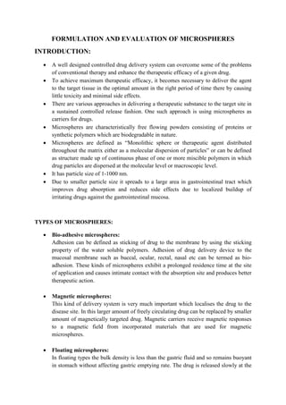
  • 9. Table 3: Calibration curve of Diclofenac in 7.5 pH buffer at 275 nm Concentation (μg/ml) Absorbance 2 0.057 4 0.106 6 0.151 8 0.185 10 0.218 Figure 1: Calibration curve of Diclofenac in 7.5 pH buffer at 275 nm y = 0.020x + 0.023 0.25 0.2 0.15 0.1 0.05 Evaluation of microbeads (Sodium Alginate beads) Table 4: Assay Evaluation parameter Sodium alginate beads Diclofenac loaded Ethyl cellulose microsphere F1 F2 F3 Assay (%w/w) 92.5 R² = 0.991 0 0 2 4 6 8 10 12 Series1 Linear (Series1)
  • 10. Table 5: Dissolution profile of Diclofenac sodium alginate microbeads Time (hrs) Absorbance Conc. Conc. * DF (a) a*5 ml Cum. in 5ml a*900 ml Cum. in 900 ml Amount (mg) Cum. % Drug Release 0.5 0.006 -0.85 -0.85 -4.25 -4.25 -765 -765 -0.76 -7.6 1 0.067 2.2 2.2 11 11 1980 1980 1.98 19.8 2 0.098 3.75 3.75 18.75 29.75 3375 3386 3.386 33.86 3 0.118 4.75 4.75 23.75 53.5 4275 4304.75 4.30475 43.0475 4 0.156 6.65 6.65 33.25 86.75 5985 6038.5 6.0385 60.385 5 0.181 7.9 7.9 39.5 126.25 7110 7196.75 7.19675 71.9675 6 0.198 8.75 8.75 43.75 170 7875 8001.25 8.00125 80.0125 Figure 2: Dissolution profile of Diclofenac sodium alginate microbeads 90 80 70 60 50 40 30 20 10 0 0 1 2 3 4 5 6 7 Series1