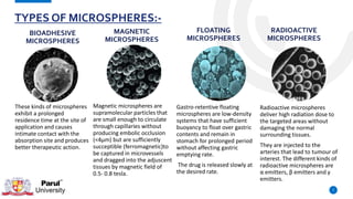
MICROSPHERES
PRESENTED BY:- UDDESHYA UPADHYAY
ENROLLMENT NO.:- 200821211012
M-PHARM ( PHARMACEUTICS)
PARUL INSTITUTE OF PHARMACY,
PARUL UNIVERSITY
CONTENTS :-
• Introduction
• Advantages
• Materials used in the preparation of
Microsphere
• Types of Microspheres
• Method of Preparation
• Characterization or Evaluation of Microspheres
• Applications
• References
2
INTRODUCTION:-
DEFINITION :-
• Microspheres are characteristically free
flowing powders consisting of proteins or
synthetic polymers, which are
biodegradable in nature and ideally
having a particle size less than 200μm.
• This is the important approach in
delivering therapeutic substance to the
target site in sustained and controlled
release drug delivery.
3
4
1. They facilitate accurate delivery of small quantities of potent drug and reduced
concentration of drug at site other than the target organ or tissue.
2. They provide protection for unstable drug before and after administration, prior
to their availability at the site of action.
3. They provide the ability to manipulate the in vivo action of the drug,
pharmacokinetic profile, tissue distribution and cellular interaction of the drug.
4. They enable controlled release of drug.
Examples: Narcotic, Antagonist, Steroid hormones
ADVANTAGES OF MICROSPHERES:-
5
• Natural polymers obtained from different
sources Like carbohydrates proteins and
chemically modified Carbohydrates.
• Carbohydrates: Agarose, Carrageenan, Chitosan,
Starch
• Proteins: Albumin, Collagen and Gelatin
Chemically modified carbohydrates: Poly
dextran, Poly starch
• Synthetic polymers are divided into two
types.
• Biodegradable polymers
E.g. Lactides, Glycolides & their co-polymers,
Poly anhydrides, Poly alkyl cyano acrylates
• Non-biodegradable polymers
E.g. Poly methyl methacrylate (PMMA),
Glycidyl methacrylate, Acrolein,
Epoxy polymers
MATERIALS USED INTHE PREPARATION OF MICROSPHERE:-
MICROSPHERES USED USUALLYARE POLYMERS.THEY ARE CLASSIFIED INTOTWOTYPES:
1. NATURAL POLYMERS 2. SYNTHETIC POLYMERS
.
6
TYPES OF MICROSPHERES:-
7
These kinds of microspheres
exhibit a prolonged
residence time at the site of
application and causes
intimate contact with the
absorption site and produces
better therapeutic action.
BIOADHESIVE
MICROSPHERES
Magnetic microspheres are
supramolecular particles that
are small enough to circulate
through capillaries without
producing embolic occlusion
(<4μm) but are sufficiently
succeptible (ferromagnetic)to
be captured in microvessels
and dragged into the adjuscent
tissues by magnetic field of
0.5- 0.8 tesla.
MAGNETIC
MICROSPHERES
Gastro-retentive floating
microspheres are low-density
systems that have sufficient
buoyancy to float over gastric
contents and remain in
stomach for prolonged period
without affecting gastric
emptying rate.
The drug is released slowly at
the desired rate.
FLOATING
MICROSPHERES
Radioactive microspheres
deliver high radiation dose to
the targeted areas without
damaging the normal
surrounding tissues.
They are injected to the
arteries that lead to tumour of
interest. The different kinds of
radioactive microspheres are
α emitters, β emitters and γ
emitters.
RADIOACTIVE
MICROSPHERES
POLYMERIC MICROSPHERE
The different types of polymeric microspheres classified as :-
I) Biodegradable polymeric microspheres
Natural polymers such as starch are used as concept that they are
biodegradable, biocompatible, and also Bioadhesive in nature. This
polymers prolongs the residence time when contact with mucous
membrane due to its high degree of swelling property with aqueous
medium, results get gel formation.
ll) Synthetic polymeric microspheres
Synthetic polymeric microspheres are widely used in clinical application,
that are also used as bulking agent, fillers, embolic particles and drug
delivery vehicles etc. and proved to be safe and biocompatible but the
disadvantage of these kind of microspheres, are tend to migrate away
from injection site and lead to potential risk, embolism , further organ
damage.
1. Spray Drying
2. Emulsion Solvent Evaporation
3. Single emulsion technique
4. Double emulsion technique
5. Phase separation coacervation
technique
6. Spray drying and spray congealing
7. Solvent extraction
8. Quassi emulsion solvent diffusion
9
METHOD OF PREPRATION:-
SPRAY DRYING:-
• In Spray Drying technique, polymer is first
dissolved in volatile organic solvent such as
dichloromethane, acetone, etc.
• The drug in solid form is then dispersed in to
polymeric solution with the high-speed
homogenization.
• This dispersion is then atomized in hot air stream.
The atomization leads to the form the small
droplets from which the solvent evaporates
instantly leads the formation of the microspheres
in the size range 1-100μm.
• Micro particles are separated from hot air by the
cyclone separator while the trace of solvent is
removed by vacuum drying. major advantages of
this process is feasibility of operation under
aseptic conditions.
10
EMULSION SOLVENT EVAPORATION:-
• In this technique the drug is dissolved in
polymer which was previously dissolved in
chloroform and the resulting solution is
added to aqueous phase containing 0.2 %
sodium of PVP as emulsifying agent.
• The above mixture was agitated at 500
rpm then the drug and polymer (eudragit)
was transformed into fine droplet which
solidified into rigid microspheres by
solvent evaporation and then collected by
filtration and washed with demineralised
water and desiccated at room
temperature for 24 hrs.
11
SINGLE EMULSIONTECHNIQUE :-
• The microparticulate carriers of natural
polymers i.e. those of proteins and
carbohydrates are prepared by single
emulsion technique.
• The natural polymers are
dissolved/dispersed in aqueous medium
followed by dispersion in the non-aqueous
medium e.g. oil.
• In the second step of preparation, cross-
linking of dispersed globule is carried out.
• The cross linking is achieved by two methods
i.e. either by heat or by means of chemical
cross linking agents including glutaraldehyde,
formaldehyde, diacid chloride etc.
12
DOUBLE EMULSIONTECHNIQUE :-
• This method of microspheres preparation
involves formation of multiple emulsions or
double emulsion of type w/o/w and is best
suited to the water soluble drugs, peptides,
proteins and vaccines.
• This method can be used with the both
natural as well as synthetic polymers.
• The aqueous protein solution is dispersed in
the lipophilic organic continuous phase.
• This protein solution may contain the active
constituents.
13
14
.
PHASE SEPARATION COACERVATIONTECHNIQUE:-
• Phase separation method is mainly
designed for preparing the reservoir type of
the system.
• This method is used to encapsulate water
soluble drugs e.g. peptides, proteins and
some of preparations having matrix type
particular, when the drug is hydrophobic in
nature e.g. steroids.
• In this technique the polymer is first
dissolved in a suitable solvent and then
drug is dispersed by making its aqueous
solution, if hydrophobic or dissolved in
polymer solution itself, If hydrophobic.
• Phase separation is then accomplished by
changing the solution conditions by the salt
addition, on-solvent addition, addition of
the incompatible polymer or change in pH
15
SPRAY DRYING AND SPRAY CONGEALING :-
• These methods are based on the drying
of the mist of polymer and drug in the
air.
• Depending upon removal of the solvent
or cooling of the solution, this two
processes are named spray drying and
spray congealing.
16
SOLVENT EXTRACTION :-
• Solvent extraction method is used for
the manufacturing of microparticles and
involves removal of the organic phase by
extraction of the non-aqueous solvent.
• This method involves the water miscible
organic solvent which is isopropanol.
• Organic phase is removed by extraction
with water. This process decreases the
hardening time for the microspheres.
• The process involves direct addition of
the drug or protein to polymer organic
solution.
17
18
1. % yield of microspheres
-Thoroughly dried microspheres were collected and weighed accurately.
-The percentage yield was then calculated using formula given below,
% Yield = mass of microsphere obtained / total weight of
drug & polymer X 100
-Particle size analysis, Particle size distribution and Particle size determination was done by sieving
method. Size distribution plays an important role in determining the release characteristics of the
microspheres.
CHARECTERIZATION/ EVALUATION OF MICROSPHERES :-
2. SHAPE AND SURFACE CHARACTERIZATION :-
• The shape and surface characterization of
microspheres were observed under a
Scanning Electron Microscope (SEM).
• The instrument used for this study was
ZEOL JSM – 5610 scanning electron
microscope.
• The microspheres were mounted directly
on the SEM sample stub, using double-
sided sticking tape and coated with gold
film (thickness 200 nm) under reduced
pressure (0.001 torr) and photographed.
19
3. DETERMINATION OF DRUG CONTENT :-
• Accurately weighed 100 mg microspheres
were crushed in glass mortar and pestle,
powder microspheres were suspended in
100 ml of suitable solvent.
• After 12 hours the solution was filtered
and the filtrate was analysed for the drug
content using UV-Visible
spectrophotometer.
20
4. ENCAPSULATION EFFICIENCY :-
• Encapsulation efficiency was calculated
using the following formula;
E = Qp / Qt X 100
Where, E = percentage of encapsulation of
microspheres
Qp = quantity of drug encapsulated
in microspheres
Qt = quantity of the drug added for
encapsulation
21
5. SWELLING STUDIES :-
• A known weight (50 mg) of microspheres was
placed in a glass vial containing 10 ml of distilled
water at 37 ± 0.50C in incubator with occasional
shaking.
• The microspheres were periodically removed,
blotted with filter paper and their changes in
weights were measured during the swelling until
equilibrium was attained.
• Finally, the weight of the swollen microspheres
was recorded after a period of 3 hours, and the
swelling ratio (SR) was then calculated from the
following formula.
• The studies were carried out in triplicate.
SR=We-Wo / Wo
Where, Wo = Initial weight of the dry microspheres,
We = Weight of the swollen microspheres at
equilibrium swelling in the media.
22
23
TYPE OF MICROSPHERES APPLICATIONS
Bioadhesive Microspheres
Buccal, oral, ocular, nasal, colonic drug delivery
Nasal - Gentamicin, Insulin,
GI - Glipizide
Colonic - Insulin
Ocular - Methyl prednisolone(22)
Magnetic Microspheres
Used in DNA analysis, cell isolation, protein purification
and targetting drugs to tumour sites( Doxorubicin)
Floating Microspheres
Carriers for drugs like antiviral, antifungal and antibiotic
agents (so called absorption windows), non-steroidal
anti inflammatory drugs, Prednisolone, Lansoprazole
APPLICATIONS :-
24
TYPE OF MICROSPHERES APPLICATIONS
Radioactive Microspheres
For diagnostic purpose - Diagnostic radioembolization:
99mTc-macro aggregated human serum albumin (MAA),
Thrombus imaging in deep vein thrombosis : 99mTc-
sulfur colloid
For therapeutic purpose - Radioembolization of liver
and spleen tumours: 90Y microspheres,
Local radiotherapy: 212Pb-sulfur colloid.
Polymeric Microspheres
Vaccine delivery : Hepatitis, Influenza , Pertussis,
Diptheria toxoid,
Oral drug delivery of easily degraded drugs : Gene
therapy with DNA plasmids; delivery of insulin, LHRH
Controlled drug delivery after local application : Release
of proteins, hormones and peptides over extended
times
APPLICATIONS :-
25
• Vyas and Khar. Targeted and Controlled drug delivery (CBS Publishers and distributors, 2001).
• Ramteke K.H, Jadhav V.B, Dhole S.N. “Microspheres: as carrieres used for novel drug delivery system”
IOSR Journal of Pharmacy (IOSRPHR) ISSN: 2250-3013, Vol. 2, Issue 4 (July2012), PP 44-48
• N.R. Patel, D.A. Patel, P.D Bharadia, V. Pandya, V. Modi, Microsphere as a novel drug deliver, Int J of
Pharm & Life Sci, 2(8),2011,992-997.
• V.V. Prasanth, A.C. Moy, S.T. Mathew, R. Mathapan, Microspheres: an overview, Int J of Pharm &
Biomedical Sci, 2(2),2011,332-338.
• Fischer S., Foreg C., Merkle P.H., Gander B., Chitosan Coated PLGA-Microspheres-A Modular System
for Targetting Drug Delivery, European Cells and Materials. 2004; 7:11-12.
REFERENCES :-
STAY HOME. SAVE LIVES.
26
HELP STOP CORONAVIRUS !

Microspheres

  • 1.
    MICROSPHERES PRESENTED BY:- UDDESHYAUPADHYAY ENROLLMENT NO.:- 200821211012 M-PHARM ( PHARMACEUTICS) PARUL INSTITUTE OF PHARMACY, PARUL UNIVERSITY
  • 2.
    CONTENTS :- • Introduction •Advantages • Materials used in the preparation of Microsphere • Types of Microspheres • Method of Preparation • Characterization or Evaluation of Microspheres • Applications • References 2
  • 3.
    INTRODUCTION:- DEFINITION :- • Microspheresare characteristically free flowing powders consisting of proteins or synthetic polymers, which are biodegradable in nature and ideally having a particle size less than 200μm. • This is the important approach in delivering therapeutic substance to the target site in sustained and controlled release drug delivery. 3
  • 4.
    4 1. They facilitateaccurate delivery of small quantities of potent drug and reduced concentration of drug at site other than the target organ or tissue. 2. They provide protection for unstable drug before and after administration, prior to their availability at the site of action. 3. They provide the ability to manipulate the in vivo action of the drug, pharmacokinetic profile, tissue distribution and cellular interaction of the drug. 4. They enable controlled release of drug. Examples: Narcotic, Antagonist, Steroid hormones ADVANTAGES OF MICROSPHERES:-
  • 5.
    5 • Natural polymersobtained from different sources Like carbohydrates proteins and chemically modified Carbohydrates. • Carbohydrates: Agarose, Carrageenan, Chitosan, Starch • Proteins: Albumin, Collagen and Gelatin Chemically modified carbohydrates: Poly dextran, Poly starch • Synthetic polymers are divided into two types. • Biodegradable polymers E.g. Lactides, Glycolides & their co-polymers, Poly anhydrides, Poly alkyl cyano acrylates • Non-biodegradable polymers E.g. Poly methyl methacrylate (PMMA), Glycidyl methacrylate, Acrolein, Epoxy polymers MATERIALS USED INTHE PREPARATION OF MICROSPHERE:- MICROSPHERES USED USUALLYARE POLYMERS.THEY ARE CLASSIFIED INTOTWOTYPES: 1. NATURAL POLYMERS 2. SYNTHETIC POLYMERS
  • 6.
  • 7.
    TYPES OF MICROSPHERES:- 7 Thesekinds of microspheres exhibit a prolonged residence time at the site of application and causes intimate contact with the absorption site and produces better therapeutic action. BIOADHESIVE MICROSPHERES Magnetic microspheres are supramolecular particles that are small enough to circulate through capillaries without producing embolic occlusion (<4μm) but are sufficiently succeptible (ferromagnetic)to be captured in microvessels and dragged into the adjuscent tissues by magnetic field of 0.5- 0.8 tesla. MAGNETIC MICROSPHERES Gastro-retentive floating microspheres are low-density systems that have sufficient buoyancy to float over gastric contents and remain in stomach for prolonged period without affecting gastric emptying rate. The drug is released slowly at the desired rate. FLOATING MICROSPHERES Radioactive microspheres deliver high radiation dose to the targeted areas without damaging the normal surrounding tissues. They are injected to the arteries that lead to tumour of interest. The different kinds of radioactive microspheres are α emitters, β emitters and γ emitters. RADIOACTIVE MICROSPHERES
  • 8.
    POLYMERIC MICROSPHERE The differenttypes of polymeric microspheres classified as :- I) Biodegradable polymeric microspheres Natural polymers such as starch are used as concept that they are biodegradable, biocompatible, and also Bioadhesive in nature. This polymers prolongs the residence time when contact with mucous membrane due to its high degree of swelling property with aqueous medium, results get gel formation. ll) Synthetic polymeric microspheres Synthetic polymeric microspheres are widely used in clinical application, that are also used as bulking agent, fillers, embolic particles and drug delivery vehicles etc. and proved to be safe and biocompatible but the disadvantage of these kind of microspheres, are tend to migrate away from injection site and lead to potential risk, embolism , further organ damage.
  • 9.
    1. Spray Drying 2.Emulsion Solvent Evaporation 3. Single emulsion technique 4. Double emulsion technique 5. Phase separation coacervation technique 6. Spray drying and spray congealing 7. Solvent extraction 8. Quassi emulsion solvent diffusion 9 METHOD OF PREPRATION:-
  • 10.
    SPRAY DRYING:- • InSpray Drying technique, polymer is first dissolved in volatile organic solvent such as dichloromethane, acetone, etc. • The drug in solid form is then dispersed in to polymeric solution with the high-speed homogenization. • This dispersion is then atomized in hot air stream. The atomization leads to the form the small droplets from which the solvent evaporates instantly leads the formation of the microspheres in the size range 1-100μm. • Micro particles are separated from hot air by the cyclone separator while the trace of solvent is removed by vacuum drying. major advantages of this process is feasibility of operation under aseptic conditions. 10
  • 11.
    EMULSION SOLVENT EVAPORATION:- •In this technique the drug is dissolved in polymer which was previously dissolved in chloroform and the resulting solution is added to aqueous phase containing 0.2 % sodium of PVP as emulsifying agent. • The above mixture was agitated at 500 rpm then the drug and polymer (eudragit) was transformed into fine droplet which solidified into rigid microspheres by solvent evaporation and then collected by filtration and washed with demineralised water and desiccated at room temperature for 24 hrs. 11
  • 12.
    SINGLE EMULSIONTECHNIQUE :- •The microparticulate carriers of natural polymers i.e. those of proteins and carbohydrates are prepared by single emulsion technique. • The natural polymers are dissolved/dispersed in aqueous medium followed by dispersion in the non-aqueous medium e.g. oil. • In the second step of preparation, cross- linking of dispersed globule is carried out. • The cross linking is achieved by two methods i.e. either by heat or by means of chemical cross linking agents including glutaraldehyde, formaldehyde, diacid chloride etc. 12
  • 13.
    DOUBLE EMULSIONTECHNIQUE :- •This method of microspheres preparation involves formation of multiple emulsions or double emulsion of type w/o/w and is best suited to the water soluble drugs, peptides, proteins and vaccines. • This method can be used with the both natural as well as synthetic polymers. • The aqueous protein solution is dispersed in the lipophilic organic continuous phase. • This protein solution may contain the active constituents. 13
  • 14.
  • 15.
    PHASE SEPARATION COACERVATIONTECHNIQUE:- •Phase separation method is mainly designed for preparing the reservoir type of the system. • This method is used to encapsulate water soluble drugs e.g. peptides, proteins and some of preparations having matrix type particular, when the drug is hydrophobic in nature e.g. steroids. • In this technique the polymer is first dissolved in a suitable solvent and then drug is dispersed by making its aqueous solution, if hydrophobic or dissolved in polymer solution itself, If hydrophobic. • Phase separation is then accomplished by changing the solution conditions by the salt addition, on-solvent addition, addition of the incompatible polymer or change in pH 15
  • 16.
    SPRAY DRYING ANDSPRAY CONGEALING :- • These methods are based on the drying of the mist of polymer and drug in the air. • Depending upon removal of the solvent or cooling of the solution, this two processes are named spray drying and spray congealing. 16
  • 17.
    SOLVENT EXTRACTION :- •Solvent extraction method is used for the manufacturing of microparticles and involves removal of the organic phase by extraction of the non-aqueous solvent. • This method involves the water miscible organic solvent which is isopropanol. • Organic phase is removed by extraction with water. This process decreases the hardening time for the microspheres. • The process involves direct addition of the drug or protein to polymer organic solution. 17
  • 18.
    18 1. % yieldof microspheres -Thoroughly dried microspheres were collected and weighed accurately. -The percentage yield was then calculated using formula given below, % Yield = mass of microsphere obtained / total weight of drug & polymer X 100 -Particle size analysis, Particle size distribution and Particle size determination was done by sieving method. Size distribution plays an important role in determining the release characteristics of the microspheres. CHARECTERIZATION/ EVALUATION OF MICROSPHERES :-
  • 19.
    2. SHAPE ANDSURFACE CHARACTERIZATION :- • The shape and surface characterization of microspheres were observed under a Scanning Electron Microscope (SEM). • The instrument used for this study was ZEOL JSM – 5610 scanning electron microscope. • The microspheres were mounted directly on the SEM sample stub, using double- sided sticking tape and coated with gold film (thickness 200 nm) under reduced pressure (0.001 torr) and photographed. 19
  • 20.
    3. DETERMINATION OFDRUG CONTENT :- • Accurately weighed 100 mg microspheres were crushed in glass mortar and pestle, powder microspheres were suspended in 100 ml of suitable solvent. • After 12 hours the solution was filtered and the filtrate was analysed for the drug content using UV-Visible spectrophotometer. 20
  • 21.
    4. ENCAPSULATION EFFICIENCY:- • Encapsulation efficiency was calculated using the following formula; E = Qp / Qt X 100 Where, E = percentage of encapsulation of microspheres Qp = quantity of drug encapsulated in microspheres Qt = quantity of the drug added for encapsulation 21
  • 22.
    5. SWELLING STUDIES:- • A known weight (50 mg) of microspheres was placed in a glass vial containing 10 ml of distilled water at 37 ± 0.50C in incubator with occasional shaking. • The microspheres were periodically removed, blotted with filter paper and their changes in weights were measured during the swelling until equilibrium was attained. • Finally, the weight of the swollen microspheres was recorded after a period of 3 hours, and the swelling ratio (SR) was then calculated from the following formula. • The studies were carried out in triplicate. SR=We-Wo / Wo Where, Wo = Initial weight of the dry microspheres, We = Weight of the swollen microspheres at equilibrium swelling in the media. 22
  • 23.
    23 TYPE OF MICROSPHERESAPPLICATIONS Bioadhesive Microspheres Buccal, oral, ocular, nasal, colonic drug delivery Nasal - Gentamicin, Insulin, GI - Glipizide Colonic - Insulin Ocular - Methyl prednisolone(22) Magnetic Microspheres Used in DNA analysis, cell isolation, protein purification and targetting drugs to tumour sites( Doxorubicin) Floating Microspheres Carriers for drugs like antiviral, antifungal and antibiotic agents (so called absorption windows), non-steroidal anti inflammatory drugs, Prednisolone, Lansoprazole APPLICATIONS :-
  • 24.
    24 TYPE OF MICROSPHERESAPPLICATIONS Radioactive Microspheres For diagnostic purpose - Diagnostic radioembolization: 99mTc-macro aggregated human serum albumin (MAA), Thrombus imaging in deep vein thrombosis : 99mTc- sulfur colloid For therapeutic purpose - Radioembolization of liver and spleen tumours: 90Y microspheres, Local radiotherapy: 212Pb-sulfur colloid. Polymeric Microspheres Vaccine delivery : Hepatitis, Influenza , Pertussis, Diptheria toxoid, Oral drug delivery of easily degraded drugs : Gene therapy with DNA plasmids; delivery of insulin, LHRH Controlled drug delivery after local application : Release of proteins, hormones and peptides over extended times APPLICATIONS :-
  • 25.
    25 • Vyas andKhar. Targeted and Controlled drug delivery (CBS Publishers and distributors, 2001). • Ramteke K.H, Jadhav V.B, Dhole S.N. “Microspheres: as carrieres used for novel drug delivery system” IOSR Journal of Pharmacy (IOSRPHR) ISSN: 2250-3013, Vol. 2, Issue 4 (July2012), PP 44-48 • N.R. Patel, D.A. Patel, P.D Bharadia, V. Pandya, V. Modi, Microsphere as a novel drug deliver, Int J of Pharm & Life Sci, 2(8),2011,992-997. • V.V. Prasanth, A.C. Moy, S.T. Mathew, R. Mathapan, Microspheres: an overview, Int J of Pharm & Biomedical Sci, 2(2),2011,332-338. • Fischer S., Foreg C., Merkle P.H., Gander B., Chitosan Coated PLGA-Microspheres-A Modular System for Targetting Drug Delivery, European Cells and Materials. 2004; 7:11-12. REFERENCES :-
  • 26.
    STAY HOME. SAVELIVES. 26 HELP STOP CORONAVIRUS !