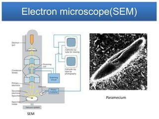
Microscope
Presented by
Dr. Avinanda Biswas
Tutor of Microbiology
D Y Patil Medical College
and Hospital
TERMS AND DEFINITIONS
Principle
Microscopy is to get a magnified image, in which structures may be
resolved which could not be resolved with the help of an unaided eye.
Magnification
• It is the ratio of the size of an object seen
under microscope to the actual size
observed with unaided eye.
• The total magnification of microscope
is calculated by multiplying the magnifying
power of the objective lens by that of eye piece.
Resolving power
• It is the ability to differentiate two close points as separate.
• The resolving power of human eye is 0.25 mm
• The light microscope can separate dots that are 0.25µm apart.
• The electron microscope can separate dots that are 0.5nm apart.
TERMS AND DEFINITIONS
Limit of resolution
It is the minimum distance between two points to identify them
separately.
It is calculated by Abbé equation.
Limit of resolution is inversely proportional to power or resolution.
If the wavelength is shorter then the resolution will be greater.
Working distance
• It is the distance between the objective
and the objective slide.
• The working distance decreases with
increasing magnification.
TERMS AND DEFINITIONS
Numerical aperture(NA)
The numerical aperture of a lens is the ratio of the diameter of the
lens to its focal length.
NA of a lens is an index of the resolving power.
NA can be decreased by decreasing the amount of light that passes through
a lens.
Diameter of the lens
Light microscope
• In 1590 F.H Janssen & Z.Janssen constructed the first simple compound
light microscope.
• In 1665 Robert Hooke developed a first laboratory
compound microscope.
• Later, Kepler and galileo developed a modern class
room microscope.
• In 1672 Leeuwenhoek developed a first simple microscope with a
magnification of 200x – 300x.
• He is called as
Father of microscopy.
• The term microscope
was coined by Faber
in 1623.
Light microscope
Light microscope
Parts of microscope
• Illuminator - This is the light source located below the specimen.
• Condenser - Focuses the ray of light through the specimen.
• Stage - The fixed stage is a horizontal platform that holds the specimen.
• Objective - The lens that is directly above the stage.
• Nosepiece - The portion of the body that holds the objectives over the
stage.
• Iris diaphragm - Regulates the amount of light into the condenser.
• Base – Base supports the microscope which is horseshoe shaped.
• Coarse focusing knob - Used to make relatively wide focusing adjustments
to the microscope.
• Fine focusing knob - Used to make relatively small adjustments to the
microscope.
• Body - The microscope body.
• Ocular eyepiece - Lens on the top of the body tube. It has a magnification of
10× normal vision.
Light microscope
Objective
PROPERTY LOW POWER HIGH POWER OIL IMMERSION
Magnification of
objective
10x 40-45x 90-100x
Magnification of 10x 10x 10x
eyepiece
Total magnification 100x 450 – 450x 900 – 1000x
Numerical aperture 0.25 – 0.30 0.55 – 0.65 1.25 – 1.4
Mirror used Concave Concave Plane
Focal length (Approx) 16 mm 4 mm 1.8 – 2 mm
Working distance 4 – 8 mm 0.5 – 0.7 mm 0.1 mm
Iris diaphragm Partially closed Partially opened Fully opened
Position of condenser Lowest Slightly raised Fully raised
Maximum
resolution(Approx)
0.9 µm 0.35µm 0.18µm
Light microscope
Baccili and cocci under light microscope
Paramecium specimen
A bright-field microscope can be adapted as a dark-field microscope by adding a
special disc called a stop to the condenser.
The stop blocks all light from entering the objective lens except peripheral light that
is reflected off the sides of the specimen itself. The resulting image is a brightly
illuminated specimens surrounded by a dark (black) field.
Dark field microscope
Uses:
This microscope is used to study spirochetes in the exudates form leptospiral or
syphilitic Infections.
Dark field microscope
Paramecium
Treponema vincenti
Volvox and Spirogyra
Phase contrast microscope
In 1935 F.Zernike produced the phase contrast microscope.
Phase-contrast microscope is also called as zernike
microscope.
Phase-contrast microscope uses a special condenser and
objective lenses.
This condenser lens on the light microscope splits a light
beam and throws the light rays slightly out of phase.
The separated beams of light then pass through and
around the specimen, and small differences in the refractive
index within the specimen show up as different degrees of
brightness and contrast.
Uses:
Phase-contrast microscopy is especially useful for studying
microbial motility, studying eukaryotic Cells, determining the
shape of living cells, and detecting bacterial components
such as endospores and Inclusion bodies that contain
poly--hydroxyalkanoates (e.g., poly-hydroxybutyrate),
polymetaphosphate, sulfur, or other substances.
Phase contrast microscope
Macronucleus
Micronucleus
Paramecium
Phase contrast microscope
Rhodospirillum rubrum
Fluorescence microscope
It was developed by Haitinger and coons
A fluorescence microscope differs from an ordinary brightfield microscope in several
respects.
It utilizes a powerful mercury vapor arc lamp for its light source.
A darkfield condenser is usually used in place of the conventional Abbé brightfield
condenser.
It employs three sets of filters to alter the light that passes up through the
instrument to the eye.
Microbiological speciemen that is to be studied must be coated with special
compounds that possess the quality of fluorescence. Such compounds are called
fluorochromes.
AuramineO, acridine orange, and fluorescein are well-known fluorochromes.
Uses:
It is used to study the substance like chlorophylls, riboflavin,
vitaminA, collagen which have the property of auto fluorescence.
Some cellular components like cellulose, starch, glycogen,
protein and Y chromosome can be made visible under this
microscope by staining them with fluorochromes.
It used to identify Y chromosome to determine sex,
determination of microbial cells in the infected tissue and to
study the structure of proteins.
Fluorescence microscope
Oral cavity
Bacillus subtilis
Electron microscope
In 1932 Knoll and Ruska invented first electron microscope.
The electron microscope uses a beam of electrons rather than visible light.
The magnified image is visible on a fluorescent screen and can be recorded on a
photographic film.
The drawback of the electron microscope is specimen are killed in order to view the
cells or organisms.
Images produced by electrons lack color, electron micrographs are always
shades of black, gray, and white.
Two general forms of EM are the transmission electron microscope (TEM) and the
scanning electron microscope (SEM).
Transmission electron microscopes are the method of choice for viewing the detailed
structure of cells and viruses.
This microscope produces its image by transmitting electrons through the
specimen.
Because electrons cannot readily penetrate thick preparations, the specimen
must be sectioned into extremely thin slices (20–100 nm thick) and stained or coated
with metals that will increase image contrast.
The darkest areas of TEM micrographs represent the thicker (denser) parts, and
the lighter areas indicate the more transparent and less dense parts.
Electron microscope(TEM)
Chlamydomonas
Electron microscope(SEM)
The specimen is placed in the vacuum chamber and covered with a thin coat of gold.
The electron beam then scans across the specimen and knocks loose showers of electrons
that are captured by a detector. An image builds line by line, as in a television receiver.
Electrons that strike a sloping surface yield fewer electrons, thereby producing a darker
contrasting spot and a sense of three dimensions.
The resolving power of the conventional SEM is about 10 nm and magnifications with the
SEM are limited to about 20,000x.
Electron microscope(SEM)
Paramecium
SEM
Light Vs Electron microscope
Uses
Thank you

Microscope 120109025117-phpapp02

  • 1.
    Microscope Presented by Dr. AvinandaBiswas Tutor of Microbiology D Y Patil Medical College and Hospital
  • 2.
    TERMS AND DEFINITIONS Principle Microscopyis to get a magnified image, in which structures may be resolved which could not be resolved with the help of an unaided eye. Magnification • It is the ratio of the size of an object seen under microscope to the actual size observed with unaided eye. • The total magnification of microscope is calculated by multiplying the magnifying power of the objective lens by that of eye piece. Resolving power • It is the ability to differentiate two close points as separate. • The resolving power of human eye is 0.25 mm • The light microscope can separate dots that are 0.25µm apart. • The electron microscope can separate dots that are 0.5nm apart.
  • 3.
    TERMS AND DEFINITIONS Limitof resolution It is the minimum distance between two points to identify them separately. It is calculated by Abbé equation. Limit of resolution is inversely proportional to power or resolution. If the wavelength is shorter then the resolution will be greater. Working distance • It is the distance between the objective and the objective slide. • The working distance decreases with increasing magnification.
  • 4.
    TERMS AND DEFINITIONS Numericalaperture(NA) The numerical aperture of a lens is the ratio of the diameter of the lens to its focal length. NA of a lens is an index of the resolving power. NA can be decreased by decreasing the amount of light that passes through a lens. Diameter of the lens
  • 5.
    Light microscope • In1590 F.H Janssen & Z.Janssen constructed the first simple compound light microscope. • In 1665 Robert Hooke developed a first laboratory compound microscope. • Later, Kepler and galileo developed a modern class room microscope. • In 1672 Leeuwenhoek developed a first simple microscope with a magnification of 200x – 300x. • He is called as Father of microscopy. • The term microscope was coined by Faber in 1623.
  • 6.
  • 7.
    Light microscope Parts ofmicroscope • Illuminator - This is the light source located below the specimen. • Condenser - Focuses the ray of light through the specimen. • Stage - The fixed stage is a horizontal platform that holds the specimen. • Objective - The lens that is directly above the stage. • Nosepiece - The portion of the body that holds the objectives over the stage. • Iris diaphragm - Regulates the amount of light into the condenser. • Base – Base supports the microscope which is horseshoe shaped. • Coarse focusing knob - Used to make relatively wide focusing adjustments to the microscope. • Fine focusing knob - Used to make relatively small adjustments to the microscope. • Body - The microscope body. • Ocular eyepiece - Lens on the top of the body tube. It has a magnification of 10× normal vision.
  • 8.
    Light microscope Objective PROPERTY LOWPOWER HIGH POWER OIL IMMERSION Magnification of objective 10x 40-45x 90-100x Magnification of 10x 10x 10x eyepiece Total magnification 100x 450 – 450x 900 – 1000x Numerical aperture 0.25 – 0.30 0.55 – 0.65 1.25 – 1.4 Mirror used Concave Concave Plane Focal length (Approx) 16 mm 4 mm 1.8 – 2 mm Working distance 4 – 8 mm 0.5 – 0.7 mm 0.1 mm Iris diaphragm Partially closed Partially opened Fully opened Position of condenser Lowest Slightly raised Fully raised Maximum resolution(Approx) 0.9 µm 0.35µm 0.18µm
  • 9.
    Light microscope Baccili andcocci under light microscope Paramecium specimen
  • 10.
    A bright-field microscopecan be adapted as a dark-field microscope by adding a special disc called a stop to the condenser. The stop blocks all light from entering the objective lens except peripheral light that is reflected off the sides of the specimen itself. The resulting image is a brightly illuminated specimens surrounded by a dark (black) field. Dark field microscope Uses: This microscope is used to study spirochetes in the exudates form leptospiral or syphilitic Infections.
  • 11.
    Dark field microscope Paramecium Treponemavincenti Volvox and Spirogyra
  • 12.
    Phase contrast microscope In1935 F.Zernike produced the phase contrast microscope. Phase-contrast microscope is also called as zernike microscope. Phase-contrast microscope uses a special condenser and objective lenses. This condenser lens on the light microscope splits a light beam and throws the light rays slightly out of phase. The separated beams of light then pass through and around the specimen, and small differences in the refractive index within the specimen show up as different degrees of brightness and contrast. Uses: Phase-contrast microscopy is especially useful for studying microbial motility, studying eukaryotic Cells, determining the shape of living cells, and detecting bacterial components such as endospores and Inclusion bodies that contain poly--hydroxyalkanoates (e.g., poly-hydroxybutyrate), polymetaphosphate, sulfur, or other substances.
  • 13.
  • 14.
  • 15.
    Fluorescence microscope It wasdeveloped by Haitinger and coons A fluorescence microscope differs from an ordinary brightfield microscope in several respects. It utilizes a powerful mercury vapor arc lamp for its light source. A darkfield condenser is usually used in place of the conventional Abbé brightfield condenser. It employs three sets of filters to alter the light that passes up through the instrument to the eye. Microbiological speciemen that is to be studied must be coated with special compounds that possess the quality of fluorescence. Such compounds are called fluorochromes. AuramineO, acridine orange, and fluorescein are well-known fluorochromes.
  • 16.
    Uses: It is usedto study the substance like chlorophylls, riboflavin, vitaminA, collagen which have the property of auto fluorescence. Some cellular components like cellulose, starch, glycogen, protein and Y chromosome can be made visible under this microscope by staining them with fluorochromes. It used to identify Y chromosome to determine sex, determination of microbial cells in the infected tissue and to study the structure of proteins. Fluorescence microscope
  • 17.
  • 18.
    Electron microscope In 1932Knoll and Ruska invented first electron microscope. The electron microscope uses a beam of electrons rather than visible light. The magnified image is visible on a fluorescent screen and can be recorded on a photographic film. The drawback of the electron microscope is specimen are killed in order to view the cells or organisms. Images produced by electrons lack color, electron micrographs are always shades of black, gray, and white. Two general forms of EM are the transmission electron microscope (TEM) and the scanning electron microscope (SEM). Transmission electron microscopes are the method of choice for viewing the detailed structure of cells and viruses. This microscope produces its image by transmitting electrons through the specimen. Because electrons cannot readily penetrate thick preparations, the specimen must be sectioned into extremely thin slices (20–100 nm thick) and stained or coated with metals that will increase image contrast. The darkest areas of TEM micrographs represent the thicker (denser) parts, and the lighter areas indicate the more transparent and less dense parts.
  • 19.
  • 20.
  • 21.
    Electron microscope(SEM) The specimenis placed in the vacuum chamber and covered with a thin coat of gold. The electron beam then scans across the specimen and knocks loose showers of electrons that are captured by a detector. An image builds line by line, as in a television receiver. Electrons that strike a sloping surface yield fewer electrons, thereby producing a darker contrasting spot and a sense of three dimensions. The resolving power of the conventional SEM is about 10 nm and magnifications with the SEM are limited to about 20,000x.
  • 22.
  • 23.
  • 24.
  • 25.