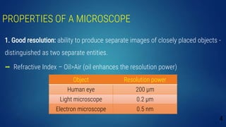
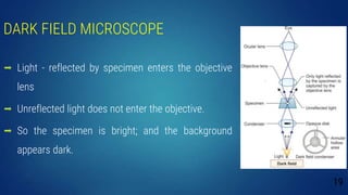
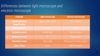
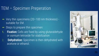
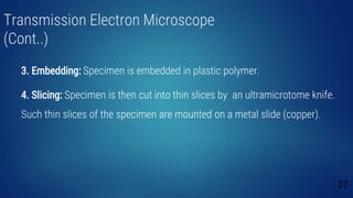
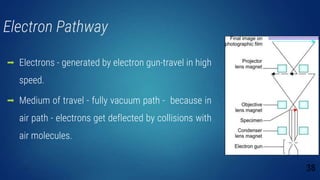
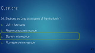
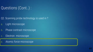
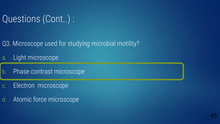
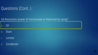
It is about microscope in details about microscope. It includes simple microscope, fluoresence microscope, electron microscope and dark field microscope. These are described in details in this presentation. This presentation has images also.
2. INTRODUCTION
Size:
Bacteria- 0.2–1.5 μm in diameter; 3–5 μm in length
Viruses- 20–300 nm in diameter
Parasites- Protozoans measured in µm; helminths - few mm to meters.
Fungi- Grow as hyphae - thread-like structures 2–10 µm in diameter and up
to several cm in length
2
3. Different types of Microscopes
Bright-field or light microscope
Dark field (or dark ground) microscope
Phase contrast microscope
Fluorescence microscope
Electron microscope.
3
4. PROPERTIES OF A MICROSCOPE
1. Good resolution: ability to produce separate images of closely placed objects -
distinguished as two separate entities.
Refractive Index – Oil>Air (oil enhances the resolution power)
4
Object Resolution power
Human eye 200 µm
Light microscope 0.2 µm
Electron microscope 0.5 nm
5. PROPERTIES OF A MICROSCOPE (Cont..)
2. Good contrast:
Improved by staining the specimen.
When stains bind to the cells, the contrast is increased
5
6. PROPERTIES OF A MICROSCOPE (Cont..)
3. Good magnification: Achieved by use of lenses.
Ocular lens - magnification power of 10x
Objective lens-scanning (4x), low power (10x), high power (40x) and oil
immersion (100x)
Total magnification = magnification of objective lens * ocular lens.
6
8. BRIGHT-FIELD OR LIGHT MICROSCOPE
Forms a dark image against a brighter
background.
Structure
The parts are divided into three groups:
Mechanical Parts
Magnifying Parts
Illuminating Parts
8
9. Mechanical Parts
Base: Holds various parts of the microscope
C-shaped arm: Holds the microscope, connects -
ocular lens to objective lens
Mechanical stage: Hold the slides.
9
11. Illuminating Parts
Condenser: Focuses light on the slide (beneath the stage)
Iris diaphragm: Controls the light that passes through the
condenser
Light source: mirror or an electric bulb
Fine and coarse adjustment knobs: Sharpen the image.
11
12. Working Principle
Rays from the light source pass through the iris and fall
on the specimen
Light rays are gathered by the objective and a magnified
image is formed
12
18. DARK FIELD MICROSCOPE
Working principle: Object appears bright against a dark
background - by use of a special dark field condenser.
o Central opaque area (blocks light from entering the
objective lens)
o Peripheral annular hollow area (allows the light to pass
through and focus on the specimen obliquely)
18
19. DARK FIELD MICROSCOPE
Light - reflected by specimen enters the objective
lens
Unreflected light does not enter the objective.
So the specimen is bright; and the background
appears dark.
19
20. Applications
Used to identify the living, unstained cells and thin bacteria like spirochetes
which cannot be visualized by light microscopy.
20
22. PHASE CONTRAST MICROSCOPE
Contrast is enhanced.
Visualizes the unstained living cells by creating difference in contrast
between cells and water.
22
23. Working principle
Condenser is similar to dark field microscope.
Cone of light passes through a cell
some light rays are bent due to variations in density and refractive
index within the specimen and are retarded by 1/4th of a wavelength
23
24. Working principle (Cont..)
Undeviated light rays strike a phase ring
Deviated rays miss the ring and pass through the rest of the plate
Background is bright, while the unstained object appears dark
24
25. Working principle (Cont..)
The light rays go through → condenser → specimen (e.g. bacteria) →
phase ring → objective lens → ocular lens.
25
28. FLUORESCENCE MICROSCOPE
Working principle:
Fluorescent dyes - exposed to UV rays, become excited and fluoresce
i.e. they convert short wavelength rays into longer wavelengths
Mercury lamp emits rays that pass through an excitation filter
28
29. Working principle (Cont..)
Excitation filter allows only short wavelength
UV light to pass through
Exciting rays get reflected by a dichromatic
mirror - fall on the specimen stained by
fluorescent dye.
Barrier filter – removes remaining UV light or
blue and violet light
29
30. Applications
Epifluorescence microscope: Simplest form of fluorescence microscope. Has
following applications:
Auto fluorescence (eg. Cyclospora)
Microbes coated with fluorescent dye (eg. Acridine orange dye)
Immunofluorescence
30
33. ELECTRON MICROSCOPE
Invented by German physicist - Ernst Ruska in 1931.
Source of illumination - Accelerated electrons
Wavelength of electrons 100,000 times shorter than visible light photons
- better resolving power than a light microscope.
33
34. ELECTRON MICROSCOPE (Cont..)
It is of two types:
Transmission electron microscope (TEM, most common type)
Scanning electron microscope (SEM)
34
35. Differences between light microscope and
electron microscope
35
Features Light microscope Electron microscope
Highest practical
magnification
About 1,000–1,500 Over 100,000
Best resolution 0.2 µm 0.5 nm
Radiation source Visible light Electron beam
Medium of travel Air High vacuum
Specimen mount Glass slide Metal grid (usually copper)
Type of lens Glass Electromagnet
36. TEM – Specimen Preperation
Very thin specimens (20–100 nm thickness) -
suitable for EM.
Steps to prepare thin specimen:
1. Fixation: Cells are fixed by using glutaraldehyde
or osmium tetroxide for stabilization
2. Dehydration: Specimen is then dehydrated with
acetone or ethanol.
36
37. Transmission Electron Microscope
(Cont..)
3. Embedding: Specimen is embedded in plastic polymer.
4. Slicing: Specimen is then cut into thin slices by an ultramicrotome knife.
Such thin slices of the specimen are mounted on a metal slide (copper).
37
38. Electron Pathway
Electrons - generated by electron gun-travel in high
speed.
Medium of travel - fully vacuum path - because in
air path - electrons get deflected by collisions with
air molecules.
38
39. Applications and Modifications of EM
Virus detection
The contrast of EM - increased by —
(i) Negative staining with heavy metals (phosphotungstic acid)
(ii) Shadowing.
Freeze-etching technique - alternative method for specimen preparation.
39
40. Scanning Electron Microscope
Examine the surfaces of microorganisms in detail.
Resolution of 7 nm or less.
Produces image from electrons emitted by object’s surface.
40
42. ATOMIC FORCE MICROSCOPY
Advanced microscope - uses scanning probe technology to study the
cellular structure.
Has a resolution power in fraction of nanometer.
42
43. Questions:
Q1. Electrons are used as a source of illumination in?
a. Light microscope
b. Phase contrast microscope
c. Electron microscope
d. Fluorescence microscope
43
44. Questions (Cont..) :
Q2. Scanning probe technology is used in ?
a. Light microscope
b. Phase contrast microscope
c. Electron microscope
d. Atomic force microscope
44
45. Questions (Cont..) :
Q3. Microscope used for studying microbial motility?
a. Light microscope
b. Phase contrast microscope
c. Electron microscope
d. Atomic force microscope
45