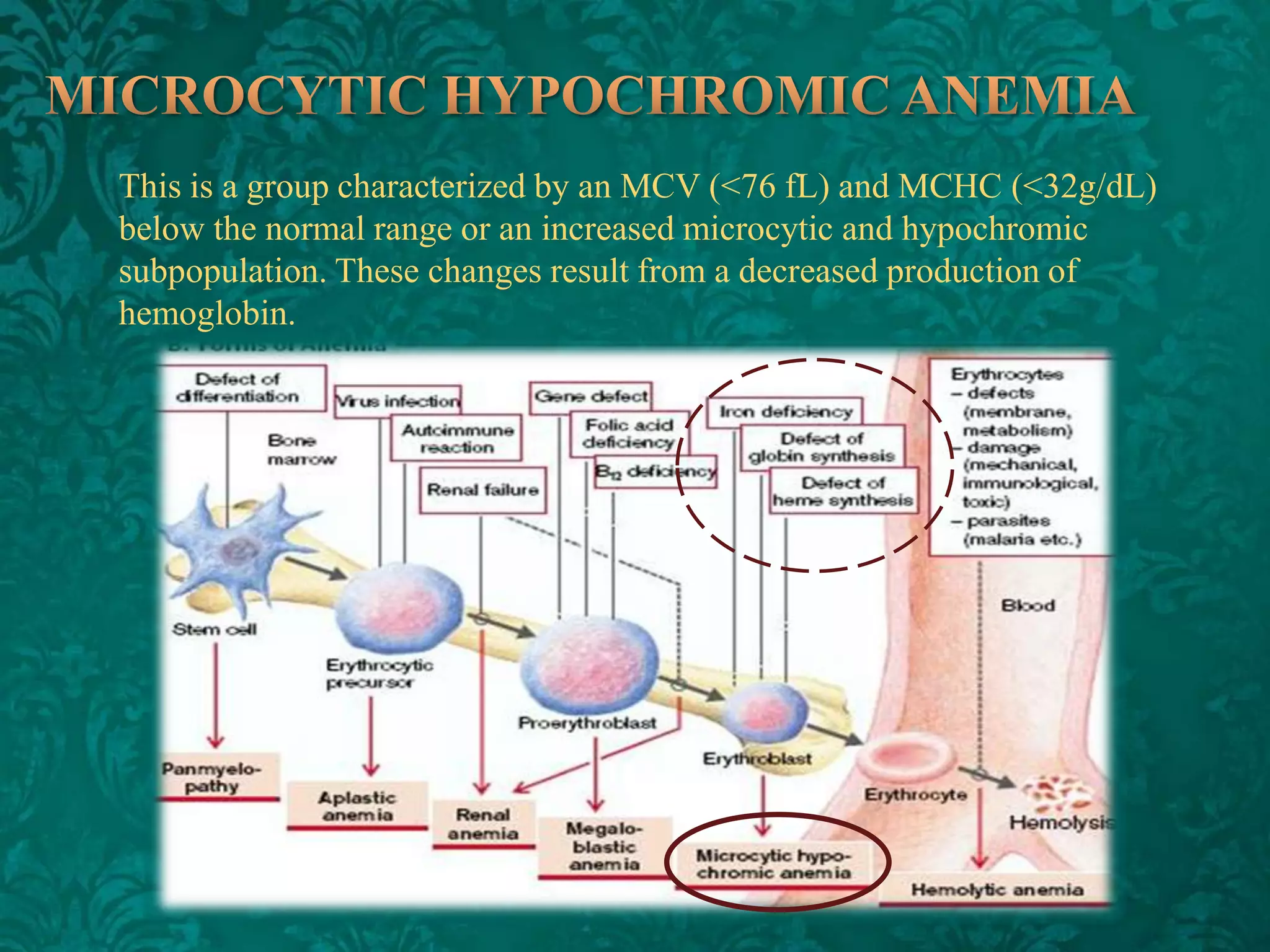
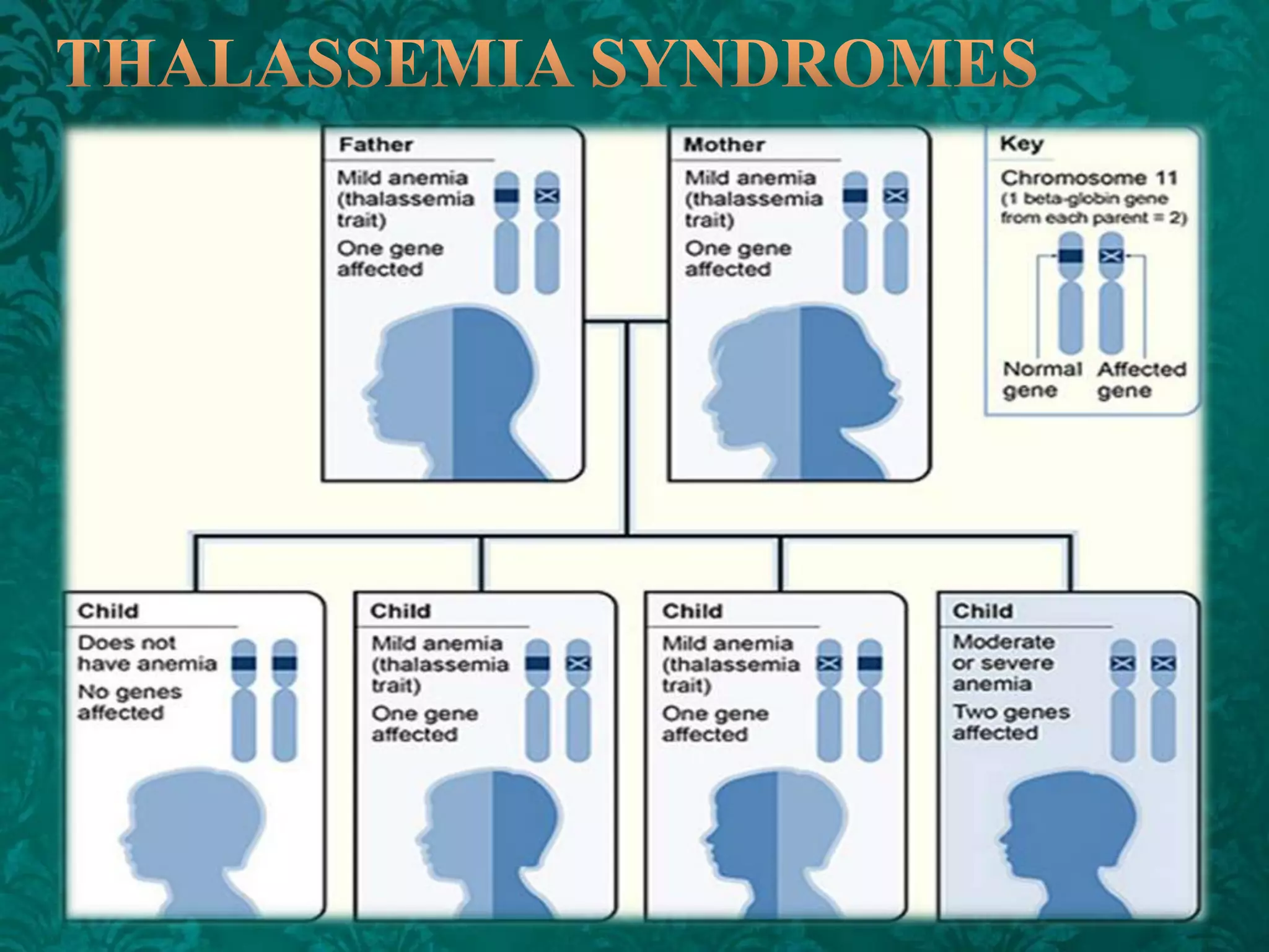
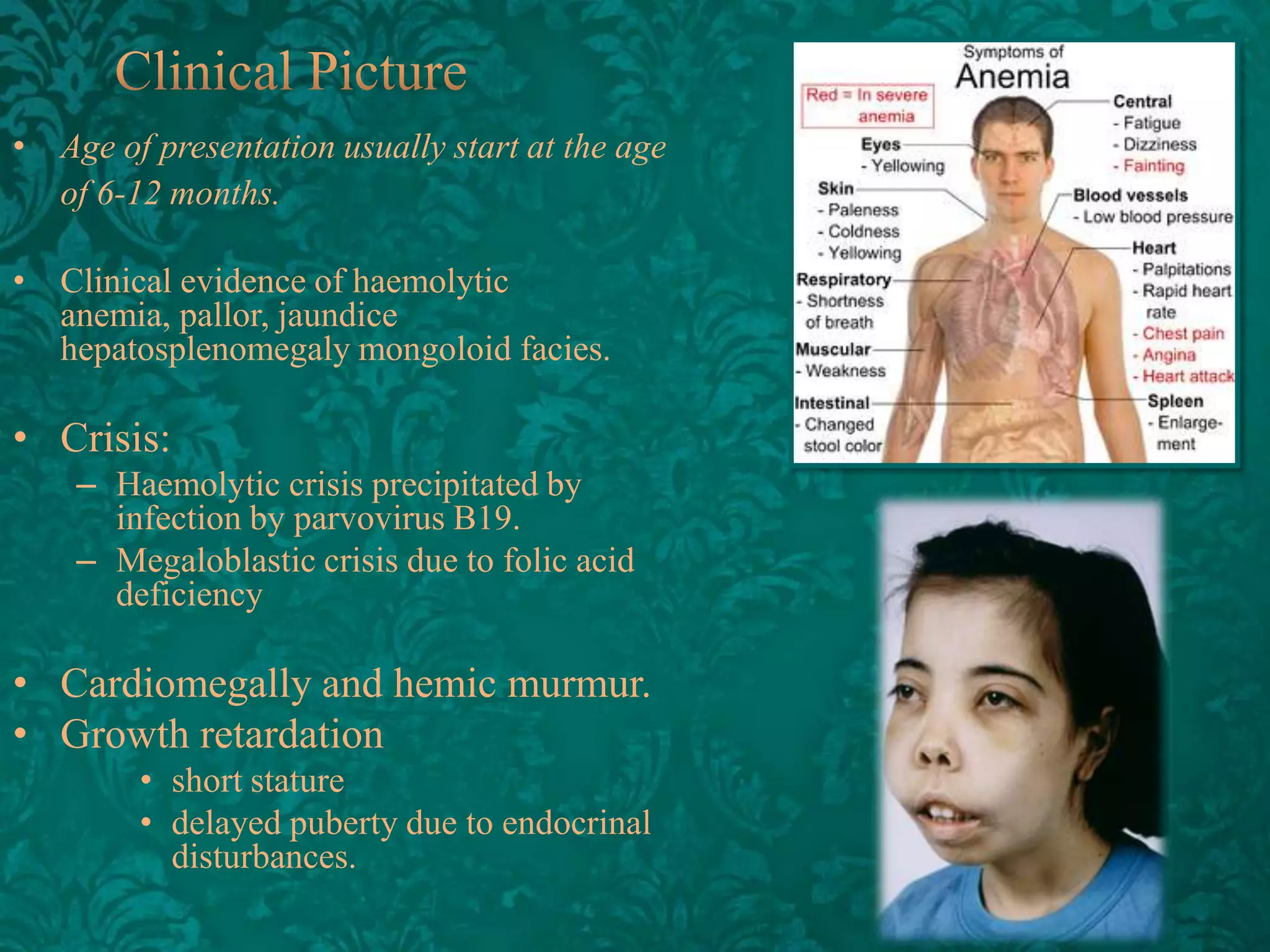
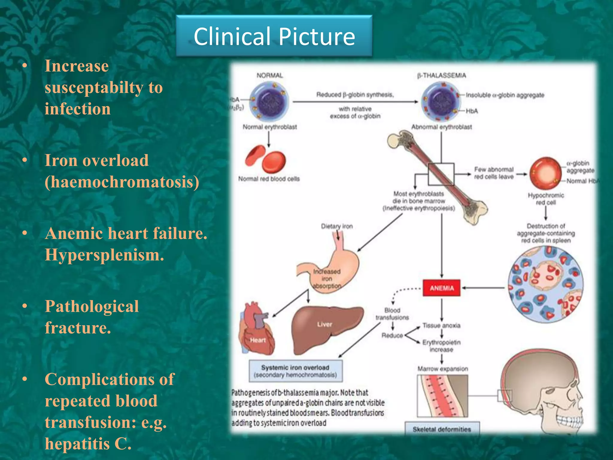
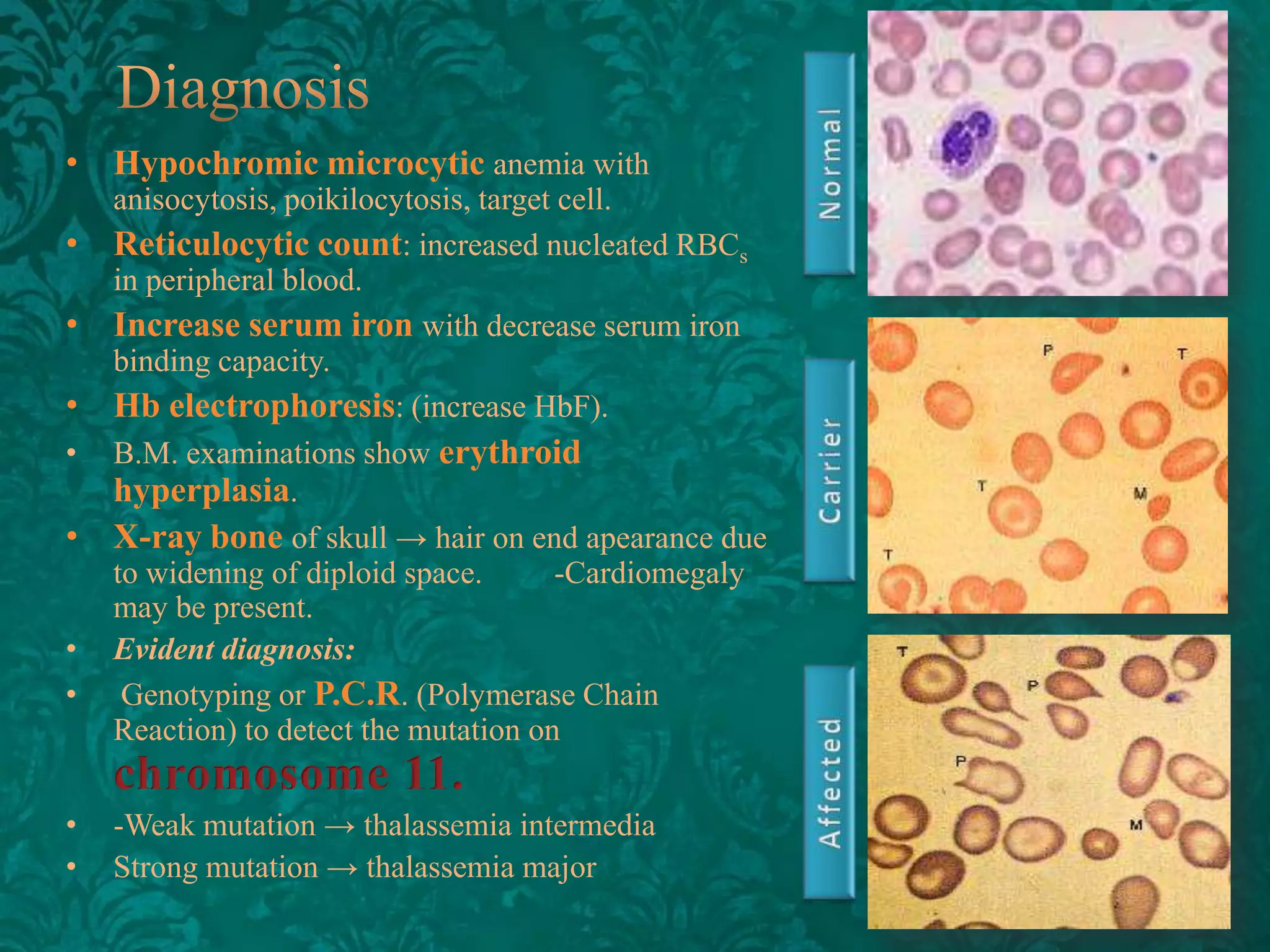
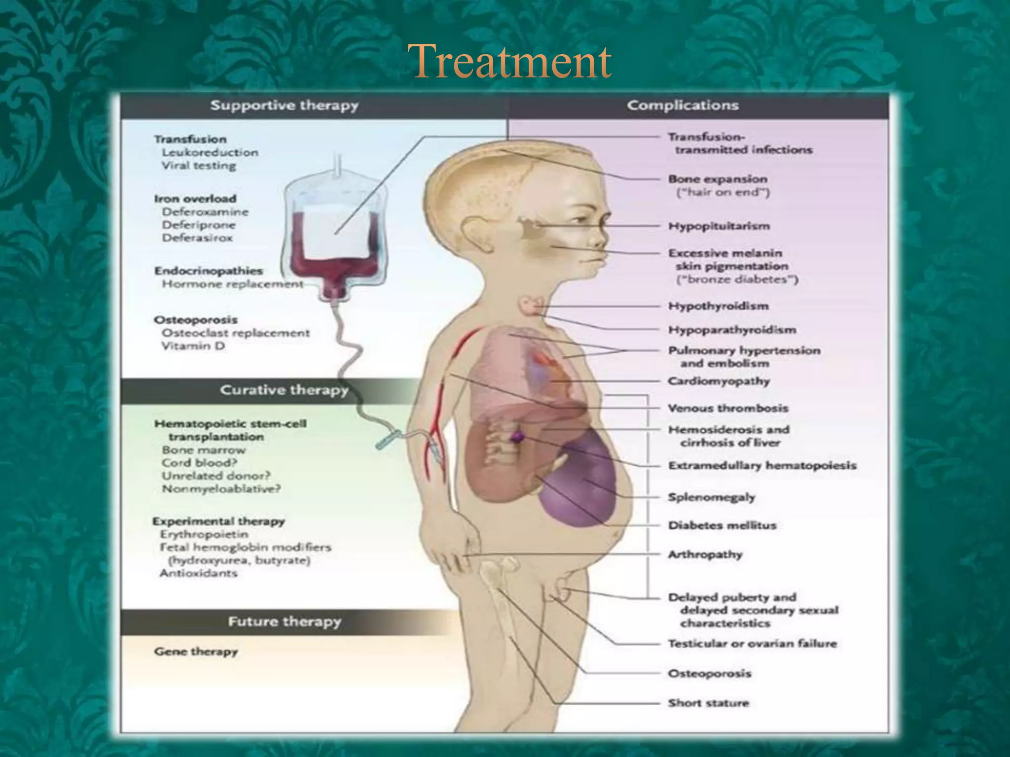
This document discusses β-thalassemia, a genetic blood disorder caused by mutations in the β-globin gene resulting in reduced or absent β-chain production and hemoglobin synthesis. It is characterized by microcytic hypochromic anemia and is most common around the Mediterranean sea. The degree of β-chain deficiency determines the severity from β° (no β-chains) to β++ (more β-chains). Clinical manifestations include anemia, jaundice, hepatosplenomegaly, skeletal abnormalities, and heart failure. Management involves blood transfusions, chelation therapy, and folic acid supplementation.
Molecular testing and tumor testing. Have you ever been asked about it? Have you wondered the importance of it, as it relates to your particular cancer? Have you ever wondered if you should or shouldn't have your tumor tested, and what that involves? Dr. Bekaii-Saab, MD will discuss the importance of testing the molecular biology of an individual patients tumor. How they do that and why it may or may not be important to have done. He will talk about how this is playing an even bigger role in choice of treatment options for patients now more than ever. And about the way physicians are making treatment choices based on each individuals molecular biology of their tumor.
Dr. Bekaii-Saab is the Section Chief, Gastrointestinal Oncology, James Cancer Hospital and Solove Research Institute. Dr. Bekaii-Saab is one of America’s Best Doctors. Additionally, he has been listed in U.S. News and World Report’s Top Doctors for multiple consecutive years. His research interests include experimental therapeutics/translational research focused on molecularly-targeted and immune-mediated therapies in gastrointestinal (GI) cancers. He is the principal investigator on numerous clinical trials, including studies supported through research grants from the National Cancer Institute (NCI) and the National Comprehensive Cancer Network (NCCN). Dr. Bekaii-Saab is the recipient of the prestigious NCI clinical investigator team leadership award and the ASCO leadership program development award.
Molecular genetics: it deals with the structure, composition, function and replication of chromosomes and genes, representing genetics material like DNA and RNA.
Case presentation of a patient with autoimmune hemolytic anemia. Interventions are embedded in a detailed graph displaying the clinical course over 4 years.
Please scroll through using Go: Next Item in Adobe. This was originally presented in Keynote.
Iron Chelation Therapy
Ashutosh Lal, MD.
January 18, 2014
Thalassemia Patient and Family Conference
Northern California Comprehensive Thalassemia Center
Children's Hospital Oakland
Anemia - Types, Pathophysiology, Clinical Manifestations, Etiology, TreatmentMd Altamash Ahmad
Anaemia can be defined as a reduction from normal of the quantity of haemoglobin in the blood.
It is not one disease, but a condition that results from a number of different pathologies.
The World Health Organisation defines anaemia in adults as haemoglobin levels less than 13g/dL for males and less than 12g/dL for females.
The low haemoglobin level results in a corresponding decrease in the oxygen-carrying capacity of the blood.
Anaemia is possibly one of the most common conditions in the world and results in significant morbidity and mortality, particularly in the developing world.
Title: Understanding Anemia: Causes, Types, Clinical Features, and Diagnostic Investigations
Anemia is a condition characterized by a deficiency in the number or quality of red blood cells (RBCs) or hemoglobin in the blood, leading to reduced oxygen-carrying capacity. It is a prevalent global health issue affecting people of all ages, genders, and socioeconomic backgrounds. Understanding the causes, types, clinical features, and diagnostic investigations of anemia is crucial for effective management and treatment.
**Causes of Anemia:**
Anemia can result from various factors that disrupt the production, lifespan, or function of red blood cells. Some common causes include:
1. **Iron Deficiency:** Insufficient intake or absorption of iron, essential for hemoglobin synthesis, is a primary cause of anemia globally. It can stem from poor dietary intake, chronic blood loss (e.g., menstruation, gastrointestinal bleeding), or increased demand during pregnancy.
2. **Vitamin Deficiencies:** Deficiencies in vitamins such as vitamin B12 (cobalamin) or folate (vitamin B9) can impair RBC production, leading to megaloblastic anemia.
3. **Chronic Diseases:** Conditions like chronic kidney disease, inflammatory disorders (e.g., rheumatoid arthritis), and infections can disrupt erythropoiesis (RBC production) or accelerate RBC destruction, causing anemia.
4. **Hemolytic Disorders:** Inherited or acquired conditions that increase the breakdown (hemolysis) of red blood cells, such as sickle cell disease, thalassemia, or autoimmune hemolytic anemia, can result in anemia.
5. **Bone Marrow Disorders:** Diseases affecting the bone marrow, including leukemia, myelodysplastic syndromes, and aplastic anemia, can lead to decreased RBC production and anemia.
**Types of Anemia:**
Anemia is classified based on the underlying mechanism or etiology, leading to several types:
1. **Iron-Deficiency Anemia:** Characterized by low iron levels, resulting in decreased hemoglobin synthesis and microcytic (small-sized) RBCs.
2. **Megaloblastic Anemia:** Caused by impaired DNA synthesis in RBC precursors due to deficiencies in vitamin B12 or folate, leading to macrocytic (large-sized) RBCs.
3. **Hemolytic Anemia:** Occurs due to increased RBC destruction, either intravascularly (within blood vessels) or extravascularly (outside blood vessels), leading to various subtypes like autoimmune hemolytic anemia, hereditary spherocytosis, and sickle cell disease.
4. **Anemia of Chronic Disease:** Associated with chronic inflammation, infections, or malignancies, leading to impaired iron metabolism and decreased RBC production.
5. **Aplastic Anemia:** Results from bone marrow failure, leading to decreased production of all blood cell types, including RBCs.
**Clinical Features of Anemia:**
The clinical presentation of anemia can vary depending on its severity, underlying cause, and individual factors. Common clinical features include:
A condition in which the blood doesn't have enough healthy red blood cells.
Anaemia results from a lack of red blood cells or dysfunctional red blood cells in the body. This leads to reduced oxygen flow to the body's organs.
Utility of routine surveillance blood cultures in asymptomatic allogeneic he...Ahmed Abdelhakeem
Utility of routine surveillance blood cultures in asymptomatic allogeneic hematopoietic stem cell transplant recipients with indwelling central venous catheters at a comprehensive cancer center
Title: Sense of Smell
Presenter: Dr. Faiza, Assistant Professor of Physiology
Qualifications:
MBBS (Best Graduate, AIMC Lahore)
FCPS Physiology
ICMT, CHPE, DHPE (STMU)
MPH (GC University, Faisalabad)
MBA (Virtual University of Pakistan)
Learning Objectives:
Describe the primary categories of smells and the concept of odor blindness.
Explain the structure and location of the olfactory membrane and mucosa, including the types and roles of cells involved in olfaction.
Describe the pathway and mechanisms of olfactory signal transmission from the olfactory receptors to the brain.
Illustrate the biochemical cascade triggered by odorant binding to olfactory receptors, including the role of G-proteins and second messengers in generating an action potential.
Identify different types of olfactory disorders such as anosmia, hyposmia, hyperosmia, and dysosmia, including their potential causes.
Key Topics:
Olfactory Genes:
3% of the human genome accounts for olfactory genes.
400 genes for odorant receptors.
Olfactory Membrane:
Located in the superior part of the nasal cavity.
Medially: Folds downward along the superior septum.
Laterally: Folds over the superior turbinate and upper surface of the middle turbinate.
Total surface area: 5-10 square centimeters.
Olfactory Mucosa:
Olfactory Cells: Bipolar nerve cells derived from the CNS (100 million), with 4-25 olfactory cilia per cell.
Sustentacular Cells: Produce mucus and maintain ionic and molecular environment.
Basal Cells: Replace worn-out olfactory cells with an average lifespan of 1-2 months.
Bowman’s Gland: Secretes mucus.
Stimulation of Olfactory Cells:
Odorant dissolves in mucus and attaches to receptors on olfactory cilia.
Involves a cascade effect through G-proteins and second messengers, leading to depolarization and action potential generation in the olfactory nerve.
Quality of a Good Odorant:
Small (3-20 Carbon atoms), volatile, water-soluble, and lipid-soluble.
Facilitated by odorant-binding proteins in mucus.
Membrane Potential and Action Potential:
Resting membrane potential: -55mV.
Action potential frequency in the olfactory nerve increases with odorant strength.
Adaptation Towards the Sense of Smell:
Rapid adaptation within the first second, with further slow adaptation.
Psychological adaptation greater than receptor adaptation, involving feedback inhibition from the central nervous system.
Primary Sensations of Smell:
Camphoraceous, Musky, Floral, Pepperminty, Ethereal, Pungent, Putrid.
Odor Detection Threshold:
Examples: Hydrogen sulfide (0.0005 ppm), Methyl-mercaptan (0.002 ppm).
Some toxic substances are odorless at lethal concentrations.
Characteristics of Smell:
Odor blindness for single substances due to lack of appropriate receptor protein.
Behavioral and emotional influences of smell.
Transmission of Olfactory Signals:
From olfactory cells to glomeruli in the olfactory bulb, involving lateral inhibition.
Primitive, less old, and new olfactory systems with different path
These simplified slides by Dr. Sidra Arshad present an overview of the non-respiratory functions of the respiratory tract.
Learning objectives:
1. Enlist the non-respiratory functions of the respiratory tract
2. Briefly explain how these functions are carried out
3. Discuss the significance of dead space
4. Differentiate between minute ventilation and alveolar ventilation
5. Describe the cough and sneeze reflexes
Study Resources:
1. Chapter 39, Guyton and Hall Textbook of Medical Physiology, 14th edition
2. Chapter 34, Ganong’s Review of Medical Physiology, 26th edition
3. Chapter 17, Human Physiology by Lauralee Sherwood, 9th edition
4. Non-respiratory functions of the lungs https://academic.oup.com/bjaed/article/13/3/98/278874
Best Ayurvedic medicine for Gas and IndigestionSwastikAyurveda
Here is the updated list of Top Best Ayurvedic medicine for Gas and Indigestion and those are Gas-O-Go Syp for Dyspepsia | Lavizyme Syrup for Acidity | Yumzyme Hepatoprotective Capsules etc
These lecture slides, by Dr Sidra Arshad, offer a quick overview of the physiological basis of a normal electrocardiogram.
Learning objectives:
1. Define an electrocardiogram (ECG) and electrocardiography
2. Describe how dipoles generated by the heart produce the waveforms of the ECG
3. Describe the components of a normal electrocardiogram of a typical bipolar lead (limb II)
4. Differentiate between intervals and segments
5. Enlist some common indications for obtaining an ECG
6. Describe the flow of current around the heart during the cardiac cycle
7. Discuss the placement and polarity of the leads of electrocardiograph
8. Describe the normal electrocardiograms recorded from the limb leads and explain the physiological basis of the different records that are obtained
9. Define mean electrical vector (axis) of the heart and give the normal range
10. Define the mean QRS vector
11. Describe the axes of leads (hexagonal reference system)
12. Comprehend the vectorial analysis of the normal ECG
13. Determine the mean electrical axis of the ventricular QRS and appreciate the mean axis deviation
14. Explain the concepts of current of injury, J point, and their significance
Study Resources:
1. Chapter 11, Guyton and Hall Textbook of Medical Physiology, 14th edition
2. Chapter 9, Human Physiology - From Cells to Systems, Lauralee Sherwood, 9th edition
3. Chapter 29, Ganong’s Review of Medical Physiology, 26th edition
4. Electrocardiogram, StatPearls - https://www.ncbi.nlm.nih.gov/books/NBK549803/
5. ECG in Medical Practice by ABM Abdullah, 4th edition
6. Chapter 3, Cardiology Explained, https://www.ncbi.nlm.nih.gov/books/NBK2214/
7. ECG Basics, http://www.nataliescasebook.com/tag/e-c-g-basics
263778731218 Abortion Clinic /Pills In Harare ,sisternakatoto
263778731218 Abortion Clinic /Pills In Harare ,ABORTION WOMEN’S CLINIC +27730423979 IN women clinic we believe that every woman should be able to make choices in her pregnancy. Our job is to provide compassionate care, safety,affordable and confidential services. That’s why we have won the trust from all generations of women all over the world. we use non surgical method(Abortion pills) to terminate…Dr.LISA +27730423979women Clinic is committed to providing the highest quality of obstetrical and gynecological care to women of all ages. Our dedicated staff aim to treat each patient and her health concerns with compassion and respect.Our dedicated group ABORTION WOMEN’S CLINIC +27730423979 IN women clinic we believe that every woman should be able to make choices in her pregnancy. Our job is to provide compassionate care, safety,affordable and confidential services. That’s why we have won the trust from all generations of women all over the world. we use non surgical method(Abortion pills) to terminate…Dr.LISA +27730423979women Clinic is committed to providing the highest quality of obstetrical and gynecological care to women of all ages. Our dedicated staff aim to treat each patient and her health concerns with compassion and respect.Our dedicated group of receptionists, nurses, and physicians have worked together as a teamof receptionists, nurses, and physicians have worked together as a team wwww.lisywomensclinic.co.za/
New Drug Discovery and Development .....NEHA GUPTA
The "New Drug Discovery and Development" process involves the identification, design, testing, and manufacturing of novel pharmaceutical compounds with the aim of introducing new and improved treatments for various medical conditions. This comprehensive endeavor encompasses various stages, including target identification, preclinical studies, clinical trials, regulatory approval, and post-market surveillance. It involves multidisciplinary collaboration among scientists, researchers, clinicians, regulatory experts, and pharmaceutical companies to bring innovative therapies to market and address unmet medical needs.
Ozempic: Preoperative Management of Patients on GLP-1 Receptor Agonists Saeid Safari
Preoperative Management of Patients on GLP-1 Receptor Agonists like Ozempic and Semiglutide
ASA GUIDELINE
NYSORA Guideline
2 Case Reports of Gastric Ultrasound
Local Advanced Lung Cancer: Artificial Intelligence, Synergetics, Complex Sys...Oleg Kshivets
Overall life span (LS) was 1671.7±1721.6 days and cumulative 5YS reached 62.4%, 10 years – 50.4%, 20 years – 44.6%. 94 LCP lived more than 5 years without cancer (LS=2958.6±1723.6 days), 22 – more than 10 years (LS=5571±1841.8 days). 67 LCP died because of LC (LS=471.9±344 days). AT significantly improved 5YS (68% vs. 53.7%) (P=0.028 by log-rank test). Cox modeling displayed that 5YS of LCP significantly depended on: N0-N12, T3-4, blood cell circuit, cell ratio factors (ratio between cancer cells-CC and blood cells subpopulations), LC cell dynamics, recalcification time, heparin tolerance, prothrombin index, protein, AT, procedure type (P=0.000-0.031). Neural networks, genetic algorithm selection and bootstrap simulation revealed relationships between 5YS and N0-12 (rank=1), thrombocytes/CC (rank=2), segmented neutrophils/CC (3), eosinophils/CC (4), erythrocytes/CC (5), healthy cells/CC (6), lymphocytes/CC (7), stick neutrophils/CC (8), leucocytes/CC (9), monocytes/CC (10). Correct prediction of 5YS was 100% by neural networks computing (error=0.000; area under ROC curve=1.0).
Flu Vaccine Alert in Bangalore Karnatakaaddon Scans
As flu season approaches, health officials in Bangalore, Karnataka, are urging residents to get their flu vaccinations. The seasonal flu, while common, can lead to severe health complications, particularly for vulnerable populations such as young children, the elderly, and those with underlying health conditions.
Dr. Vidisha Kumari, a leading epidemiologist in Bangalore, emphasizes the importance of getting vaccinated. "The flu vaccine is our best defense against the influenza virus. It not only protects individuals but also helps prevent the spread of the virus in our communities," he says.
This year, the flu season is expected to coincide with a potential increase in other respiratory illnesses. The Karnataka Health Department has launched an awareness campaign highlighting the significance of flu vaccinations. They have set up multiple vaccination centers across Bangalore, making it convenient for residents to receive their shots.
To encourage widespread vaccination, the government is also collaborating with local schools, workplaces, and community centers to facilitate vaccination drives. Special attention is being given to ensuring that the vaccine is accessible to all, including marginalized communities who may have limited access to healthcare.
Residents are reminded that the flu vaccine is safe and effective. Common side effects are mild and may include soreness at the injection site, mild fever, or muscle aches. These side effects are generally short-lived and far less severe than the flu itself.
Healthcare providers are also stressing the importance of continuing COVID-19 precautions. Wearing masks, practicing good hand hygiene, and maintaining social distancing are still crucial, especially in crowded places.
Protect yourself and your loved ones by getting vaccinated. Together, we can help keep Bangalore healthy and safe this flu season. For more information on vaccination centers and schedules, residents can visit the Karnataka Health Department’s official website or follow their social media pages.
Stay informed, stay safe, and get your flu shot today!
- Video recording of this lecture in English language: https://youtu.be/lK81BzxMqdo
- Video recording of this lecture in Arabic language: https://youtu.be/Ve4P0COk9OI
- Link to download the book free: https://nephrotube.blogspot.com/p/nephrotube-nephrology-books.html
- Link to NephroTube website: www.NephroTube.com
- Link to NephroTube social media accounts: https://nephrotube.blogspot.com/p/join-nephrotube-on-social-media.html
Muktapishti is a traditional Ayurvedic preparation made from Shoditha Mukta (Purified Pearl), is believed to help regulate thyroid function and reduce symptoms of hyperthyroidism due to its cooling and balancing properties. Clinical evidence on its efficacy remains limited, necessitating further research to validate its therapeutic benefits.
2. Ahmed Salah
Eldin (42)
Ahmed Adel
Emara (49)
Ahmed Salah
Eldin Maarof
(43)
Ahmed Adel
Fouad (48)
Ahmed Tarek
Abd Elnabi
(45)
Ahmed Adel
Abd Abd
Elrahman (46)
3. This is a group characterized by an MCV (<76 fL) and MCHC (<32g/dL)
below the normal range or an increased microcytic and hypochromic
subpopulation. These changes result from a decreased production of
hemoglobin.
4.
5.
6. β - Most common around meditranian sea.
In β - thalassemia there is impaired production of β -chain due to
mutation of one gene "heterozygous" or both genes homozygous".
According to degree of β – chain deficiency β thalassmia is divided
into:
• Βo no β-chain synthesis no HbA only HbF and HbA2.
• β+ little β-chain synthesis, so HbA is scanty and more HbF and
HbA2.
• β++ more β chain synthesis → thalassemia intermediate.
7. • Age of presentation usually start at the age
of 6-12 months.
• Clinical evidence of haemolytic
anemia, pallor, jaundice
hepatosplenomegaly mongoloid facies.
• Crisis:
– Haemolytic crisis precipitated by
infection by parvovirus B19.
– Megaloblastic crisis due to folic acid
deficiency
• Cardiomegally and hemic murmur.
• Growth retardation
• short stature
• delayed puberty due to endocrinal
disturbances.
8. Clinical Picture
• Increase
susceptabilty to
infection
• Iron overload
(haemochromatosis)
• Anemic heart failure.
Hypersplenism.
• Pathological
fracture.
• Complications of
repeated blood
transfusion: e.g.
hepatitis C.
9. • Hypochromic microcytic anemia with
•
•
•
•
•
•
•
•
•
anisocytosis, poikilocytosis, target cell.
Reticulocytic count: increased nucleated RBCs
in peripheral blood.
Increase serum iron with decrease serum iron
binding capacity.
Hb electrophoresis: (increase HbF).
B.M. examinations show erythroid
hyperplasia.
X-ray bone of skull → hair on end apearance due
to widening of diploid space.
-Cardiomegaly
may be present.
Evident diagnosis:
Genotyping or P.C.R. (Polymerase Chain
Reaction) to detect the mutation on
-Weak mutation → thalassemia intermedia
Strong mutation → thalassemia major
10.
11. (Heterozygous ) It may be asymptomatic or symptomatic:
-Mild anemia (Hb 9-11 g/dl) accentuated during infections.
-Anemia is microcytic hypochromic and does not respond to iron
therapy.
-No jaundice or other signs of hemolysis.
-Hb electrophoresis: Hb A is predominant, Hb A2 is increase
( 4%), Hb F may be slightly increase.
12. : Affects 2-10 % of homozygous patients and is characterized by:
• Patients have higher ability to produce β –chain and high production of γ-chains.
• Genetic, morphologic and biochemical features: like β-thalassemia major.
• Anemia is milder (Hg level 7-10g/dl).
• Pallor, jaundice, facial bony changes, splenomegaly and hepatomegaly are present.
• No growth retardation and no hypogonadism.
Treatment: -Blood transfusion is usually not needed and given only when required.
-Decrease iron absorption from GIT (e.g. drink a cup of tea after each meal).
-Chelation therapy and folic acid supplementation.
13. •
•
•
Hb E is the second most common Hb variant.
The clinical course of E β-thalassemia is punctuated by acute and chronic
complications that may cause serious morbidity and mortality.
The marked expansion of erythropoiesis is responsible for much of the pathology of
the disease, including:
1. hepatosplenomegaly
2. extramedullary hematopoietic masses,
3. growth retardation,
4. delayed sexual maturation
5. bone deformities.
6. Cardiopulmonary disease is the most common cause of death in Hb E βThalassemia.
16. 1-Dietary factors->Lack of breast feeding with unsupplemented
cow milk or formula and delayed weaning-Substances that
diminish the absorption of iron e.g. phytates...
2-Storage defect->iron deficient mother- preterm child-LBWIUGR.
3-Infection->reduces appetite, absorption-increases
loss(vomiting..)-depresses BM.
4-Hemorrhage->chronic blood loss e.g. hookworm infestationsmilk protein intolerance.
5-Hemosiderinuria, hemoglobinuria, and pulmonary
hemosiderosis->loss of iron.
6-Malabsorption of iron->Prolonged achlorhydria.
17. Clinical Picture:
In addition to general manifestations
of anemia there are decrease
mental concentration, atrophy of
mucosa,anorexia,immune
suppretion,muscular weakness… .
Investigations:
Serum ferritin is low, Decreased
serum iron, increased iron binding
capacity, Microcytosis, hypochromi
a, anisocytosis, Increased red cell
distribution width (RDW).
18. Treatment:
Prophylactic->adequate fe supply for pregnantsupplementation starting from 4th month.
Curative-> 1-6 mg/kg/day of elemental iron.
2-Diet adjustments- increase amount of Fe in the diet.
3-Transfusion in severe cases Hb<6.
4-parentral is not preferred.
19. Atransferrinemia
Definition:
Atranferrinemia is a genetic disorder wherein the plasma protein that carries iron
(transferrin) in the blood stream is lacking.
Pathophysiology:
This disease is characterized by a microcytic hypochromic anemia, and an iron
deposition in the heart and liver.
This iron damage to the heart can also be the cause of heart
failure while anemia is typically hypochromic and microcytic.
Death may occur due to heart failure or pneumonia.
20. Clinical Picture:
Iron overload occurs mainly in the
liver, eye, pancreas, thyroid, kidney and
joints, leading to moderate to serious symptoms of
liver and eye, arthropathy and hypothyroidism.
Treatment:
plasma infusion of transferrin->If this therapy is
repeated once or twice monthly, the patient becomes
hematologically normal within a few months.
21.
22. •
•
•
•
A heterogeneous group of disorders
Variable red cell hypochromia, anemia
The ringed sideroblasts should total 10% or more of the
nucleated red cell
Failure to utilize iron properly during heme synthesis in the
mitochondria
24. Defect in mitochondrial function
and heme synthesis
mutant ALAS
enzymes
Ferrochelatase
defects
copro-oxidase
defects
25. •History of failure of growth
•Family history of mitochondrial disease and anemia
•General symptoms of anemia,
•Vital signs – Hypothermia
•Skin - Photosensitivity (porphyria)
•Cardiovascular – Fatigue
•Respiratory – Dyspnea
•Musculoskeletal - Muscular weakness
•Genitourinary - Pink staining of diapers from porphyrins in
urine
26. • Complete blood count (CBC),
• peripheral smear,
• iron studies,
• bone marrow aspiration and biopsy.
Microcytosis
• variable degree of anemia
• Serum iron and ferritin are elevated
• TIBC reduced.
partially treated iron deficiency anemia.
acquired disorders associated with the
presence of excess ringed sideroblasts
in the marrow
27. • Infants developing in the womb of a woman, who have elevated
blood lead level, are susceptible to lead poisoning. There is also a
risk as the baby comes closer to full term, as the woman is more
likely to have a premature, or with a low birth weight.
• Children are more at risk for lead poisoning because their smaller
bodies are in a continuous state of growth and development.
• The classic signs and symptoms in children are loss of appetite,
abdominal pain, vomiting, weight loss, constipation, anemia,
kidney failure, irritability, lethargy, learning disabilities, and
behavioral problems.
28.
29.
Copper deficiency is a very rare
hematological and neurological disorder.
Copper deficiency is only seen in
malnourished premature babies and in
patients receiving long-term parenteral
nutrition with inadequate copper
supplementation.
Copper is absorbed almost exclusively
during the last trimester and deficiency in
a premature infant may be accentuated if
the child is given copper-deficient feeds
such as cows’ milk at an early age.
30. •
Surgery : Bariatric surgery, such as gastric bypass surgery.The
disruption of the intestines and stomach from the surgery can cause
absorption difficulties.
• Zinc Toxicity
• Hereditary Disorders : Menkes disease is a hereditary condition
caused by a defective gene involved with the metabolism of copper
in the body. Menkes disease is usually a fatal disease with most
children dying within the first ten years of life.
• Other : It is rarely suggested that excess iron supplementation
causes copper deficiency myelopathy. celiac disease, probably due
to malabsorption in the intestines.Still, a large percentage, around
20%, of cases have unknown causes.
31. •
Iron Transportation: Hephaestin is a copper containing ferroxidase
enzyme located in the duodenal muscosa that oxidizes iron and
facilitates its transfer across the basolateral membrane into circulation.
• Ceruloplasmin,This enzyme is required to mobilize iron from the
reticuloendothelial cell to plasma. It also oxidizes iron from its ferrous
state to the ferric form that is required for iron binding.
• Impairment in these copper dependent enzymes that transport iron
may cause the secondary iron deficiency anemia.
• Another speculation for the cause of anemia is involving the
mitochondrial enzyme cytochrome c oxidase. The lower rate of the
enzyme might cause the excess iron to clump, giving the heme an
unusual pattern known as ringed sideroblastic anemia cells.
32. Hematological Findings:
• tiredness, fatigue, and light headedness.
These are all common symptoms of
anemia.
• All types of anemia including microcytic
, macrocytic and normocytic manifest.
Around half of the patients displayed
“leukopenia".
• In addition to leukopenia, many patients
are deficient in neutrophils (neutropenia).
• Usually prolonged copper deficiency has
to persist to manifest thrombocytopenia.
33. Neurological presentation:
• Myelopathy : difficulty in walking caused by sensory
ataxia due to dorsal column dysfunction or degeneration
of the spinal cord
• Peripheral neuropathy : numbness or tingling that can start
in the extremities .
• Optic neuropathy : signs of vision and color loss. The
vision is usually lost in the peripheral views of the eye.
34. • The diagnosis is made by demonstrating a low serum copper and
ceruloplasmin.
• Examination of the marrow reveals decreased granulocyte
maturation, vacuolization of red blood cell precursors, and ringed
sideroblastic cells are present .
• Neutropenia has become a hematological hallmark, enabling
physicians to investigate copper deficiency as a diagnosis.
35.
36. Lead poisoning is a medical condition
caused by increased levels of the heavy
metal lead in the body.
Routes of exposure to lead includes
contaminated air, water, soil, food,and
consumer products. Occupational exposure
is a common cause of lead poisoning in
adults.
37. • Infants developing in the
womb of a woman, who have
elevated blood lead level, are
susceptible to lead poisoning.
There is also a risk to have a
premature, or with a low birth
weight.
• Children are more at risk for
lead poisoning because their
smaller bodies are in a
continuous state of growth
and development.
38. •
One of the main causes for the pathology of lead is that it
interferes with the activity of an essential enzyme called deltaaminolevulinic acid dehydratase, or ALAD, which is important in
the biosynthesis of heme, the cofactor found in hemoglobin.
• Lead also inhibits the enzyme ferrochelatase, another enzyme
involved in the formation of heme.
• Lead's interference with heme synthesis results in production of
zinc protoporphyrin and the development of anemia.
• Another effect of lead's interference with heme synthesis is the
buildup of heme precursors, such as aminolevulinic acid, which
may be directly or indirectly harmful to neurons.
39. • The main tool in diagnosing and assessing the severity of lead
poisoning is laboratory analysis of the blood lead level (BLL).
• Blood film examination may reveal basophilic stippling of red
blood cells (dots in red blood cells visible through a microscope),
as well as the changes normally associated with iron-deficiency
anemia (microcytosis and hypochromasia).
40. • Anemia is the commonest hematologic manifestation of
systemic disease and is the result of a number of contributing
factors. It occurs in patients with a variety of chronic
inflammatory and malignant conditions and can be
complicated by bleeding, immune phenomena, nutritional
deficiencies and other organ- or disease-specific features.
41.
42. The anemia is usually mild, 2–3 g/dL below the lower normal limit for
a given individual, and nonprogressive with the severity being related
to the severity of the disease process.
•
Associated features indicative of chronic inflammation such as
neutrophilia, thrombocytosis and rouleaux formation may be present.
• In addition to hematologic tests, there may be low albumin, elevated
fibrinogen and γ-globulin concentrations.
In anemia of chronic disease without iron deficiency, ferritin levels
should be normal or high, reflecting the fact that iron is stored within
cells. In iron deficiency anemia ferritin should be low.
Examination of the bone marrow to look for the absence or presence
of iron, or a trial of iron can provide more definitive diagnoses.
43.
44. • Vitamin b6 deficiency results in inadequate heme production and, thus,
lowers concentrations of hemoglobin in red blood cells. This condition,
called microcytic, hypochromic anemia, interferes with the ability of red
blood cells to deliver oxygen to tissues and impairs their ability to produce
ATP via anaerobic metabolism.
46. • Vitamin B6 deficiency is rare when food is abundant, however,
the damaging effect of high temperature on Vitamin B6 was
not well understood, and Vitamin B6 added to infant formula
was destroyed during heat processing, thus many formula-fed
infants developed Vitamin B6 deficiency. This unfortunate
“epidemic” of Vitamin B6 caused serious complications such
as seizures and convulsions. Manufacturing procedures have
changed so that infant formula now contains sufficient
amounts of this essential nutrient.