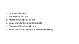
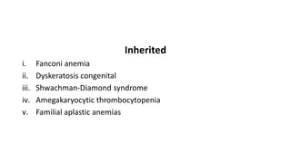
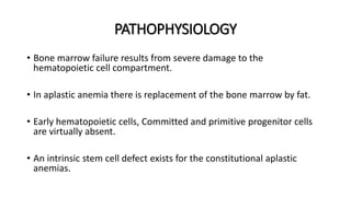
- Dr Rahul Arya
- Assistant Professor
- Department of Medicine
DEFINATION
• Aplastic anemia is pancytopenia with bone marrow hypocellularity.
CAUSES
Acquired
1. Secondary
i. Radiation
ii. Drugs and chemicals- Benzene, allopurinol, leflunomide, gold, etc
iii. Viruses
a. Epstein-Barr virus
b. Hepatitis (non-A, non-B, non-C hepatitis)
c. Parvovirus B19
d. HIV-1 (AIDS)
iv. Immune diseases
a. Eosinophilic fasciitis
b. Hyperimmunoglobulinemia
c. Large granular lymphocytosis (LGL)
d. Thymoma/thymic carcinoma
e. Graft-versus-host disease in immunodeficiency
v. Paroxysmal nocturnal
vi. hemoglobinuria (PNH)
vii. Pregnancy
viii. Idiopathic
Inherited
i. Fanconi anemia
ii. Dyskeratosis congenital
iii. Shwachman-Diamond syndrome
iv. Amegakaryocytic thrombocytopenia
v. Familial aplastic anemias
EPIDEMIOLOGY
• Men and women are affected with equal frequency.
• The age distribution is biphasic, with the major peak in the teens and
twenties and a second rise in older adults.
PATHOPHYSIOLOGY
• Bone marrow failure results from severe damage to the
hematopoietic cell compartment.
• In aplastic anemia there is replacement of the bone marrow by fat.
• Early hematopoietic cells, Committed and primitive progenitor cells
are virtually absent.
• An intrinsic stem cell defect exists for the constitutional aplastic
anemias.
CLINICAL FEATURES- HISTORY
• Aplastic anemia can appear abruptly or insidiously.
• Bleeding is the most common early symptom
• Easy bruising, oozing from the gums, nose bleeds, heavy menstrual flow,
petechiae.
• Symptoms of anemia are also frequent, including lassitude, weakness, shortness
of breath, and a pounding sensation in the ears.
• Patients often feel and look remarkably well despite drastically reduced blood
counts.
• Prior drug use, chemical exposure, and preceding viral illnesses must
often be elicited.
Physical Examination
• Pallor
• Petechiae, ecchymoses, retinal hemorrhages.
• Lymphadenopathy and splenomegaly are highly atypical of aplastic
anemia.
LABORATORY STUDIES
 Blood
• large erythrocytes and a paucity of platelets and granulocytes.
• Mean corpuscular volume (MCV) is commonly increased.
• Reticulocytes are absent or few, and lymphocyte numbers may be
normal or reduced.
 Bone Marrow
• The bone marrow is usually readily aspirated but dilute on smear, and
the fatty biopsy specimen may be grossly pale on withdrawal.
• In severe aplasia, the smear of the aspirated specimen shows only red
cells, residual lymphocytes, and stromal cells.
• Biopsy shows mainly fat under the microscope, with hematopoietic
cells occupying <25% of the marrow space.
TREATMENT
 HEMATOPOIETIC STEM CELL TRANSPLANTATION
• This is the best therapy for the younger patient with a fully
histocompatible sibling donor.
• For allogeneic transplant from fully matched siblings, long-term
survival rates for children are approximately 90%.
 IMMUNOSUPPRESSION
• Antithymocyte globulin (ATG) in combination with cyclosporine.
• It induces hematologic recovery in 60–70% of patients.
• Relapse is frequent.
• Horse ATG is administered as intravenous infusions over 4 days.
• Methylprednisolone is administered with ATG to ameliorate the immune
consequences of heterologous protein infusion.
• Cyclosporine is administered orally at an initial high dose, with subsequent
adjustment according to blood levels obtained every 2 weeks.
SUPPORTIVE CARE
• Infection in the presence of severe neutropenia must be aggressively
treated by prompt institution of parenteral, broad-spectrum antibiotics.
• Both platelet and erythrocyte numbers can be maintained by transfusion.
• RBCs should be transfused to maintain a normal level of activity, usually at
a hemoglobin value of 7 g/dL.
• A regimen of 2 units every 2 weeks will replace normal losses in a patient
without a functioning bone marrow.
• In chronic anemia, the iron chelators, deferoxamine and deferasirox, should
be added at approximately the fiftieth transfusion to avoid secondary
hemochromatosis.
PROGNOSIS
• The natural history of severe aplastic anemia is rapid deterioration and
death.
• The major prognostic determinant is the blood count.
• Severe disease has been defined by the presence of two of three
parameters:
- absolute neutrophil count <500/Μl
- platelet count <20,000/μL
- corrected reticulocyte count <1%.
Aplastic anemia

Aplastic anemia

  • 1.
    - Dr RahulArya - Assistant Professor - Department of Medicine
  • 2.
    DEFINATION • Aplastic anemiais pancytopenia with bone marrow hypocellularity.
  • 3.
    CAUSES Acquired 1. Secondary i. Radiation ii.Drugs and chemicals- Benzene, allopurinol, leflunomide, gold, etc iii. Viruses a. Epstein-Barr virus b. Hepatitis (non-A, non-B, non-C hepatitis) c. Parvovirus B19 d. HIV-1 (AIDS)
  • 4.
    iv. Immune diseases a.Eosinophilic fasciitis b. Hyperimmunoglobulinemia c. Large granular lymphocytosis (LGL) d. Thymoma/thymic carcinoma e. Graft-versus-host disease in immunodeficiency
  • 5.
    v. Paroxysmal nocturnal vi.hemoglobinuria (PNH) vii. Pregnancy viii. Idiopathic
  • 6.
    Inherited i. Fanconi anemia ii.Dyskeratosis congenital iii. Shwachman-Diamond syndrome iv. Amegakaryocytic thrombocytopenia v. Familial aplastic anemias
  • 7.
    EPIDEMIOLOGY • Men andwomen are affected with equal frequency. • The age distribution is biphasic, with the major peak in the teens and twenties and a second rise in older adults.
  • 8.
    PATHOPHYSIOLOGY • Bone marrowfailure results from severe damage to the hematopoietic cell compartment. • In aplastic anemia there is replacement of the bone marrow by fat. • Early hematopoietic cells, Committed and primitive progenitor cells are virtually absent. • An intrinsic stem cell defect exists for the constitutional aplastic anemias.
  • 9.
    CLINICAL FEATURES- HISTORY •Aplastic anemia can appear abruptly or insidiously. • Bleeding is the most common early symptom • Easy bruising, oozing from the gums, nose bleeds, heavy menstrual flow, petechiae. • Symptoms of anemia are also frequent, including lassitude, weakness, shortness of breath, and a pounding sensation in the ears. • Patients often feel and look remarkably well despite drastically reduced blood counts.
  • 10.
    • Prior druguse, chemical exposure, and preceding viral illnesses must often be elicited.
  • 11.
    Physical Examination • Pallor •Petechiae, ecchymoses, retinal hemorrhages. • Lymphadenopathy and splenomegaly are highly atypical of aplastic anemia.
  • 12.
    LABORATORY STUDIES  Blood •large erythrocytes and a paucity of platelets and granulocytes. • Mean corpuscular volume (MCV) is commonly increased. • Reticulocytes are absent or few, and lymphocyte numbers may be normal or reduced.
  • 13.
     Bone Marrow •The bone marrow is usually readily aspirated but dilute on smear, and the fatty biopsy specimen may be grossly pale on withdrawal. • In severe aplasia, the smear of the aspirated specimen shows only red cells, residual lymphocytes, and stromal cells. • Biopsy shows mainly fat under the microscope, with hematopoietic cells occupying <25% of the marrow space.
  • 14.
    TREATMENT  HEMATOPOIETIC STEMCELL TRANSPLANTATION • This is the best therapy for the younger patient with a fully histocompatible sibling donor. • For allogeneic transplant from fully matched siblings, long-term survival rates for children are approximately 90%.
  • 15.
     IMMUNOSUPPRESSION • Antithymocyteglobulin (ATG) in combination with cyclosporine. • It induces hematologic recovery in 60–70% of patients. • Relapse is frequent. • Horse ATG is administered as intravenous infusions over 4 days. • Methylprednisolone is administered with ATG to ameliorate the immune consequences of heterologous protein infusion. • Cyclosporine is administered orally at an initial high dose, with subsequent adjustment according to blood levels obtained every 2 weeks.
  • 16.
    SUPPORTIVE CARE • Infectionin the presence of severe neutropenia must be aggressively treated by prompt institution of parenteral, broad-spectrum antibiotics. • Both platelet and erythrocyte numbers can be maintained by transfusion. • RBCs should be transfused to maintain a normal level of activity, usually at a hemoglobin value of 7 g/dL. • A regimen of 2 units every 2 weeks will replace normal losses in a patient without a functioning bone marrow. • In chronic anemia, the iron chelators, deferoxamine and deferasirox, should be added at approximately the fiftieth transfusion to avoid secondary hemochromatosis.
  • 17.
    PROGNOSIS • The naturalhistory of severe aplastic anemia is rapid deterioration and death. • The major prognostic determinant is the blood count. • Severe disease has been defined by the presence of two of three parameters: - absolute neutrophil count <500/Μl - platelet count <20,000/μL - corrected reticulocyte count <1%.