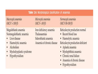
Anemia
• reduction in the HB concentration below
normal range(12 to 15 g/dl-females , 13 to
17g/dl –males) .may or may not be
accompanied by decrease in the red cell
mass .
ANEMIA OF BLOOD LOSS:
acute blood loss
• > 20% of blood volume: hypovolemic shock rather
than anemia.
• The anemia is normocytic normochromic
• plasma is restored within 2 to 3 days;
• reticulocytosis : 5 to 7 days.
chronic blood loss
• iron stores are gradually depleted.
• Iron deficiency anemia
• The anemia is microcytic hypochromic
Iron Deficiency Anemia
• Very common
• 25% to 50% of people in developing countries .
• Causes:
1. Chronic blood loss is the most important
cause :peptic ulcers, colonic cancer,
hemorrhoids,menorrhagia, metrorrhagia,
cancers).
2. low intake
3. Increased demands (pregnancy and infancy.)
4. Malabsorption (celiac disease or after
gastrectomy)
• iron deficiency develops insidiously.
• Iron stores are depleted fist, marked by :
1. a decline in serum ferritin
2. and the absence of stainable iron in the
bone marrow.
3. decrease in serum iron and
4. a rise in the serum transferrin.
5. Microcytic hypochromic anemia,
6. impaired work and cognitive performance,
7. reduced immunocompetence.
• Clinical Features
• weakness, and pallor,
• figernails, thinning, flttening, and “spooning,”
• pica, the compunction to consume non food stuffs such as ice
or clay.
• In peripheral smears red cells are microcytic and
hypochromic
• Diagnostic criteria
1. anemia, hypochromic and microcytic
2. low serum ferritin and iron levels,
3. low transferrin saturation,
4. increased total iron-binding capacity,
5. For unclear reasons, the platelet count often is elevated.
6. Erythropoietin levels are increased, but marrow cellularity
usually is only slightly increased.
Normal peripheral blood film
Microcytosis hypochromia
There is also increased anisocytosis (variation in size)
and poikilocytosis (variation in shape).
Anemia of Chronic Disease
• Most common form of anemia in hospitalized
patients.
• Arises from the suppression of erythropoiesis
by systemic inflammation.
1. Chronic microbial infections: osteomyelitis,
bacterial endocarditis&lung abscess
2. Chronic immune disorders, such as
rheumatoid arthritis and regional enteritis
3. Neoplasms, such as Hodgkin lymphoma
and carcinomas of the lung and breast
• PATHOGENESIS:
• high levels of plasma hepcidin, which blocks
the transfer of
iron to erythroid precursors by downregulating
ferroportin in macrophages.
• The elevated hepcidin levels are caused by pro-
inflmmatory cytokines such as IL-6, which
increase hepatic hepcidin synthesis.
• chronic inflammation blunts erythropoietin
synthesis by the kidney,
• the serum iron levels usually are low
• red cells may even be slightly
hypochromic and microcytic.
• storage iron in the bone marrow is
increased, the serum ferritin concentration
is elevated, and the total iron-binding
capacity is reduced.
Megaloblastic Anemias
• Folate deficiency and vitamin B12 deficiency.
• Both vitamins are required for DNA synthesis .
• The morphologic hallmark of is the presence of megaloblasts, enlarged
erythroid precursors that give rise to abnormally large red cells
(macrocytes).
• Granulocyte precursors are also increased in size
• Underlying this cellular gigantism is a defect in DNA synthesis that impairs
nuclear maturation and cell division
• the hematopoietic precursors show nuclear-cytoplasmic asynchrony
• ineffective hematopoiesis
• pancytopenia (anemia, thrombocytopenia, and
granulocytopenia
• The bone marrow is markedly hypercellular and
contains numerous megaloblastic erythroid
progenitors.
• Megaloblasts are larger than normal erythroid
progenitors (normoblasts)
• giant metamyelocytes.
• Megakaryocytes may also be abnormally large and
have bizarre multilobed nuclei.
Folate (Folic Acid) Deficiency
Anemia
• Not common,
1. poor diet (vegetables and fruits)
2. increased metabolic needs (pregnant women and ,chronic
hemolytic anemias).
3. Phenytoin and a few other drugs also inhibit folate
absorption, while
others, such as methotrexate, inhibit folate metabolism.
4. site of absorption:upper third of the small intestine;,
malabsorptive disorders that
affect this level of the gut, such as celiac disease and
tropical sprue,
• PATHOGENESIS:
• after absorption folate is transported in the blood mainly as a
monoglutamate.
• Within cells it is converted from dihydrofolate to
tetrahydrofolate by dihydrofolate reductase
• Tetrahydrofolate acts as an acceptor and donor of one-
carbon units in several reactions that are required for the
synthesis of purines and thymidylate, the building blocks of
DNA,
• its deficiency accounts for the defect in DNA replication
that underlies megaloblastic anemia.
Clinical Features:
The onset is insidious,
• weakness and easy fatigability.
• sore tongue
• The diagnosis is by measuring serum and
red cell folate and vitamin B12 levels
Vitamin B12 (Cobalamin)
Deficiency Anemia (Pernicious
Anemia)
• result in a megaloblastic anemia identical
to folate deficiency.
• However, vitamin B12 deficiency can
also cause a demyelinating disorder of the
peripheral nerves and the spinal cord.
• PATHOGENESIS:
• Long-standing malabsorption underlies the vast majority
of cases of vitamin B12 deficiency.
•
• Pernicious anemia is the most frequent cause of vitamin
B12 deficiency.
• autoimmune reaction against parietal cells and intrinsic factor
itself, which produces gastric mucosal atrophy
• Autoantibodies are present in the serum and gastric juice of
most patients.
• blocking antibodies, which disrupt the binding of vitamin
B12 to intrinsic factor;
• intrinsic factor–B12 complex antibodies,
• Chronic vitamin B12 malabsorption is also seen:
1. after gastrectomy (owing to loss of intrinsic
factor–producing cells)
2. ileal resection (owing to loss of intrinsic factor–
B12 complex–absorbing cells),
3. and in disorders that disrupt the function of the
distal ileum (such as Crohn disease, tropical
sprue, and Whipple disease).
4. Particularly in older persons, gastric atrophy and
achlorhydria may interfere with the production of
acid and pepsin, which are needed to release the
vitamin B12 from its bound form in food.
• The main neurologic lesions associated with
vitamin B12 deficiency are:
• demyelination of the posterior and lateral
columns of the spinal cord,
• sometimes beginning in the peripheral
nerves. In time, axonal degeneration may
supervene.
• The severity of the neurologic manifestations
is not related to the degree of anemia.
• the neurologic disease may occur in the
absence of overt megaloblastic anemia.
• Clinical Features:
• pallor, easy fatigability, and,
• in severe cases, dyspnea and even congestive
heart failure.
• The increased destruction of erythroid progenitors
may give rise to mild jaundice.
• symmetric numbness, tingling, and burning in feet
or hands,
• followed by unsteadiness of gait and loss of
position sense, particularly in the toes.
• Patients with pernicious anemia have an
increased risk for the development of gastric
carcinoma.
• The diagnostic features of pernicious anemia
include :
• (1)low serum vitamin B12 levels,
• (2) normal or elevated serum folate levels,
• (3) serum antibodies to intrinsic factor,
• (4) moderate to severe megaloblastic anemia,
• (5) leukopenia with hypersegmented
granulocytes, and
• (6) a dramatic reticulocytic response (within 2 to
3 days) to parenteral
administration of vitamin B12
Anemia.pptx
Anemia.pptx

Anemia.pptx

  • 1.
  • 2.
    • reduction inthe HB concentration below normal range(12 to 15 g/dl-females , 13 to 17g/dl –males) .may or may not be accompanied by decrease in the red cell mass .
  • 6.
    ANEMIA OF BLOODLOSS: acute blood loss • > 20% of blood volume: hypovolemic shock rather than anemia. • The anemia is normocytic normochromic • plasma is restored within 2 to 3 days; • reticulocytosis : 5 to 7 days. chronic blood loss • iron stores are gradually depleted. • Iron deficiency anemia • The anemia is microcytic hypochromic
  • 7.
    Iron Deficiency Anemia •Very common • 25% to 50% of people in developing countries . • Causes: 1. Chronic blood loss is the most important cause :peptic ulcers, colonic cancer, hemorrhoids,menorrhagia, metrorrhagia, cancers). 2. low intake 3. Increased demands (pregnancy and infancy.) 4. Malabsorption (celiac disease or after gastrectomy)
  • 8.
    • iron deficiencydevelops insidiously. • Iron stores are depleted fist, marked by : 1. a decline in serum ferritin 2. and the absence of stainable iron in the bone marrow. 3. decrease in serum iron and 4. a rise in the serum transferrin. 5. Microcytic hypochromic anemia, 6. impaired work and cognitive performance, 7. reduced immunocompetence.
  • 9.
    • Clinical Features •weakness, and pallor, • figernails, thinning, flttening, and “spooning,” • pica, the compunction to consume non food stuffs such as ice or clay. • In peripheral smears red cells are microcytic and hypochromic • Diagnostic criteria 1. anemia, hypochromic and microcytic 2. low serum ferritin and iron levels, 3. low transferrin saturation, 4. increased total iron-binding capacity, 5. For unclear reasons, the platelet count often is elevated. 6. Erythropoietin levels are increased, but marrow cellularity usually is only slightly increased.
  • 10.
  • 11.
    Microcytosis hypochromia There isalso increased anisocytosis (variation in size) and poikilocytosis (variation in shape).
  • 13.
    Anemia of ChronicDisease • Most common form of anemia in hospitalized patients. • Arises from the suppression of erythropoiesis by systemic inflammation. 1. Chronic microbial infections: osteomyelitis, bacterial endocarditis&lung abscess 2. Chronic immune disorders, such as rheumatoid arthritis and regional enteritis 3. Neoplasms, such as Hodgkin lymphoma and carcinomas of the lung and breast
  • 14.
    • PATHOGENESIS: • highlevels of plasma hepcidin, which blocks the transfer of iron to erythroid precursors by downregulating ferroportin in macrophages. • The elevated hepcidin levels are caused by pro- inflmmatory cytokines such as IL-6, which increase hepatic hepcidin synthesis. • chronic inflammation blunts erythropoietin synthesis by the kidney,
  • 15.
    • the serumiron levels usually are low • red cells may even be slightly hypochromic and microcytic. • storage iron in the bone marrow is increased, the serum ferritin concentration is elevated, and the total iron-binding capacity is reduced.
  • 16.
    Megaloblastic Anemias • Folatedeficiency and vitamin B12 deficiency. • Both vitamins are required for DNA synthesis . • The morphologic hallmark of is the presence of megaloblasts, enlarged erythroid precursors that give rise to abnormally large red cells (macrocytes). • Granulocyte precursors are also increased in size • Underlying this cellular gigantism is a defect in DNA synthesis that impairs nuclear maturation and cell division • the hematopoietic precursors show nuclear-cytoplasmic asynchrony
  • 17.
    • ineffective hematopoiesis •pancytopenia (anemia, thrombocytopenia, and granulocytopenia • The bone marrow is markedly hypercellular and contains numerous megaloblastic erythroid progenitors. • Megaloblasts are larger than normal erythroid progenitors (normoblasts) • giant metamyelocytes. • Megakaryocytes may also be abnormally large and have bizarre multilobed nuclei.
  • 18.
    Folate (Folic Acid)Deficiency Anemia • Not common, 1. poor diet (vegetables and fruits) 2. increased metabolic needs (pregnant women and ,chronic hemolytic anemias). 3. Phenytoin and a few other drugs also inhibit folate absorption, while others, such as methotrexate, inhibit folate metabolism. 4. site of absorption:upper third of the small intestine;, malabsorptive disorders that affect this level of the gut, such as celiac disease and tropical sprue,
  • 19.
    • PATHOGENESIS: • afterabsorption folate is transported in the blood mainly as a monoglutamate. • Within cells it is converted from dihydrofolate to tetrahydrofolate by dihydrofolate reductase • Tetrahydrofolate acts as an acceptor and donor of one- carbon units in several reactions that are required for the synthesis of purines and thymidylate, the building blocks of DNA, • its deficiency accounts for the defect in DNA replication that underlies megaloblastic anemia.
  • 20.
    Clinical Features: The onsetis insidious, • weakness and easy fatigability. • sore tongue • The diagnosis is by measuring serum and red cell folate and vitamin B12 levels
  • 21.
    Vitamin B12 (Cobalamin) DeficiencyAnemia (Pernicious Anemia) • result in a megaloblastic anemia identical to folate deficiency. • However, vitamin B12 deficiency can also cause a demyelinating disorder of the peripheral nerves and the spinal cord.
  • 22.
    • PATHOGENESIS: • Long-standingmalabsorption underlies the vast majority of cases of vitamin B12 deficiency. • • Pernicious anemia is the most frequent cause of vitamin B12 deficiency. • autoimmune reaction against parietal cells and intrinsic factor itself, which produces gastric mucosal atrophy • Autoantibodies are present in the serum and gastric juice of most patients. • blocking antibodies, which disrupt the binding of vitamin B12 to intrinsic factor; • intrinsic factor–B12 complex antibodies,
  • 23.
    • Chronic vitaminB12 malabsorption is also seen: 1. after gastrectomy (owing to loss of intrinsic factor–producing cells) 2. ileal resection (owing to loss of intrinsic factor– B12 complex–absorbing cells), 3. and in disorders that disrupt the function of the distal ileum (such as Crohn disease, tropical sprue, and Whipple disease). 4. Particularly in older persons, gastric atrophy and achlorhydria may interfere with the production of acid and pepsin, which are needed to release the vitamin B12 from its bound form in food.
  • 24.
    • The mainneurologic lesions associated with vitamin B12 deficiency are: • demyelination of the posterior and lateral columns of the spinal cord, • sometimes beginning in the peripheral nerves. In time, axonal degeneration may supervene. • The severity of the neurologic manifestations is not related to the degree of anemia. • the neurologic disease may occur in the absence of overt megaloblastic anemia.
  • 25.
    • Clinical Features: •pallor, easy fatigability, and, • in severe cases, dyspnea and even congestive heart failure. • The increased destruction of erythroid progenitors may give rise to mild jaundice. • symmetric numbness, tingling, and burning in feet or hands, • followed by unsteadiness of gait and loss of position sense, particularly in the toes. • Patients with pernicious anemia have an increased risk for the development of gastric carcinoma.
  • 26.
    • The diagnosticfeatures of pernicious anemia include : • (1)low serum vitamin B12 levels, • (2) normal or elevated serum folate levels, • (3) serum antibodies to intrinsic factor, • (4) moderate to severe megaloblastic anemia, • (5) leukopenia with hypersegmented granulocytes, and • (6) a dramatic reticulocytic response (within 2 to 3 days) to parenteral administration of vitamin B12