SlideShare a Scribd company logo
1 of 10
Download to read offline
Journal of Cranio-Maxillofacial Surgery (1996) 24, 195-204
© 1996European Associationfor Cranio-MaxinofacialSurgery
Historical development of orthognathic surgery
E. W. Steinh~tuser
Department of Oraland Maxillofacial Surgery, (Professor Emeritus: Emil W. Steinhiiuser,DDS, MD),
UniversityErlangen, Nuremberg, Germany
Undoubtedly, the origin of orthognathic surgery,
which was at that time limited to mandibular surgery,
was in the United States of America. The first oper-
ation for the correction of malocclusion was
Hullihen's procedure which was carried out in 1849.
He, like most surgeons who operated on the jaw
bones and who performed a remarkable variety of
operative procedures, was basically a general surgeon,
but he had also had a dental training (Fig. 1). Other
examples of general surgeons of the 19th century who
reported on different maxillofacial operations were:
von Langenbeck, Cheever, Billroth, Dufourmentel
and others. The cradle of early orthognathic surgery,
however, was in St. Louis where the orthodontist
Edward Angle (1898) and the surgeon Vilray Blair
(1906) worked together. Both were involved in the
first described ostectomy of the horizontal ramus for
the correction of a case of mandibular prognathism,
which was reported in the literature by Whipple
(1898). After several complications, an acceptable
result was finally achieved. The result of this so-called
'St Louis operation', which was carried out in 1897,
was questioned by another American surgeon, Dr
Talbot (1907) from Chicago. He even claimed priority
for suggesting this type of operation many years
earlier, but finally the St. Louis group of Angle, Blair
and Whipple succeeded in the 'Battle of Priority'.
The background to this scientific dispute is elaborated
upon in the interesting publication by Biederman
(1956) who called it 'The strange story of the Angle
operation'.
There is no question, however, that Blair was the
dominant figure in early orthognathic surgery
~';..Z~ 2: < ,
'~,z~,. k,.
Fig. 1 - (a) Photograph of S. R. Hullihan, the first surgeon who
performed orthognathic surgery (courtesy Anthony Wolfe).
(b) Hullihan's operation in a case of distortion of the face with
protrusion of the mandibular alveolar segment caused by a burn.
1This paper is based on the Converse Lecture presented at the
63rd Annual Scientific Meeting of the American Society of Plastic
and Reconstructive Surgeons, San Diego 1994.
195
Fig. 2 - (a) Photograph of V. P. Blair who was the first to describe
several operative techniques for the correction of maxillofacial
deformities (courtesy Anthony Wolfe), (b) Ostectomy of the
mandibular body as performed by Blair in 1897 in the so-called 'St
Louis operation'.
196 Journal of Cranio-MaxillofacialSurgery
(Fig. 2a). Before he published his first textbook in
1912, he described several methods for the correction
of maxillofacial deformities in an excellent article on
'Operations on the Jaw-Bone and Face' in 1907. Blair
emphasized how important it is to consider racial
differences also in treatment planning, in order to
achieve a harmonious face. He was also the first to
divide jaw deformities into five classes: mandibular
prognathism, mandibular retrognathism, alveolar
mandibular and maxillary protrusion and open bite.
He advocated several operations for corrective jaw
surgery, the ostectomy of the mandibular body
(Fig. 2b), the horizontal osteotomy of the ramus and
the v-shaped osteotomy for open bite closure. He
also made a clear statement when he wrote: 'An
approximately ideal occlusion would rarely be
accompanied with the best facial result.' This sen-
tence, which is so true, is still not understood by
many of our dental colleagues. Blair was also the
first to realize the benefits of the cooperation between
orthodontists and surgeons. A citation of the last
sentence of his article published in 1907 is a distinct
recommendation for such cooperation. He wrote:
'Treating of skeletal deformities is really surgical
work, but the earlier a competent, congenial ortho-
dontist is associated with the case, the better it will
be for both the surgeon and the patient.'
During WW I, Blair was chief consultant of the
American Military Forces and after the war he estab-
lished, together with Robert Ivy who was the first
professor of plastic surgery at an American univeristy,
a number of military hospital centres for the treat-
ment of face and jaw injuries. Thus, the first period
of orthognathic surgery in the USA came to an end
and it would take a long time before the speciality
was rediscovered in this country again.
There is not much to report on orthognathic surgi-
cal procedures which were carried out in Europe in
the 19th century. An exception is a report by Berger
(1897) from Lyon, France who described a condylar
osteotomy for the correction of prognathism (Fig. 3).
This method had been practiced in France until 1950,
when Dufourmontel and Mouly (1959) reported good
results with this technique. Babcock (1909) in the
USA, and several years later, Bruhn and Lindemann
(1921) in Germany, described an almost identical
method to the one introduced by Blair in 1907: a
horizontal osteotomy just between the sigmoid notch
and the mandibular foramen (Fig. 4). This operative
technique was amended a few years later by Kostecka
(1931) who worked in Prague. He described his
technique as a 'blind procedure', with the osteotomy
Fig. 3 - The techniqueofcondylectomyforthe correctionof
mandibularprognathismas describedby Berger,fromFrance,in
1897.
a
-<,,, Il
Fig.4 - (a) Horizontalosteotomyofthe ramuswhichwas
describedby Blairin 1907,by Babcockin 1909and laterby Bruhn
in 1921,(b) blindtechniqueofhorizontalosteotomyoftheramus
accordingto Bruhn-Lindemann.
t
a

0 J
Fig.5- Horizontalosteotomywitha Giglisawas describedbyKosteckain 1931.
Historical developmentof orthognathic surgery 197
being carried out with a Gigli saw through a stab
incision (Fig. 5). The horizontal osteotomy of the
ramus, as well as the condylar osteotomy were rather
easy procedures, but the results were not satisfactory.
Too much relapse and open bite problems occurred
due to the small bony contact areas and the fragment
dislocations which resulted from the pull of the
inserted muscles. This was the reason why many
proposals for the improvement of mandibular oste-
otomies were published between 1920 and 1940.
Several renowned maxillofacial surgeons invented
new methods and new bone cuts for the correction
of skeletal mandibular deformities. In Austria and
Germany these were: Perthes(1922) from Tt~bingen,
Pichler (1928) from Vienna, Wassmund(1935) from
Berlin and Hofer (1936) from Linz. During this
period not much progress in orthognathic surgery
was reported from the USA or from other European
countries. Only Kazanjian (1932)and Dingman(1944)
from the US must be mentioned, because both
described new techniques or improvements for the
correction of mandibular deformities. Also, Limberg
(1928) from Russia, who had had part of his training
with Ivy in the United States, added some new
operative procedures to the treatment of jaw deform-
ities. Most of these maxillofacial surgeons from the
'early days' are listed in the chapter on 'Dramatis
personae' in Anthony Wolfe's book on Plastic
Surgery of the FacialSkeleton (Wolfe and Berkowitz,
1989). It is really a delight to read these highly
interesting comments on the pioneers of our specialty.
Again, there was a halt in the development of
orthognathic surgery during WW II. The few maxillo-
facial surgeons who were able to perform corrective
surgery on the jaw bones were totally committed to
the treatment of facial injuries and later on over-
loaded with reconstructive procedures. It was thus
until the beginning of the 1950s, when orthognathic
surgery as a true specialty had its origins, which led
to tremendous success all over the world.
The cradle of modern orthognathic surgery was
now central Europe, in particular Vienna and Graz,
and further north Berlin and Hamburg. The founder
of the 'Vienna School' of maxillofacial surgery was
Pichler, succeeded by his pupil Trauner (1955) who
later moved on to Graz (Fig. 6). Trauner was the
inaugurator of several orthognathic surgical pro-
cedures, but his main claim to fame was that he
trained Heinz K61e and Hugo Obwegeser, who really
gave the decisive boost to orthognathic surgery. In
Berlin, Martin Wassmund who started the 'German
Fig. 6 - (a) Photograph of R. Trauner who was a pupil ofPichler,
the founder ofthe 'ViennaSchool'ofmaxillofacialsurgery Fig.7- (a) Photograph of M. Wassmund,the founder ofthe
(courtesyHeinzK61e),(b) Inverted L osteotomyof the ramus for 'German School'ofmaxillofacialsurgery,(b) Wassmund's
the correctionofmandibular prognathismadvocatedby Trauner procedure
for the correctionofmaxillaryprotrusion whichwas
in 1955. publishedin 1935.
198 Journal of Cranio-Maxillofacial Surgery
School' was an important figure in maxillofacial
surgery. It was he who developed the anterior maxil-
lary osteotomy.(Fig. 7) which is still utilized today,
in addition to other corrective procedures on the
facial skeleton. His famous pupil was Karl
Schuchhardt (Fig. 8) who developed the posterior
maxillary osteotomy (Schuchhardt, 1955), as well as
an oblique sagittal osteotomy of the mandibular
ramus. However, the main input to orthognathic
surgery came from the two associates of Trauner,
Heinz Krte (who succeeded Trauner in Graz) and
Hugo Obwegeser who was appointed to the chair of
oral and maxillofacial surgery in Zurich in 1956.
The main innovations which came from Krle
(1959) were several new methods for changing the
position of the alveolar process. To the best of our
knowledge, he was the first to describe bimaxillary
alveolar surgery for the correction of protrusion
(Fig. 9), but also for deep bite or short face deformit-
ies. He also produced a new technique for open bite
closure and for genioplasty. His particular genioplasty
procedure proved to be very successful, because the
chin could be advanced and shortened in height at
Fig. 8 - (a) Photograph of K. Schuchhardt who was a pupil of
Wassmund (courtesy Gerhard Pfeifer), (b) Outline of the posterior
maxillary osteotomy which was described by Schuchhardt in 1955
as a two-stage procedure.
C ~7 -I b
Fig. 9 - (a) Photograph of H. K61e who was a pupil of R. Trauner
(courtesy Heinz Krle), (b) Bimaxillary alveolar osteotomy for the
correction of protrusion or open bite which was introduced by
K61e in 1959, (c) Genioplasty with ostectomy and advancement of
the lower border as recommended by KOle in 1968.
Historical developmentof orthognathic surgery 199
the same time (KOle, 1968). K61e contributed numer-
ous articles to the literature and in 1964 published,
together with Reichenbach and Br~ickl (1964), the
first textbook in the literature on 'Surgical
Orthodontics'. This book is still a standard work
today (Reichenbach et al., 1964). The other famous
pupil of Trauner, Hugo Obwegeser, started his career
i!•'
't
Fig. 10 - (a) Photograph of H. Obwegeserwho trained together
with H, K61ein Graz in Austria; theircommonteacherwas
R. Trauner (courtesyH, Obwegeser),(b) Originaldrawingofthe
sagittal splittingtechniquewhichwasfirstpublishedbyObwegeser
in 1955.
as an assistant in Graz and in 1955 published the
world-reknowned method of the 'intraoral sagittal
split of the mandible' (Fig. 10) This method which
was improved by the Italian surgeon Dal-Pont in
1958, opening new dimensions in mandibular surgery.
In particular, for the advancement of the mandible
this technique was ideal as no bone grafting was
necessary (Fig. 11). But in addition to the sagittal
split operation, Obwegeser started on maxillary sur-
gery in 1960. He was the first to present a large series
of Le Fort I osteotomies (Obwegeser, 1969), at the
beginning in non-cleft patients, but a short time later
also in cleft patients (Fig. 12). Prior to this, maxillary
surgery for the correction of deformities was done
only in a limited number of cases. Cohn-Stock (1921)
in Berlin started with anterior maxillary osteotomies,
followed by Wassmund (1935) who described this
technique as a one-stage procedure. Wassmund was
probably also the first to perform a total maxillary
osteotomy in a patient with an open bite, in 1927.
Axhausen (1939) who also worked at that time in
Berlin, was the first to mobilize and advance a
malunited maxillary fracture by a Le Fort I osteotomy
and an additional vertical osteotomy (Fig. 13). In
the 1960s, Hogeman and Wilmar (1967) from Sweden
demonstrated Le Fort I osteotomies in cleft patients,
but the real boost to maxillary surgery came from
Obwegeser's unit in Switzerland. In the 1960s and
Pig. 12- Total maxillaryadvancementby a Le Fort I osteotomyin
a cleftpalate patient as publishedby Obwegeserin 1969.
. • I~
a b
Fig. 11- Modificationofthe sagjttal splittingofthe mandibleby Fig. 13- Le Fort I osteotomywithadditional verticalbone cut for
Dal Pont in 1958.(a) the bone cut on the lingual,(b) on the buccal the correctionof a malunitedfracture;this techniquewasfirst
side. describedbyAxhausenin 1934.
200 Journal of Cranio-Maxillofacial Surgery
1970s oral and maxillofacial surgeons from all over
the world, in particular from the USA, travelled to
Zurich to observe the 'Master' performing ortho-
gnathic and other types of maxillofacial surgery.
On the other side of the Atlantic, in the United
States, where orthognathic surgery originally started,
there was, as yet, not much interest in this particular
field. John Marquis Converse was the exception; at
that time he published several methods for corrections
of jaw deformities and together with the orthodontist
Horowitz (1969) he stressed the importance of close
collaboration between surgeon and orthodontist
(Fig. 14). Converse was also one of the first in the
plastic surgery community who was interested in
facial skeletal surgery in connection with recon-
structive procedures on the soft tissues.
Another input to orthognathic surgery came from
the American oral surgeons. This group, which had
separated from the plastic surgeons shortly after
World War II, moved slowly in the direction of
orthognathic surgery in the 1950s. The military oral
surgeons CaldwelI and Letterman (1954) and several
other oral surgeons, such as Robinson (1956), Hinds
(1958) and Thorna (1961) came up with different
methods for the correction of mandibular deformities
(Fig. 15). The military oral surgeon Hunsuck (1968)
should also be mentioned, as he modified Obwegeser's
sagittal split procedure with his technique. However,
it took quite a while until the American oral-maxillo-
facial and plastic surgeons discovered the Le Fort I
and other maxillary and midfacial osteotomies. In
the meantime, their European colleagues were 10
years ahead, it was not until the late 1970s and into
the 1980s that the USA caught up, with many excel-
lent text books on orthognathic surgery being pub-
lished at this time (Bell, 1980; Bell, 1985, Epker and
Fish, 1986; Profitt and White, 1991). In all these
books, written by surgeons and orthodontists, the
close cooperation between these two specialties was
always emphasized.
Another important progress in orthognathic sur-
gery was 'two-jaw surgery' which represents the simul-
taneous mobilization of the total maxilla and
mandible. K61e had already introduced bimaxillary
alveolar surgery in 1959, but it took some time to be
universally accepted. Obwegeser published his experi-
ence in 1970 as the first to perform total maxillary
and mandibular osteotomies. This was in 1970 when
the Le Fort I osteotomy was already a 'routine
procedure' in Zurich. Obwegeser alluded already at
that time to the main advantages of this rather
extensive surgical procedure; less relapse because of
better skeletal stability and major aesthetic improve-
ment due to the harmonization of the bony facial
structures. With the improvement of surgical tech-
niques, progress in anaesthesia and better stabiliz-
ation of the osteotomized segments, two-jaw surgery
b e
Fig. 14 - (a) Photograph of J. M. Converse (courtesy Anthony
Wolfe), (b,c) step osteotomy of the mandibular body for the
correction of mandibular prognathism as describedby Converse
and Shapiro in 1952.
a b
Fig. 15 - (a,b) Technique of Caldwell and Letterman, published in
1954, for the correction of mandibular prognathism.
" ~ Z ~ ;
Fig. 16 - Stabilization of the bony segmentswith screws and plates
in two~aw surgery.
Historicaldevelopmentof orthognathicsurgery 201
is nowadays a widely used procedure in orthognathic
surgery (Fig. 16).
Meanwhile, craniofacial surgery was developed in
Europe, primarily in France by Paul Tessier (Fig. 17).
It should be mentioned, however, that according to
Wolfe (1995) the first Le Fort III osteotomy was
performed by Gillies and Harrison in London in 1942
(Fig. 18). Another case of Le Fort III osteotomy on
a cleft patient was reported by Gillies and Rowe in
1954 (Fig. 19). But these were probably single cases,
because it took several more years until Tessier dem-
onstrated for the first time the spectacular results of
cranio-maxillofacial surgery at the plastic surgery
meeting in Rome in 1967 (Tessier, 1967). Also, ortho-
gnathic surgery was involved in the procedures which
he demonstrated but most of his cases were difficult
corrections of severe orbito-craniofacial deformities.
Paul Tessier was also the person who reinterested
American plastic surgeons in facial bone structures
and thereby in orthognathic surgery.
The latest aspect in the development of ortho-
gnathic surgery is the application of rigid or semi-
rigid fixation of the bony segments with plates and
screws. This technique has its origin in traumatology,
and orthopaedic surgeons utilized this system first. It
is interesting to learn that the first bone plating in
the maxillofacial region was carried out by the
German general surgeon Soerensen in 1917. He used
a wedding ring which was transformed into a small
gold plate for the stabilization of a comminuted
mandibular fracture (Fig. 20). It took over 50 years
until this method was rediscovered again, and at the
end of the 1960s the Swiss osteosynthesis group AO
developed, for the first time, smaller bone plates for
the mandible besides a large collection of plates and
screws for the extremities. However, it took another
/
Fig. 17 - (a) PhotographofP. Tessierthe founderofcraniofacialsurgery(courtesyPaulTessier),(b,c) illustrationoftransorbitalLe
Fort III typeosteotomyas describedbyTessierin 1967.
a b
Fig. 18- (a,b) Illustrationofthe firstLeFort III typeosteotomyas carriedout by Gilliesand Harrisonin 1942.
202 Journal of Cranio-Maxillofacial Surgery
10 years until the principles of rigid osteosynthesis
were applied in orthognathic surgery.
Bernd Spiessl, a native Bavarian who later moved
to Switzerland, must be given credit for being the
first maxillofacial surgeon to apply the AO principles
to the fixation of a sagittal split osteotomy of the
mandible (Fig. 21). In 1974 he published an article
in which he described the technique of compression
screw application in the mandible. He also claimed
that with this technique the relapse, which was always
a danger in mandibular set-back or advancement
cases, was literally impossible. Many of his colleagues
did not believe that this meant progress in the treat-
ment of trauma, as well as in orthognathic surgery.
Obwegeser, too, was one of the 'non-believers' and
there were serious disputes between the two in several
conferences of the German and European Societies.
Another maxillofacial surgeon, who did a great
deal of work on the development of rigid osteosynth-
esis in trauma cases and in orthognathic surgery is
Fig. 19 - (a) Photograph of H. Gillies (courtesy British Association
of Plastic Surgeons), (b) N. Rowe (courtesy Mrs C. Rowe) who
reported the first Le Fort III osteotomy on a cleft patient in 1954.
Fig. 20 - First bone plating procedure in the mandible which was
described by Soerensen and Warnekros in 1917. A golden wedding
ring was made into a bone plate for the fixation of a comminuted
fracture of the mandible.
b
Fig. 21 - (a) Photograph of B. Spiessl who was the first to apply
rigid fixation in orthognathic surgery in 1974 (courtesy Bernd
Spiessl), (b) Screw fixation of a sagittal split osteotomy according
to Spiessl.
Historicaldevelopmentof orthognathicsurgery 203
Fig. 22 - (a)PhotographofH. Luhrwhointroducedtheprinciple
ofminiboneplatesinorthognathicsurgeryofthemidfacialbone
structuresin 1979(courtesyHansLuhr),(b) thefirstsetofLuhr
miniplateswhichwasdesignedfortheapplicationofcompression
osteosynthesisandwhichwasutilizedinorthognathicsurgery.
Hans Luhr from Germany (Fig. 22). He improved
the miniplates, which were originally developed in
France by Michelet and Festal (1972) and Peri et al.
(1973) in the early 1970s, and in 1979 introduced his
own first miniplate set which was very helpful in the
treatment of trauma, and also for the stabilization of
the small and delicate midfacial skeleton. At that
time Luhr, as well as Spiessl, insisted on the principles
of compression osteosynthesis and again a heated
discussion started about the necessity and the value
of compression for better stabilization and bone
healing. Champy and Lodde (1976) and also
Steinhi~user (1982), who developed their own minipl-
ate sets in 1976 and in 1979 respectively, felt that the
application of compression screws and plates was
more difficult and more hazardous for the adjustment
of the occlusion. Thus, they denied that compression-
osteosynthesis was necessary, and nowadays it is
generally accepted that compression is not so import-
ant for bone healing, at least not in maxillofacial and
orthognathic surgery.
What are the advantages of plate and screw appli-
cation for orthognathic surgery? The answer is clear:
the application of plates and screws is more rapid
and easier. The stabilization of the bone segments is
better and much more reliable. The convenience for
the patient is greater, because intermaxillary fixation
is no longer necessary and, most importantly, there
is less danger for the patient, because in the critical
postoperative phase after extubation the mouth can
be opened and cleaned and the airway can be easily
controlled.
There is no doubt that rigid fixation brought
tremendous progress in the treatment of trauma, and
also in orthognathic surgery. Many operations would
not be possible today, if this method of fixation and
stabilization was not available.
The latest development in orthognathic surgery is
the growing interest of plastic surgeons in this particu-
lar field. Within the last 10-15 years maxillofacial
surgeons, who are associated in the US with plastic
and reconstructive surgery, have made remarkable
contributions to this important part of corrective
facial surgery. The main input which has come from
this group was the addition of typical facial plastic
surgical procedures to the already well-established
osteotomies of the facial skeleton. The adjunct of
blepharoplasty, rhinoplasty, rhitidectomy, liposuc-
tion and lip corrections, just to name a few of these
procedures, has led again to a tremendous boost and
progress. In addition to soft tissue procedures, which
have always been the focus of interest for plastic
surgeons, a variety of corrections on the facial skel-
eton have been introduced. In this regard, aug-
mentation or reduction of the mandibular angle, the
malar bones, the supraorbital rim and the forehead
should be mentioned. And now we have probably
reached the culmination of this work, the combi-
nation of orthognathic and craniofacial surgery. It
was Paul Tessier who laid the foundations of this
fascinating part of reconstructive surgery, but the
refinements were carried out by his pupils and dis-
ciples including Wolfe (1989), Salyer (1989) and
Ousterhout (1991) who have all written excellent
textbooks on Plastic and Aesthetic Surgery of the
Craniofacial skeleton. In these books, and in many
other publications, the importance of the combination
of facial or craniofacial plastic surgery with tra-
ditional orthognathic surgical procedures is con-
stantly emphasized.
The pendulum of progress and development has
now swung back to the US where orthognathic
surgery started about 100 years ago. Most of these
combinations of aesthetic and orthognathic surgical
procedures have originated there within the last few
years. On the other hand, in Europe as well, mainly
due to the development of better techniques and
materials in osteosynthesis, a lot of progress in ortho-
204 Journal of Cranio-Maxillofacial Surgery
gnathic surgery has been achieved within the past
few years. All this leads to the conclusion that ortho-
gnathic surgery is now a grown-up specialty and an
important part of oral, maxillofacial and facial plas-
tic surgery.
References
Angle, E. H.: Double resection of the lower maxilla. Dent. Cosmos
Philadelphia 40 (1898) 635
Axhausen, G: Zur Behandlung veralteter disloziert geheilter
Oberkieferbrache. Dtsch Zahn-Mund-Kieferheilk 6 (1934)
582
Babcock, ~K W.: Surgical treatment of certain deformities of jaw-
associated with malocclusion of teeth. JAMA 53 (1909) 833
Bell, W. H.: Surgical correction of Dentofacial Deformities.
Philadelphia: W.B. Saunders, 1980 and 1985
Berger, P.: Du traitement chirurgical du prognathisme. Lyon;
Med. Th6se, 1897
Biederman, W.: The strange story of the Angle operation. Ann.
Dent. 15 (1956) 1
Blair, V. P.: Report of a case of double resection for the correction
of protrusion of the mandible. Dent. Cosmos Philadelphia 48
(1906) 817
Blair, V. P.: Operations on the Jaw-bone and Face. Surg. Gynecol.
Obstet. 4 (1907) 67
Blair, V. P.: Surgery and Diseases of the Mouth and Jaws. St.
Louis; Mosby, 1912
Bruhn, Ch.: Zum Ausgleich der Makrognathie des Unterkiefers.
Dtsch. Mschr. Zahnheilk. 39 (1921) 385
Caldwell, J. B., G. S. Letterman." Vertical osteotomy in the
mandibular rami for the correction of prognathism. J. Oral
Surg. 12 (1954) 185
Charnpy, M., J. P. Lodde: Syntheses mandibulaires. Localisation
des syntheses en function des contraintes mandibulaires. Rev.
Stomatol. Chir. Maxillofac (Paris) 77 (1976) 971
Cohn-Stoek, G.: Die chirurgische Immediatreguliernng der Kiefer,
speziell die chirurgische Behandlung der Prognathie. Vjschr.
Zahnheilk. 37 (1921) 320
Converse, J. M., H. H. Shapiro: Treatment of developmental
malformations of the jaws. Plast. Surg. 10 (1952) 473
Converse, J. M., S. L. Horowitz: The surgical orthodontic
approach to the treatment of dentofacial deformities. Am.
J. Orthodont. 55 (1969) 217
Dal Pont, G.: L'osteotomia retromolare per la correzione della
progenia. Minerva Chir. 1 (1958)
Dingman, R. 0.." Surgical correction of mandibular prognathism,
an improved method. Am. J. Orthodont. (Oral Surg. Section)
30 (1944) 683
Dufourmentel, C., R. MoulT Chirurgie plastique Paris 1959
Epker, B. N., L. C. Fish: Dentofacial Deformities. St. Louis;
Mosby, 1986
Gillies, H., S. A. Harrison: Operative correction by osteotomy of
recessed malar maxillary compound in a case of oxycephaly.
Br. J. Plast. Surg. 3 (1950) 123
Gillies, H., N. L. Rowe: L'osteotomie des maxillaire superieur
envisag6e essentiellement dans le cas de bec-de-li~vre total.
Rev. Stomatol. Chir. Maxillofac. 55 (1954) 545
Hinds, E. C: Correction of prognathism by subcondylar
osteotomy. J. Oral Surg. 16 (1958) 209
Hofer, 0.: Die vertikale Osteotomie des einseitig verkarzten
aufsteigenden Unterkieferastes. Z. Stomat. 34 (1936) 826
Hogeman, K. E., K. Wilmar: Die Vorverlagernng des Oberkiefers
zur Korrektur yon Gebiganomalien. Fortschr. Kiefer
Gesichtschir. Bd 12, Stuttgart; Thieme, 1967
Hullihen, S. P.: Case of elongation of the underjaw and distortion
of the face and neck, caused by a burn, successfully treated.
Am. J. Dent. Sci. 9 (1849) 157
Hunsuck, E. E.: A modified intraoral sagittal splitting technique
for correction of mandibular prognathism. J. Oral Surg. 26
(1968) 249
Kazanjian, K H.: Surgical correction of mandibular prognathism.
Int. J. Orthodont. 18 (1932) 1224
K6le, H.: Surgical operations on the alveolar ridge to correct
occlusal abnormalities. Oral Surg. Oral Med. Oral Path. 12
(1959) 277
Kdle, H.: Die chirurgische Behandlung von Formverfinderungen
des Kinns. Wien. Med. Wschr. 118 (1968) 331
Kosteeka, F.: Die chirurgische Therapie der Progenie. Zahn/irztl.
Rundschau 40 ( 1931) 669
Limberg, A. A.: Oblique osteotomy of the ramus for mandibular
prognathism. J. Am. Dent. Assoc. 15 (1928) 851
Luhr, H. G.: Stabile Fixation von Oberkiefer-
Mittelgesichtsfrakturen durch Mini-Kompressionsplatten.
Dtsch. Zahnfirztl. Z. 34 (1979) 851
Michelet, F X., F. Festal. Osteosynthese pur plaques visees dans
les fractures de l'etage moyen. Sci. Recherche Odonto-
Stomat. 2 (1972) 4
Obwegeser, H.: Surgical correction of small or retrodisplaced
maxillae. J. Plast. Reconstr. Surg. 43 (1969) 351
Obwegeser, H.: Die einzeitige Vorbewegung des Oberkiefers und
Rtickbewegung des Unterkiefers zur Korrektur der extremen
'Progenie'. Schweiz Mschr Zahnheilk 80 (1970) 305
Ousterhout, D. K.: Aesthetic Contouring of the Craniofacial
Skeleton.. Boston; Little Brown, 1991
Peri, G., J. Jourde, R. Menes: De trous surtout pour reconstrniere
certains segments du squelleter facial. Ann. Chit. Plast.
Esthet. 18 (1973) 170
Perthes, G.: Operative Korrektur der Progenie. Zentralbl. Chir. 49
(1922) 1540
Piehler, H.." (Jber Progenieoperationen. Wien. Klin. Wschr. 41
(1928) 1333
Proffit, W. R., R. P. White: Surgical Orthodontic Treatment. St.
Louis; Mosby-Year Book, 1991
Reichenbaeh, E., H. KOle, H. Briickl: Chirurgische
Kieferorthop/idie. Leipzig; J.A. Barth 1964
Robinson, M.: Prognathism corrected by open vertical
condylotomy. J. S. C. D. Assoc. 24 (1956) 22
Salyer, K. E.: Techniques in Aesthetic Craniofacial Surgery. New
York; Gower Medical Publishers, 1989
Schuchardt, K: Formen des offenen Bisses und ihre operativen
Behandlungsm6glichkeiten. Fortschr. Kiefer Gesichtschir.,
Bd. I Stuttgart; Thieme, 1955
Soerensen, J.., L. Warnekros: Befestigung von Goldschienen unter
dem Periost. Chirurg u. Zahnarzt, Heft 1, Berlin: Springer,
1917
Spiessl, B.: Osteosynthese bei sagittaler Osteotomie nach
Obwegeser-Dal Pont. Fortschr Kiefer Gesichtschir., Bd 18,
Stuttgart; Thieme, 1974
Steinhauser, E. W: Bone screws and plates in orthognathic
surgery. Int. J. Oral Surg. 11 (1982) 209
Talbot, W 0.: A case of double resection of the mandible. Dent.
Cosmos Philadelphia 49 (1907) 1002
Tessier, P.: Osteotomies totales de la face. Syndrome de Crouzon,
Syndrome d'Apert, Oxycephalies, Scaphocephalies,
Turricephalies. Ann. Chir. Plast. Esthet. 12 (1967) 273
Thoma, K. H.: Oblique osteotomy of the mandibular ramus. Oral
Surg. Oral Med. Oral Path. 14 (1961) Suppl. 1
Trauner, R.: Zur Progenieoperation. Ost. Z. Stomat. 52 (1955) 361
Trauner, R., H. Obwegeser: Zur Operationstechnik bei der
Progenie und anderen Unterkieferanomalien. Dtsch Zah~
Mund-Kieferheilk. 23 (1955/56) 1
Wassmund, M.: Frakturen und Luxationen des Gesichtsschgdels.
Leipzig; Meusser, 1927
Wassmund, M.: Lehrbuch der praktischen Chirurgie des Mundes
und der Kiefer. Bd. I., Leipzig; Meusser, 1935
Whipple, J. W: Double resection of the inferior maxilla for
protruding lower jaw. Dent. Cosmos Philadelphia 40 (1898)
552
Wolfe, S. A., S. Berkowitz: Plastic Surgery of the Facial Skeleton.
Boston; Little Brown, 1989
Wolfe, S. A.: personal communication 1995
E. W. Steinh/iuser, DDS, MD
University Erlangen--Nttrnberg
Klinik ft~rMund-, Kiefer-, Gesichtschirurgie
Gltickstr. 11
91054 Erlangen, Germany
Paper received 2 May 1995
Paper accepted 7 May 1996

More Related Content

What's hot

Surgery first orthognathic approach
Surgery first orthognathic approach Surgery first orthognathic approach
Surgery first orthognathic approach Dr.Lekshmi Vijayan
 
Maxillary orthognathic surgery
Maxillary orthognathic surgeryMaxillary orthognathic surgery
Maxillary orthognathic surgerydrmohitmangla
 
Mock surgery,softwares & advances orthognathic
Mock surgery,softwares & advances orthognathicMock surgery,softwares & advances orthognathic
Mock surgery,softwares & advances orthognathicArjun Shenoy
 
Naso orbito ethmoid (noe) complex fracture
Naso orbito ethmoid (noe) complex fractureNaso orbito ethmoid (noe) complex fracture
Naso orbito ethmoid (noe) complex fracturesailesh kumar
 
Mandibular osteotomy and genioplasty
Mandibular osteotomy and genioplastyMandibular osteotomy and genioplasty
Mandibular osteotomy and genioplastyDr Rohie Jawarker
 
Orthognathic Positioning System
Orthognathic Positioning System Orthognathic Positioning System
Orthognathic Positioning System Arjun Shenoy
 
Maxillary Osteotomy Procedures
Maxillary Osteotomy ProceduresMaxillary Osteotomy Procedures
Maxillary Osteotomy Proceduresdr.nikil נαιη
 
HEMIFACIAL MICROSOMIA DR VIPIN V NAIR
HEMIFACIAL MICROSOMIA     DR VIPIN V NAIRHEMIFACIAL MICROSOMIA     DR VIPIN V NAIR
HEMIFACIAL MICROSOMIA DR VIPIN V NAIRPGIMER Chandigarh
 
Costochondral graft in maxillofacial surgery
Costochondral graft in maxillofacial surgeryCostochondral graft in maxillofacial surgery
Costochondral graft in maxillofacial surgeryJamil Kifayatullah
 
surgical approaches to the mandibular condyle
surgical approaches to the mandibular condylesurgical approaches to the mandibular condyle
surgical approaches to the mandibular condyleJamil Kifayatullah
 
Conylar hyperplasia
Conylar hyperplasia Conylar hyperplasia
Conylar hyperplasia Weam Faroun
 

What's hot (20)

Lefort 1 osteotomy
Lefort 1 osteotomyLefort 1 osteotomy
Lefort 1 osteotomy
 
Surgery first orthognathic approach
Surgery first orthognathic approach Surgery first orthognathic approach
Surgery first orthognathic approach
 
Maxillary orthognathic surgery
Maxillary orthognathic surgeryMaxillary orthognathic surgery
Maxillary orthognathic surgery
 
Mock surgery,softwares & advances orthognathic
Mock surgery,softwares & advances orthognathicMock surgery,softwares & advances orthognathic
Mock surgery,softwares & advances orthognathic
 
3 approaches to the tmj
3 approaches to the tmj3 approaches to the tmj
3 approaches to the tmj
 
Naso orbito ethmoid (noe) complex fracture
Naso orbito ethmoid (noe) complex fractureNaso orbito ethmoid (noe) complex fracture
Naso orbito ethmoid (noe) complex fracture
 
Mandibular osteotomy and genioplasty
Mandibular osteotomy and genioplastyMandibular osteotomy and genioplasty
Mandibular osteotomy and genioplasty
 
Genioplasty
GenioplastyGenioplasty
Genioplasty
 
Orthognathic Positioning System
Orthognathic Positioning System Orthognathic Positioning System
Orthognathic Positioning System
 
Maxillary Osteotomy Procedures
Maxillary Osteotomy ProceduresMaxillary Osteotomy Procedures
Maxillary Osteotomy Procedures
 
Facial Asymmetry
Facial Asymmetry Facial Asymmetry
Facial Asymmetry
 
Distraction osteogenesis
Distraction osteogenesisDistraction osteogenesis
Distraction osteogenesis
 
Hemifacial microsomia2.1
Hemifacial microsomia2.1Hemifacial microsomia2.1
Hemifacial microsomia2.1
 
HEMIFACIAL MICROSOMIA DR VIPIN V NAIR
HEMIFACIAL MICROSOMIA     DR VIPIN V NAIRHEMIFACIAL MICROSOMIA     DR VIPIN V NAIR
HEMIFACIAL MICROSOMIA DR VIPIN V NAIR
 
Access osteotomy
Access osteotomyAccess osteotomy
Access osteotomy
 
Distraction osteogenesis (6)
Distraction osteogenesis (6)Distraction osteogenesis (6)
Distraction osteogenesis (6)
 
Costochondral graft in maxillofacial surgery
Costochondral graft in maxillofacial surgeryCostochondral graft in maxillofacial surgery
Costochondral graft in maxillofacial surgery
 
surgical approaches to the mandibular condyle
surgical approaches to the mandibular condylesurgical approaches to the mandibular condyle
surgical approaches to the mandibular condyle
 
Conylar hyperplasia
Conylar hyperplasia Conylar hyperplasia
Conylar hyperplasia
 
Pre-Prosthetic Surgery
Pre-Prosthetic SurgeryPre-Prosthetic Surgery
Pre-Prosthetic Surgery
 

Similar to Historical development of orthognathic surgery

Evolution of orthognathic surgery /certified fixed orthodontic courses by Ind...
Evolution of orthognathic surgery /certified fixed orthodontic courses by Ind...Evolution of orthognathic surgery /certified fixed orthodontic courses by Ind...
Evolution of orthognathic surgery /certified fixed orthodontic courses by Ind...Indian dental academy
 
Evolution of orthognathic surgery (2)
Evolution of orthognathic surgery (2)Evolution of orthognathic surgery (2)
Evolution of orthognathic surgery (2)Indian dental academy
 
100 years os orthodontics
100 years os orthodontics100 years os orthodontics
100 years os orthodonticsmpprestes
 
History of Orthopaedic Surgery
History of Orthopaedic SurgeryHistory of Orthopaedic Surgery
History of Orthopaedic SurgeryChayan Mahmud
 
Orthognathic surgery-Mid face procedures
Orthognathic surgery-Mid face proceduresOrthognathic surgery-Mid face procedures
Orthognathic surgery-Mid face proceduresIndian dental academy
 
Cephalometric history, evolotion & landmarks1
Cephalometric history, evolotion & landmarks1Cephalometric history, evolotion & landmarks1
Cephalometric history, evolotion & landmarks1Tony Pious
 
Shetty a evolução da artroplastia total de joelho. parte i
Shetty a evolução da artroplastia total de joelho. parte i  Shetty a evolução da artroplastia total de joelho. parte i
Shetty a evolução da artroplastia total de joelho. parte i Gustavo Resek Borges
 
Evolution of Intramedullary Nails
Evolution of Intramedullary NailsEvolution of Intramedullary Nails
Evolution of Intramedullary NailsPrateek Goel
 
Evolution of oral and maxillofacial surgery
Evolution of oral and maxillofacial surgeryEvolution of oral and maxillofacial surgery
Evolution of oral and maxillofacial surgeryDibya Falgoon Sarkar
 
winter-2015_exhibit-brochure_final
winter-2015_exhibit-brochure_finalwinter-2015_exhibit-brochure_final
winter-2015_exhibit-brochure_finalSylvie Huhn
 
Evolution of laparoscopic surgery and laparoscopic instruments
Evolution of laparoscopic surgery and laparoscopic instrumentsEvolution of laparoscopic surgery and laparoscopic instruments
Evolution of laparoscopic surgery and laparoscopic instrumentsAashish Chavan MBBS.DNB.FMAS
 
Chronological Advances in Minimal Access Surgery..pdf
Chronological Advances in Minimal Access Surgery..pdfChronological Advances in Minimal Access Surgery..pdf
Chronological Advances in Minimal Access Surgery..pdfAmzadHosen3
 
Functional appliances evolution and mode of action /certified fixed orthodon...
Functional appliances evolution and mode of action  /certified fixed orthodon...Functional appliances evolution and mode of action  /certified fixed orthodon...
Functional appliances evolution and mode of action /certified fixed orthodon...Indian dental academy
 
Evolution of internal fixation ravi raidurg.
Evolution of internal fixation ravi raidurg.Evolution of internal fixation ravi raidurg.
Evolution of internal fixation ravi raidurg.Ravi Raidurg
 
Ophthalmology Eponyms III !
Ophthalmology Eponyms III !Ophthalmology Eponyms III !
Ophthalmology Eponyms III !Ahmed Alsherbeny
 
Cephlometric history, evolution & landmarks
Cephlometric history, evolution & landmarksCephlometric history, evolution & landmarks
Cephlometric history, evolution & landmarksdromarmohdortho
 
History of fracture fixation part 1
History of fracture fixation part 1History of fracture fixation part 1
History of fracture fixation part 1fathi neana
 
Laparoscopy: The impact on the future
Laparoscopy: The impact on the futureLaparoscopy: The impact on the future
Laparoscopy: The impact on the futureGeorge S. Ferzli
 
Articulators-A Review Article
Articulators-A Review ArticleArticulators-A Review Article
Articulators-A Review ArticleMartha Brown
 
historia de autor jornal
historia de autor jornal historia de autor jornal
historia de autor jornal yeshenyajk
 

Similar to Historical development of orthognathic surgery (20)

Evolution of orthognathic surgery /certified fixed orthodontic courses by Ind...
Evolution of orthognathic surgery /certified fixed orthodontic courses by Ind...Evolution of orthognathic surgery /certified fixed orthodontic courses by Ind...
Evolution of orthognathic surgery /certified fixed orthodontic courses by Ind...
 
Evolution of orthognathic surgery (2)
Evolution of orthognathic surgery (2)Evolution of orthognathic surgery (2)
Evolution of orthognathic surgery (2)
 
100 years os orthodontics
100 years os orthodontics100 years os orthodontics
100 years os orthodontics
 
History of Orthopaedic Surgery
History of Orthopaedic SurgeryHistory of Orthopaedic Surgery
History of Orthopaedic Surgery
 
Orthognathic surgery-Mid face procedures
Orthognathic surgery-Mid face proceduresOrthognathic surgery-Mid face procedures
Orthognathic surgery-Mid face procedures
 
Cephalometric history, evolotion & landmarks1
Cephalometric history, evolotion & landmarks1Cephalometric history, evolotion & landmarks1
Cephalometric history, evolotion & landmarks1
 
Shetty a evolução da artroplastia total de joelho. parte i
Shetty a evolução da artroplastia total de joelho. parte i  Shetty a evolução da artroplastia total de joelho. parte i
Shetty a evolução da artroplastia total de joelho. parte i
 
Evolution of Intramedullary Nails
Evolution of Intramedullary NailsEvolution of Intramedullary Nails
Evolution of Intramedullary Nails
 
Evolution of oral and maxillofacial surgery
Evolution of oral and maxillofacial surgeryEvolution of oral and maxillofacial surgery
Evolution of oral and maxillofacial surgery
 
winter-2015_exhibit-brochure_final
winter-2015_exhibit-brochure_finalwinter-2015_exhibit-brochure_final
winter-2015_exhibit-brochure_final
 
Evolution of laparoscopic surgery and laparoscopic instruments
Evolution of laparoscopic surgery and laparoscopic instrumentsEvolution of laparoscopic surgery and laparoscopic instruments
Evolution of laparoscopic surgery and laparoscopic instruments
 
Chronological Advances in Minimal Access Surgery..pdf
Chronological Advances in Minimal Access Surgery..pdfChronological Advances in Minimal Access Surgery..pdf
Chronological Advances in Minimal Access Surgery..pdf
 
Functional appliances evolution and mode of action /certified fixed orthodon...
Functional appliances evolution and mode of action  /certified fixed orthodon...Functional appliances evolution and mode of action  /certified fixed orthodon...
Functional appliances evolution and mode of action /certified fixed orthodon...
 
Evolution of internal fixation ravi raidurg.
Evolution of internal fixation ravi raidurg.Evolution of internal fixation ravi raidurg.
Evolution of internal fixation ravi raidurg.
 
Ophthalmology Eponyms III !
Ophthalmology Eponyms III !Ophthalmology Eponyms III !
Ophthalmology Eponyms III !
 
Cephlometric history, evolution & landmarks
Cephlometric history, evolution & landmarksCephlometric history, evolution & landmarks
Cephlometric history, evolution & landmarks
 
History of fracture fixation part 1
History of fracture fixation part 1History of fracture fixation part 1
History of fracture fixation part 1
 
Laparoscopy: The impact on the future
Laparoscopy: The impact on the futureLaparoscopy: The impact on the future
Laparoscopy: The impact on the future
 
Articulators-A Review Article
Articulators-A Review ArticleArticulators-A Review Article
Articulators-A Review Article
 
historia de autor jornal
historia de autor jornal historia de autor jornal
historia de autor jornal
 

Recently uploaded

FORENSIC CHEMISTRY ARSON INVESTIGATION.pdf
FORENSIC CHEMISTRY ARSON INVESTIGATION.pdfFORENSIC CHEMISTRY ARSON INVESTIGATION.pdf
FORENSIC CHEMISTRY ARSON INVESTIGATION.pdfSuchita Rawat
 
MSC IV_Forensic medicine - Mechanical injuries.pdf
MSC IV_Forensic medicine - Mechanical injuries.pdfMSC IV_Forensic medicine - Mechanical injuries.pdf
MSC IV_Forensic medicine - Mechanical injuries.pdfSuchita Rawat
 
Molecular and Cellular Mechanism of Action of Hormones such as Growth Hormone...
Molecular and Cellular Mechanism of Action of Hormones such as Growth Hormone...Molecular and Cellular Mechanism of Action of Hormones such as Growth Hormone...
Molecular and Cellular Mechanism of Action of Hormones such as Growth Hormone...Ansari Aashif Raza Mohd Imtiyaz
 
PARENTAL CARE IN FISHES.pptx for 5th sem
PARENTAL CARE IN FISHES.pptx for 5th semPARENTAL CARE IN FISHES.pptx for 5th sem
PARENTAL CARE IN FISHES.pptx for 5th semborkhotudu123
 
dkNET Webinar: The 4DN Data Portal - Data, Resources and Tools to Help Elucid...
dkNET Webinar: The 4DN Data Portal - Data, Resources and Tools to Help Elucid...dkNET Webinar: The 4DN Data Portal - Data, Resources and Tools to Help Elucid...
dkNET Webinar: The 4DN Data Portal - Data, Resources and Tools to Help Elucid...dkNET
 
NUMERICAL Proof Of TIme Electron Theory.
NUMERICAL Proof Of TIme Electron Theory.NUMERICAL Proof Of TIme Electron Theory.
NUMERICAL Proof Of TIme Electron Theory.syedmuneemqadri
 
Film Coated Tablet and Film Coating raw materials.pdf
Film Coated Tablet and Film Coating raw materials.pdfFilm Coated Tablet and Film Coating raw materials.pdf
Film Coated Tablet and Film Coating raw materials.pdfPharmatech-rx
 
NuGOweek 2024 programme final FLYER short.pdf
NuGOweek 2024 programme final FLYER short.pdfNuGOweek 2024 programme final FLYER short.pdf
NuGOweek 2024 programme final FLYER short.pdfpablovgd
 
Heads-Up Multitasker: CHI 2024 Presentation.pdf
Heads-Up Multitasker: CHI 2024 Presentation.pdfHeads-Up Multitasker: CHI 2024 Presentation.pdf
Heads-Up Multitasker: CHI 2024 Presentation.pdfbyp19971001
 
POST TRANSCRIPTIONAL GENE SILENCING-AN INTRODUCTION.pptx
POST TRANSCRIPTIONAL GENE SILENCING-AN INTRODUCTION.pptxPOST TRANSCRIPTIONAL GENE SILENCING-AN INTRODUCTION.pptx
POST TRANSCRIPTIONAL GENE SILENCING-AN INTRODUCTION.pptxArpitaMishra69
 
Heat Units in plant physiology and the importance of Growing Degree days
Heat Units in plant physiology and the importance of Growing Degree daysHeat Units in plant physiology and the importance of Growing Degree days
Heat Units in plant physiology and the importance of Growing Degree daysBrahmesh Reddy B R
 
MSCII_ FCT UNIT 5 TOXICOLOGY.pdf
MSCII_              FCT UNIT 5 TOXICOLOGY.pdfMSCII_              FCT UNIT 5 TOXICOLOGY.pdf
MSCII_ FCT UNIT 5 TOXICOLOGY.pdfSuchita Rawat
 
Costs to heap leach gold ore tailings in Karamoja region of Uganda
Costs to heap leach gold ore tailings in Karamoja region of UgandaCosts to heap leach gold ore tailings in Karamoja region of Uganda
Costs to heap leach gold ore tailings in Karamoja region of UgandaTimothyOkuna
 
TEST BANK for Organic Chemistry 6th Edition.pdf
TEST BANK for Organic Chemistry 6th Edition.pdfTEST BANK for Organic Chemistry 6th Edition.pdf
TEST BANK for Organic Chemistry 6th Edition.pdfmarcuskenyatta275
 
GBSN - Microbiology (Unit 4) Concept of Asepsis
GBSN - Microbiology (Unit 4) Concept of AsepsisGBSN - Microbiology (Unit 4) Concept of Asepsis
GBSN - Microbiology (Unit 4) Concept of AsepsisAreesha Ahmad
 
Manganese‐RichSandstonesasanIndicatorofAncientOxic LakeWaterConditionsinGale...
Manganese‐RichSandstonesasanIndicatorofAncientOxic  LakeWaterConditionsinGale...Manganese‐RichSandstonesasanIndicatorofAncientOxic  LakeWaterConditionsinGale...
Manganese‐RichSandstonesasanIndicatorofAncientOxic LakeWaterConditionsinGale...Sérgio Sacani
 
Information science research with large language models: between science and ...
Information science research with large language models: between science and ...Information science research with large language models: between science and ...
Information science research with large language models: between science and ...Fabiano Dalpiaz
 
VILLAGE ATTACHMENT For rural agriculture PPT.pptx
VILLAGE ATTACHMENT For rural agriculture  PPT.pptxVILLAGE ATTACHMENT For rural agriculture  PPT.pptx
VILLAGE ATTACHMENT For rural agriculture PPT.pptxAQIBRASOOL4
 
Technical english Technical english.pptx
Technical english Technical english.pptxTechnical english Technical english.pptx
Technical english Technical english.pptxyoussefboujtat3
 
Electricity and Circuits for Grade 9 students
Electricity and Circuits for Grade 9 studentsElectricity and Circuits for Grade 9 students
Electricity and Circuits for Grade 9 studentslevieagacer
 

Recently uploaded (20)

FORENSIC CHEMISTRY ARSON INVESTIGATION.pdf
FORENSIC CHEMISTRY ARSON INVESTIGATION.pdfFORENSIC CHEMISTRY ARSON INVESTIGATION.pdf
FORENSIC CHEMISTRY ARSON INVESTIGATION.pdf
 
MSC IV_Forensic medicine - Mechanical injuries.pdf
MSC IV_Forensic medicine - Mechanical injuries.pdfMSC IV_Forensic medicine - Mechanical injuries.pdf
MSC IV_Forensic medicine - Mechanical injuries.pdf
 
Molecular and Cellular Mechanism of Action of Hormones such as Growth Hormone...
Molecular and Cellular Mechanism of Action of Hormones such as Growth Hormone...Molecular and Cellular Mechanism of Action of Hormones such as Growth Hormone...
Molecular and Cellular Mechanism of Action of Hormones such as Growth Hormone...
 
PARENTAL CARE IN FISHES.pptx for 5th sem
PARENTAL CARE IN FISHES.pptx for 5th semPARENTAL CARE IN FISHES.pptx for 5th sem
PARENTAL CARE IN FISHES.pptx for 5th sem
 
dkNET Webinar: The 4DN Data Portal - Data, Resources and Tools to Help Elucid...
dkNET Webinar: The 4DN Data Portal - Data, Resources and Tools to Help Elucid...dkNET Webinar: The 4DN Data Portal - Data, Resources and Tools to Help Elucid...
dkNET Webinar: The 4DN Data Portal - Data, Resources and Tools to Help Elucid...
 
NUMERICAL Proof Of TIme Electron Theory.
NUMERICAL Proof Of TIme Electron Theory.NUMERICAL Proof Of TIme Electron Theory.
NUMERICAL Proof Of TIme Electron Theory.
 
Film Coated Tablet and Film Coating raw materials.pdf
Film Coated Tablet and Film Coating raw materials.pdfFilm Coated Tablet and Film Coating raw materials.pdf
Film Coated Tablet and Film Coating raw materials.pdf
 
NuGOweek 2024 programme final FLYER short.pdf
NuGOweek 2024 programme final FLYER short.pdfNuGOweek 2024 programme final FLYER short.pdf
NuGOweek 2024 programme final FLYER short.pdf
 
Heads-Up Multitasker: CHI 2024 Presentation.pdf
Heads-Up Multitasker: CHI 2024 Presentation.pdfHeads-Up Multitasker: CHI 2024 Presentation.pdf
Heads-Up Multitasker: CHI 2024 Presentation.pdf
 
POST TRANSCRIPTIONAL GENE SILENCING-AN INTRODUCTION.pptx
POST TRANSCRIPTIONAL GENE SILENCING-AN INTRODUCTION.pptxPOST TRANSCRIPTIONAL GENE SILENCING-AN INTRODUCTION.pptx
POST TRANSCRIPTIONAL GENE SILENCING-AN INTRODUCTION.pptx
 
Heat Units in plant physiology and the importance of Growing Degree days
Heat Units in plant physiology and the importance of Growing Degree daysHeat Units in plant physiology and the importance of Growing Degree days
Heat Units in plant physiology and the importance of Growing Degree days
 
MSCII_ FCT UNIT 5 TOXICOLOGY.pdf
MSCII_              FCT UNIT 5 TOXICOLOGY.pdfMSCII_              FCT UNIT 5 TOXICOLOGY.pdf
MSCII_ FCT UNIT 5 TOXICOLOGY.pdf
 
Costs to heap leach gold ore tailings in Karamoja region of Uganda
Costs to heap leach gold ore tailings in Karamoja region of UgandaCosts to heap leach gold ore tailings in Karamoja region of Uganda
Costs to heap leach gold ore tailings in Karamoja region of Uganda
 
TEST BANK for Organic Chemistry 6th Edition.pdf
TEST BANK for Organic Chemistry 6th Edition.pdfTEST BANK for Organic Chemistry 6th Edition.pdf
TEST BANK for Organic Chemistry 6th Edition.pdf
 
GBSN - Microbiology (Unit 4) Concept of Asepsis
GBSN - Microbiology (Unit 4) Concept of AsepsisGBSN - Microbiology (Unit 4) Concept of Asepsis
GBSN - Microbiology (Unit 4) Concept of Asepsis
 
Manganese‐RichSandstonesasanIndicatorofAncientOxic LakeWaterConditionsinGale...
Manganese‐RichSandstonesasanIndicatorofAncientOxic  LakeWaterConditionsinGale...Manganese‐RichSandstonesasanIndicatorofAncientOxic  LakeWaterConditionsinGale...
Manganese‐RichSandstonesasanIndicatorofAncientOxic LakeWaterConditionsinGale...
 
Information science research with large language models: between science and ...
Information science research with large language models: between science and ...Information science research with large language models: between science and ...
Information science research with large language models: between science and ...
 
VILLAGE ATTACHMENT For rural agriculture PPT.pptx
VILLAGE ATTACHMENT For rural agriculture  PPT.pptxVILLAGE ATTACHMENT For rural agriculture  PPT.pptx
VILLAGE ATTACHMENT For rural agriculture PPT.pptx
 
Technical english Technical english.pptx
Technical english Technical english.pptxTechnical english Technical english.pptx
Technical english Technical english.pptx
 
Electricity and Circuits for Grade 9 students
Electricity and Circuits for Grade 9 studentsElectricity and Circuits for Grade 9 students
Electricity and Circuits for Grade 9 students
 

Historical development of orthognathic surgery

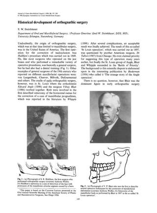
  • 1. Journal of Cranio-Maxillofacial Surgery (1996) 24, 195-204 © 1996European Associationfor Cranio-MaxinofacialSurgery Historical development of orthognathic surgery E. W. Steinh~tuser Department of Oraland Maxillofacial Surgery, (Professor Emeritus: Emil W. Steinhiiuser,DDS, MD), UniversityErlangen, Nuremberg, Germany Undoubtedly, the origin of orthognathic surgery, which was at that time limited to mandibular surgery, was in the United States of America. The first oper- ation for the correction of malocclusion was Hullihen's procedure which was carried out in 1849. He, like most surgeons who operated on the jaw bones and who performed a remarkable variety of operative procedures, was basically a general surgeon, but he had also had a dental training (Fig. 1). Other examples of general surgeons of the 19th century who reported on different maxillofacial operations were: von Langenbeck, Cheever, Billroth, Dufourmentel and others. The cradle of early orthognathic surgery, however, was in St. Louis where the orthodontist Edward Angle (1898) and the surgeon Vilray Blair (1906) worked together. Both were involved in the first described ostectomy of the horizontal ramus for the correction of a case of mandibular prognathism, which was reported in the literature by Whipple (1898). After several complications, an acceptable result was finally achieved. The result of this so-called 'St Louis operation', which was carried out in 1897, was questioned by another American surgeon, Dr Talbot (1907) from Chicago. He even claimed priority for suggesting this type of operation many years earlier, but finally the St. Louis group of Angle, Blair and Whipple succeeded in the 'Battle of Priority'. The background to this scientific dispute is elaborated upon in the interesting publication by Biederman (1956) who called it 'The strange story of the Angle operation'. There is no question, however, that Blair was the dominant figure in early orthognathic surgery ~';..Z~ 2: < , '~,z~,. k,. Fig. 1 - (a) Photograph of S. R. Hullihan, the first surgeon who performed orthognathic surgery (courtesy Anthony Wolfe). (b) Hullihan's operation in a case of distortion of the face with protrusion of the mandibular alveolar segment caused by a burn. 1This paper is based on the Converse Lecture presented at the 63rd Annual Scientific Meeting of the American Society of Plastic and Reconstructive Surgeons, San Diego 1994. 195 Fig. 2 - (a) Photograph of V. P. Blair who was the first to describe several operative techniques for the correction of maxillofacial deformities (courtesy Anthony Wolfe), (b) Ostectomy of the mandibular body as performed by Blair in 1897 in the so-called 'St Louis operation'.
  • 2. 196 Journal of Cranio-MaxillofacialSurgery (Fig. 2a). Before he published his first textbook in 1912, he described several methods for the correction of maxillofacial deformities in an excellent article on 'Operations on the Jaw-Bone and Face' in 1907. Blair emphasized how important it is to consider racial differences also in treatment planning, in order to achieve a harmonious face. He was also the first to divide jaw deformities into five classes: mandibular prognathism, mandibular retrognathism, alveolar mandibular and maxillary protrusion and open bite. He advocated several operations for corrective jaw surgery, the ostectomy of the mandibular body (Fig. 2b), the horizontal osteotomy of the ramus and the v-shaped osteotomy for open bite closure. He also made a clear statement when he wrote: 'An approximately ideal occlusion would rarely be accompanied with the best facial result.' This sen- tence, which is so true, is still not understood by many of our dental colleagues. Blair was also the first to realize the benefits of the cooperation between orthodontists and surgeons. A citation of the last sentence of his article published in 1907 is a distinct recommendation for such cooperation. He wrote: 'Treating of skeletal deformities is really surgical work, but the earlier a competent, congenial ortho- dontist is associated with the case, the better it will be for both the surgeon and the patient.' During WW I, Blair was chief consultant of the American Military Forces and after the war he estab- lished, together with Robert Ivy who was the first professor of plastic surgery at an American univeristy, a number of military hospital centres for the treat- ment of face and jaw injuries. Thus, the first period of orthognathic surgery in the USA came to an end and it would take a long time before the speciality was rediscovered in this country again. There is not much to report on orthognathic surgi- cal procedures which were carried out in Europe in the 19th century. An exception is a report by Berger (1897) from Lyon, France who described a condylar osteotomy for the correction of prognathism (Fig. 3). This method had been practiced in France until 1950, when Dufourmontel and Mouly (1959) reported good results with this technique. Babcock (1909) in the USA, and several years later, Bruhn and Lindemann (1921) in Germany, described an almost identical method to the one introduced by Blair in 1907: a horizontal osteotomy just between the sigmoid notch and the mandibular foramen (Fig. 4). This operative technique was amended a few years later by Kostecka (1931) who worked in Prague. He described his technique as a 'blind procedure', with the osteotomy Fig. 3 - The techniqueofcondylectomyforthe correctionof mandibularprognathismas describedby Berger,fromFrance,in 1897. a -<,,, Il Fig.4 - (a) Horizontalosteotomyofthe ramuswhichwas describedby Blairin 1907,by Babcockin 1909and laterby Bruhn in 1921,(b) blindtechniqueofhorizontalosteotomyoftheramus accordingto Bruhn-Lindemann. t a 0 J Fig.5- Horizontalosteotomywitha Giglisawas describedbyKosteckain 1931.
  • 3. Historical developmentof orthognathic surgery 197 being carried out with a Gigli saw through a stab incision (Fig. 5). The horizontal osteotomy of the ramus, as well as the condylar osteotomy were rather easy procedures, but the results were not satisfactory. Too much relapse and open bite problems occurred due to the small bony contact areas and the fragment dislocations which resulted from the pull of the inserted muscles. This was the reason why many proposals for the improvement of mandibular oste- otomies were published between 1920 and 1940. Several renowned maxillofacial surgeons invented new methods and new bone cuts for the correction of skeletal mandibular deformities. In Austria and Germany these were: Perthes(1922) from Tt~bingen, Pichler (1928) from Vienna, Wassmund(1935) from Berlin and Hofer (1936) from Linz. During this period not much progress in orthognathic surgery was reported from the USA or from other European countries. Only Kazanjian (1932)and Dingman(1944) from the US must be mentioned, because both described new techniques or improvements for the correction of mandibular deformities. Also, Limberg (1928) from Russia, who had had part of his training with Ivy in the United States, added some new operative procedures to the treatment of jaw deform- ities. Most of these maxillofacial surgeons from the 'early days' are listed in the chapter on 'Dramatis personae' in Anthony Wolfe's book on Plastic Surgery of the FacialSkeleton (Wolfe and Berkowitz, 1989). It is really a delight to read these highly interesting comments on the pioneers of our specialty. Again, there was a halt in the development of orthognathic surgery during WW II. The few maxillo- facial surgeons who were able to perform corrective surgery on the jaw bones were totally committed to the treatment of facial injuries and later on over- loaded with reconstructive procedures. It was thus until the beginning of the 1950s, when orthognathic surgery as a true specialty had its origins, which led to tremendous success all over the world. The cradle of modern orthognathic surgery was now central Europe, in particular Vienna and Graz, and further north Berlin and Hamburg. The founder of the 'Vienna School' of maxillofacial surgery was Pichler, succeeded by his pupil Trauner (1955) who later moved on to Graz (Fig. 6). Trauner was the inaugurator of several orthognathic surgical pro- cedures, but his main claim to fame was that he trained Heinz K61e and Hugo Obwegeser, who really gave the decisive boost to orthognathic surgery. In Berlin, Martin Wassmund who started the 'German Fig. 6 - (a) Photograph of R. Trauner who was a pupil ofPichler, the founder ofthe 'ViennaSchool'ofmaxillofacialsurgery Fig.7- (a) Photograph of M. Wassmund,the founder ofthe (courtesyHeinzK61e),(b) Inverted L osteotomyof the ramus for 'German School'ofmaxillofacialsurgery,(b) Wassmund's the correctionofmandibular prognathismadvocatedby Trauner procedure for the correctionofmaxillaryprotrusion whichwas in 1955. publishedin 1935.
  • 4. 198 Journal of Cranio-Maxillofacial Surgery School' was an important figure in maxillofacial surgery. It was he who developed the anterior maxil- lary osteotomy.(Fig. 7) which is still utilized today, in addition to other corrective procedures on the facial skeleton. His famous pupil was Karl Schuchhardt (Fig. 8) who developed the posterior maxillary osteotomy (Schuchhardt, 1955), as well as an oblique sagittal osteotomy of the mandibular ramus. However, the main input to orthognathic surgery came from the two associates of Trauner, Heinz Krte (who succeeded Trauner in Graz) and Hugo Obwegeser who was appointed to the chair of oral and maxillofacial surgery in Zurich in 1956. The main innovations which came from Krle (1959) were several new methods for changing the position of the alveolar process. To the best of our knowledge, he was the first to describe bimaxillary alveolar surgery for the correction of protrusion (Fig. 9), but also for deep bite or short face deformit- ies. He also produced a new technique for open bite closure and for genioplasty. His particular genioplasty procedure proved to be very successful, because the chin could be advanced and shortened in height at Fig. 8 - (a) Photograph of K. Schuchhardt who was a pupil of Wassmund (courtesy Gerhard Pfeifer), (b) Outline of the posterior maxillary osteotomy which was described by Schuchhardt in 1955 as a two-stage procedure. C ~7 -I b Fig. 9 - (a) Photograph of H. K61e who was a pupil of R. Trauner (courtesy Heinz Krle), (b) Bimaxillary alveolar osteotomy for the correction of protrusion or open bite which was introduced by K61e in 1959, (c) Genioplasty with ostectomy and advancement of the lower border as recommended by KOle in 1968.
  • 5. Historical developmentof orthognathic surgery 199 the same time (KOle, 1968). K61e contributed numer- ous articles to the literature and in 1964 published, together with Reichenbach and Br~ickl (1964), the first textbook in the literature on 'Surgical Orthodontics'. This book is still a standard work today (Reichenbach et al., 1964). The other famous pupil of Trauner, Hugo Obwegeser, started his career i!•' 't Fig. 10 - (a) Photograph of H. Obwegeserwho trained together with H, K61ein Graz in Austria; theircommonteacherwas R. Trauner (courtesyH, Obwegeser),(b) Originaldrawingofthe sagittal splittingtechniquewhichwasfirstpublishedbyObwegeser in 1955. as an assistant in Graz and in 1955 published the world-reknowned method of the 'intraoral sagittal split of the mandible' (Fig. 10) This method which was improved by the Italian surgeon Dal-Pont in 1958, opening new dimensions in mandibular surgery. In particular, for the advancement of the mandible this technique was ideal as no bone grafting was necessary (Fig. 11). But in addition to the sagittal split operation, Obwegeser started on maxillary sur- gery in 1960. He was the first to present a large series of Le Fort I osteotomies (Obwegeser, 1969), at the beginning in non-cleft patients, but a short time later also in cleft patients (Fig. 12). Prior to this, maxillary surgery for the correction of deformities was done only in a limited number of cases. Cohn-Stock (1921) in Berlin started with anterior maxillary osteotomies, followed by Wassmund (1935) who described this technique as a one-stage procedure. Wassmund was probably also the first to perform a total maxillary osteotomy in a patient with an open bite, in 1927. Axhausen (1939) who also worked at that time in Berlin, was the first to mobilize and advance a malunited maxillary fracture by a Le Fort I osteotomy and an additional vertical osteotomy (Fig. 13). In the 1960s, Hogeman and Wilmar (1967) from Sweden demonstrated Le Fort I osteotomies in cleft patients, but the real boost to maxillary surgery came from Obwegeser's unit in Switzerland. In the 1960s and Pig. 12- Total maxillaryadvancementby a Le Fort I osteotomyin a cleftpalate patient as publishedby Obwegeserin 1969. . • I~ a b Fig. 11- Modificationofthe sagjttal splittingofthe mandibleby Fig. 13- Le Fort I osteotomywithadditional verticalbone cut for Dal Pont in 1958.(a) the bone cut on the lingual,(b) on the buccal the correctionof a malunitedfracture;this techniquewasfirst side. describedbyAxhausenin 1934.
  • 6. 200 Journal of Cranio-Maxillofacial Surgery 1970s oral and maxillofacial surgeons from all over the world, in particular from the USA, travelled to Zurich to observe the 'Master' performing ortho- gnathic and other types of maxillofacial surgery. On the other side of the Atlantic, in the United States, where orthognathic surgery originally started, there was, as yet, not much interest in this particular field. John Marquis Converse was the exception; at that time he published several methods for corrections of jaw deformities and together with the orthodontist Horowitz (1969) he stressed the importance of close collaboration between surgeon and orthodontist (Fig. 14). Converse was also one of the first in the plastic surgery community who was interested in facial skeletal surgery in connection with recon- structive procedures on the soft tissues. Another input to orthognathic surgery came from the American oral surgeons. This group, which had separated from the plastic surgeons shortly after World War II, moved slowly in the direction of orthognathic surgery in the 1950s. The military oral surgeons CaldwelI and Letterman (1954) and several other oral surgeons, such as Robinson (1956), Hinds (1958) and Thorna (1961) came up with different methods for the correction of mandibular deformities (Fig. 15). The military oral surgeon Hunsuck (1968) should also be mentioned, as he modified Obwegeser's sagittal split procedure with his technique. However, it took quite a while until the American oral-maxillo- facial and plastic surgeons discovered the Le Fort I and other maxillary and midfacial osteotomies. In the meantime, their European colleagues were 10 years ahead, it was not until the late 1970s and into the 1980s that the USA caught up, with many excel- lent text books on orthognathic surgery being pub- lished at this time (Bell, 1980; Bell, 1985, Epker and Fish, 1986; Profitt and White, 1991). In all these books, written by surgeons and orthodontists, the close cooperation between these two specialties was always emphasized. Another important progress in orthognathic sur- gery was 'two-jaw surgery' which represents the simul- taneous mobilization of the total maxilla and mandible. K61e had already introduced bimaxillary alveolar surgery in 1959, but it took some time to be universally accepted. Obwegeser published his experi- ence in 1970 as the first to perform total maxillary and mandibular osteotomies. This was in 1970 when the Le Fort I osteotomy was already a 'routine procedure' in Zurich. Obwegeser alluded already at that time to the main advantages of this rather extensive surgical procedure; less relapse because of better skeletal stability and major aesthetic improve- ment due to the harmonization of the bony facial structures. With the improvement of surgical tech- niques, progress in anaesthesia and better stabiliz- ation of the osteotomized segments, two-jaw surgery b e Fig. 14 - (a) Photograph of J. M. Converse (courtesy Anthony Wolfe), (b,c) step osteotomy of the mandibular body for the correction of mandibular prognathism as describedby Converse and Shapiro in 1952. a b Fig. 15 - (a,b) Technique of Caldwell and Letterman, published in 1954, for the correction of mandibular prognathism. " ~ Z ~ ; Fig. 16 - Stabilization of the bony segmentswith screws and plates in two~aw surgery.
  • 7. Historicaldevelopmentof orthognathicsurgery 201 is nowadays a widely used procedure in orthognathic surgery (Fig. 16). Meanwhile, craniofacial surgery was developed in Europe, primarily in France by Paul Tessier (Fig. 17). It should be mentioned, however, that according to Wolfe (1995) the first Le Fort III osteotomy was performed by Gillies and Harrison in London in 1942 (Fig. 18). Another case of Le Fort III osteotomy on a cleft patient was reported by Gillies and Rowe in 1954 (Fig. 19). But these were probably single cases, because it took several more years until Tessier dem- onstrated for the first time the spectacular results of cranio-maxillofacial surgery at the plastic surgery meeting in Rome in 1967 (Tessier, 1967). Also, ortho- gnathic surgery was involved in the procedures which he demonstrated but most of his cases were difficult corrections of severe orbito-craniofacial deformities. Paul Tessier was also the person who reinterested American plastic surgeons in facial bone structures and thereby in orthognathic surgery. The latest aspect in the development of ortho- gnathic surgery is the application of rigid or semi- rigid fixation of the bony segments with plates and screws. This technique has its origin in traumatology, and orthopaedic surgeons utilized this system first. It is interesting to learn that the first bone plating in the maxillofacial region was carried out by the German general surgeon Soerensen in 1917. He used a wedding ring which was transformed into a small gold plate for the stabilization of a comminuted mandibular fracture (Fig. 20). It took over 50 years until this method was rediscovered again, and at the end of the 1960s the Swiss osteosynthesis group AO developed, for the first time, smaller bone plates for the mandible besides a large collection of plates and screws for the extremities. However, it took another / Fig. 17 - (a) PhotographofP. Tessierthe founderofcraniofacialsurgery(courtesyPaulTessier),(b,c) illustrationoftransorbitalLe Fort III typeosteotomyas describedbyTessierin 1967. a b Fig. 18- (a,b) Illustrationofthe firstLeFort III typeosteotomyas carriedout by Gilliesand Harrisonin 1942.
  • 8. 202 Journal of Cranio-Maxillofacial Surgery 10 years until the principles of rigid osteosynthesis were applied in orthognathic surgery. Bernd Spiessl, a native Bavarian who later moved to Switzerland, must be given credit for being the first maxillofacial surgeon to apply the AO principles to the fixation of a sagittal split osteotomy of the mandible (Fig. 21). In 1974 he published an article in which he described the technique of compression screw application in the mandible. He also claimed that with this technique the relapse, which was always a danger in mandibular set-back or advancement cases, was literally impossible. Many of his colleagues did not believe that this meant progress in the treat- ment of trauma, as well as in orthognathic surgery. Obwegeser, too, was one of the 'non-believers' and there were serious disputes between the two in several conferences of the German and European Societies. Another maxillofacial surgeon, who did a great deal of work on the development of rigid osteosynth- esis in trauma cases and in orthognathic surgery is Fig. 19 - (a) Photograph of H. Gillies (courtesy British Association of Plastic Surgeons), (b) N. Rowe (courtesy Mrs C. Rowe) who reported the first Le Fort III osteotomy on a cleft patient in 1954. Fig. 20 - First bone plating procedure in the mandible which was described by Soerensen and Warnekros in 1917. A golden wedding ring was made into a bone plate for the fixation of a comminuted fracture of the mandible. b Fig. 21 - (a) Photograph of B. Spiessl who was the first to apply rigid fixation in orthognathic surgery in 1974 (courtesy Bernd Spiessl), (b) Screw fixation of a sagittal split osteotomy according to Spiessl.
  • 9. Historicaldevelopmentof orthognathicsurgery 203 Fig. 22 - (a)PhotographofH. Luhrwhointroducedtheprinciple ofminiboneplatesinorthognathicsurgeryofthemidfacialbone structuresin 1979(courtesyHansLuhr),(b) thefirstsetofLuhr miniplateswhichwasdesignedfortheapplicationofcompression osteosynthesisandwhichwasutilizedinorthognathicsurgery. Hans Luhr from Germany (Fig. 22). He improved the miniplates, which were originally developed in France by Michelet and Festal (1972) and Peri et al. (1973) in the early 1970s, and in 1979 introduced his own first miniplate set which was very helpful in the treatment of trauma, and also for the stabilization of the small and delicate midfacial skeleton. At that time Luhr, as well as Spiessl, insisted on the principles of compression osteosynthesis and again a heated discussion started about the necessity and the value of compression for better stabilization and bone healing. Champy and Lodde (1976) and also Steinhi~user (1982), who developed their own minipl- ate sets in 1976 and in 1979 respectively, felt that the application of compression screws and plates was more difficult and more hazardous for the adjustment of the occlusion. Thus, they denied that compression- osteosynthesis was necessary, and nowadays it is generally accepted that compression is not so import- ant for bone healing, at least not in maxillofacial and orthognathic surgery. What are the advantages of plate and screw appli- cation for orthognathic surgery? The answer is clear: the application of plates and screws is more rapid and easier. The stabilization of the bone segments is better and much more reliable. The convenience for the patient is greater, because intermaxillary fixation is no longer necessary and, most importantly, there is less danger for the patient, because in the critical postoperative phase after extubation the mouth can be opened and cleaned and the airway can be easily controlled. There is no doubt that rigid fixation brought tremendous progress in the treatment of trauma, and also in orthognathic surgery. Many operations would not be possible today, if this method of fixation and stabilization was not available. The latest development in orthognathic surgery is the growing interest of plastic surgeons in this particu- lar field. Within the last 10-15 years maxillofacial surgeons, who are associated in the US with plastic and reconstructive surgery, have made remarkable contributions to this important part of corrective facial surgery. The main input which has come from this group was the addition of typical facial plastic surgical procedures to the already well-established osteotomies of the facial skeleton. The adjunct of blepharoplasty, rhinoplasty, rhitidectomy, liposuc- tion and lip corrections, just to name a few of these procedures, has led again to a tremendous boost and progress. In addition to soft tissue procedures, which have always been the focus of interest for plastic surgeons, a variety of corrections on the facial skel- eton have been introduced. In this regard, aug- mentation or reduction of the mandibular angle, the malar bones, the supraorbital rim and the forehead should be mentioned. And now we have probably reached the culmination of this work, the combi- nation of orthognathic and craniofacial surgery. It was Paul Tessier who laid the foundations of this fascinating part of reconstructive surgery, but the refinements were carried out by his pupils and dis- ciples including Wolfe (1989), Salyer (1989) and Ousterhout (1991) who have all written excellent textbooks on Plastic and Aesthetic Surgery of the Craniofacial skeleton. In these books, and in many other publications, the importance of the combination of facial or craniofacial plastic surgery with tra- ditional orthognathic surgical procedures is con- stantly emphasized. The pendulum of progress and development has now swung back to the US where orthognathic surgery started about 100 years ago. Most of these combinations of aesthetic and orthognathic surgical procedures have originated there within the last few years. On the other hand, in Europe as well, mainly due to the development of better techniques and materials in osteosynthesis, a lot of progress in ortho-
  • 10. 204 Journal of Cranio-Maxillofacial Surgery gnathic surgery has been achieved within the past few years. All this leads to the conclusion that ortho- gnathic surgery is now a grown-up specialty and an important part of oral, maxillofacial and facial plas- tic surgery. References Angle, E. H.: Double resection of the lower maxilla. Dent. Cosmos Philadelphia 40 (1898) 635 Axhausen, G: Zur Behandlung veralteter disloziert geheilter Oberkieferbrache. Dtsch Zahn-Mund-Kieferheilk 6 (1934) 582 Babcock, ~K W.: Surgical treatment of certain deformities of jaw- associated with malocclusion of teeth. JAMA 53 (1909) 833 Bell, W. H.: Surgical correction of Dentofacial Deformities. Philadelphia: W.B. Saunders, 1980 and 1985 Berger, P.: Du traitement chirurgical du prognathisme. Lyon; Med. Th6se, 1897 Biederman, W.: The strange story of the Angle operation. Ann. Dent. 15 (1956) 1 Blair, V. P.: Report of a case of double resection for the correction of protrusion of the mandible. Dent. Cosmos Philadelphia 48 (1906) 817 Blair, V. P.: Operations on the Jaw-bone and Face. Surg. Gynecol. Obstet. 4 (1907) 67 Blair, V. P.: Surgery and Diseases of the Mouth and Jaws. St. Louis; Mosby, 1912 Bruhn, Ch.: Zum Ausgleich der Makrognathie des Unterkiefers. Dtsch. Mschr. Zahnheilk. 39 (1921) 385 Caldwell, J. B., G. S. Letterman." Vertical osteotomy in the mandibular rami for the correction of prognathism. J. Oral Surg. 12 (1954) 185 Charnpy, M., J. P. Lodde: Syntheses mandibulaires. Localisation des syntheses en function des contraintes mandibulaires. Rev. Stomatol. Chir. Maxillofac (Paris) 77 (1976) 971 Cohn-Stoek, G.: Die chirurgische Immediatreguliernng der Kiefer, speziell die chirurgische Behandlung der Prognathie. Vjschr. Zahnheilk. 37 (1921) 320 Converse, J. M., H. H. Shapiro: Treatment of developmental malformations of the jaws. Plast. Surg. 10 (1952) 473 Converse, J. M., S. L. Horowitz: The surgical orthodontic approach to the treatment of dentofacial deformities. Am. J. Orthodont. 55 (1969) 217 Dal Pont, G.: L'osteotomia retromolare per la correzione della progenia. Minerva Chir. 1 (1958) Dingman, R. 0.." Surgical correction of mandibular prognathism, an improved method. Am. J. Orthodont. (Oral Surg. Section) 30 (1944) 683 Dufourmentel, C., R. MoulT Chirurgie plastique Paris 1959 Epker, B. N., L. C. Fish: Dentofacial Deformities. St. Louis; Mosby, 1986 Gillies, H., S. A. Harrison: Operative correction by osteotomy of recessed malar maxillary compound in a case of oxycephaly. Br. J. Plast. Surg. 3 (1950) 123 Gillies, H., N. L. Rowe: L'osteotomie des maxillaire superieur envisag6e essentiellement dans le cas de bec-de-li~vre total. Rev. Stomatol. Chir. Maxillofac. 55 (1954) 545 Hinds, E. C: Correction of prognathism by subcondylar osteotomy. J. Oral Surg. 16 (1958) 209 Hofer, 0.: Die vertikale Osteotomie des einseitig verkarzten aufsteigenden Unterkieferastes. Z. Stomat. 34 (1936) 826 Hogeman, K. E., K. Wilmar: Die Vorverlagernng des Oberkiefers zur Korrektur yon Gebiganomalien. Fortschr. Kiefer Gesichtschir. Bd 12, Stuttgart; Thieme, 1967 Hullihen, S. P.: Case of elongation of the underjaw and distortion of the face and neck, caused by a burn, successfully treated. Am. J. Dent. Sci. 9 (1849) 157 Hunsuck, E. E.: A modified intraoral sagittal splitting technique for correction of mandibular prognathism. J. Oral Surg. 26 (1968) 249 Kazanjian, K H.: Surgical correction of mandibular prognathism. Int. J. Orthodont. 18 (1932) 1224 K6le, H.: Surgical operations on the alveolar ridge to correct occlusal abnormalities. Oral Surg. Oral Med. Oral Path. 12 (1959) 277 Kdle, H.: Die chirurgische Behandlung von Formverfinderungen des Kinns. Wien. Med. Wschr. 118 (1968) 331 Kosteeka, F.: Die chirurgische Therapie der Progenie. Zahn/irztl. Rundschau 40 ( 1931) 669 Limberg, A. A.: Oblique osteotomy of the ramus for mandibular prognathism. J. Am. Dent. Assoc. 15 (1928) 851 Luhr, H. G.: Stabile Fixation von Oberkiefer- Mittelgesichtsfrakturen durch Mini-Kompressionsplatten. Dtsch. Zahnfirztl. Z. 34 (1979) 851 Michelet, F X., F. Festal. Osteosynthese pur plaques visees dans les fractures de l'etage moyen. Sci. Recherche Odonto- Stomat. 2 (1972) 4 Obwegeser, H.: Surgical correction of small or retrodisplaced maxillae. J. Plast. Reconstr. Surg. 43 (1969) 351 Obwegeser, H.: Die einzeitige Vorbewegung des Oberkiefers und Rtickbewegung des Unterkiefers zur Korrektur der extremen 'Progenie'. Schweiz Mschr Zahnheilk 80 (1970) 305 Ousterhout, D. K.: Aesthetic Contouring of the Craniofacial Skeleton.. Boston; Little Brown, 1991 Peri, G., J. Jourde, R. Menes: De trous surtout pour reconstrniere certains segments du squelleter facial. Ann. Chit. Plast. Esthet. 18 (1973) 170 Perthes, G.: Operative Korrektur der Progenie. Zentralbl. Chir. 49 (1922) 1540 Piehler, H.." (Jber Progenieoperationen. Wien. Klin. Wschr. 41 (1928) 1333 Proffit, W. R., R. P. White: Surgical Orthodontic Treatment. St. Louis; Mosby-Year Book, 1991 Reichenbaeh, E., H. KOle, H. Briickl: Chirurgische Kieferorthop/idie. Leipzig; J.A. Barth 1964 Robinson, M.: Prognathism corrected by open vertical condylotomy. J. S. C. D. Assoc. 24 (1956) 22 Salyer, K. E.: Techniques in Aesthetic Craniofacial Surgery. New York; Gower Medical Publishers, 1989 Schuchardt, K: Formen des offenen Bisses und ihre operativen Behandlungsm6glichkeiten. Fortschr. Kiefer Gesichtschir., Bd. I Stuttgart; Thieme, 1955 Soerensen, J.., L. Warnekros: Befestigung von Goldschienen unter dem Periost. Chirurg u. Zahnarzt, Heft 1, Berlin: Springer, 1917 Spiessl, B.: Osteosynthese bei sagittaler Osteotomie nach Obwegeser-Dal Pont. Fortschr Kiefer Gesichtschir., Bd 18, Stuttgart; Thieme, 1974 Steinhauser, E. W: Bone screws and plates in orthognathic surgery. Int. J. Oral Surg. 11 (1982) 209 Talbot, W 0.: A case of double resection of the mandible. Dent. Cosmos Philadelphia 49 (1907) 1002 Tessier, P.: Osteotomies totales de la face. Syndrome de Crouzon, Syndrome d'Apert, Oxycephalies, Scaphocephalies, Turricephalies. Ann. Chir. Plast. Esthet. 12 (1967) 273 Thoma, K. H.: Oblique osteotomy of the mandibular ramus. Oral Surg. Oral Med. Oral Path. 14 (1961) Suppl. 1 Trauner, R.: Zur Progenieoperation. Ost. Z. Stomat. 52 (1955) 361 Trauner, R., H. Obwegeser: Zur Operationstechnik bei der Progenie und anderen Unterkieferanomalien. Dtsch Zah~ Mund-Kieferheilk. 23 (1955/56) 1 Wassmund, M.: Frakturen und Luxationen des Gesichtsschgdels. Leipzig; Meusser, 1927 Wassmund, M.: Lehrbuch der praktischen Chirurgie des Mundes und der Kiefer. Bd. I., Leipzig; Meusser, 1935 Whipple, J. W: Double resection of the inferior maxilla for protruding lower jaw. Dent. Cosmos Philadelphia 40 (1898) 552 Wolfe, S. A., S. Berkowitz: Plastic Surgery of the Facial Skeleton. Boston; Little Brown, 1989 Wolfe, S. A.: personal communication 1995 E. W. Steinh/iuser, DDS, MD University Erlangen--Nttrnberg Klinik ft~rMund-, Kiefer-, Gesichtschirurgie Gltickstr. 11 91054 Erlangen, Germany Paper received 2 May 1995 Paper accepted 7 May 1996