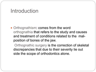
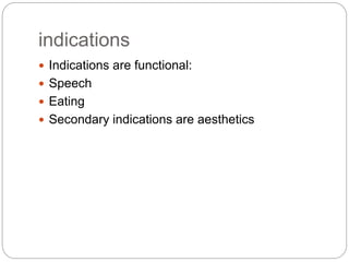
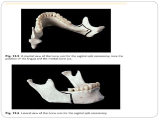
PRESENTERS
MUGABE LAURENCE
NAKIMERA BETTY
MALE JUDE
ORTHOGNATHIC SURGERY
OUTLINE
 Introduction
 Indications
 Diagnosis And Treatment Planning
 Sequence Of Treatment
 Mandibular procedure
 Maxillary procedure
Introduction
 Orthognathism: comes from the word
orthognathia that refers to the study and causes
and treatment of conditions related to the mal-
position of bones of the jaw.
Orthognathic surgery is the correction of skeletal
discrepancies that due to their severity lie out
side the scope of orthodontics alone.
indications
 Indications are functional:
 Speech
 Eating
 Secondary indications are aesthetics
Diagnosis and treatment
planning.
 This is best done jointly by an orthodontist
and a maxillofacial surgeon and ideally with
psychology input. The following information is
required:
 Patient’s perception of problem Are they
concerned with appearance of jaws or teeth,
speech, or problems with eating? Are their
expectations realistic?
 Clinical examination Assessment of the
balance and proportions of full face and
profile.
 Study models Needed to assess coordination of
arches.
 Radiographs Require DPT and lateral skull, plus
PA skull for asymmetries.
 Photographs Required as pre-Rx record and can
also be manipulated with lateral skull for visual
computer predictions
 It is important to correlate desired facial changes
with the patient’s occlusion
 Pre-surgical orthodontics will be required to
decompensate teeth so that a full surgical
correction is possible.
 It is important (in order to obtain informed
consent) that patients are fully informed of the
risks of surgery, particularly mandibular
procedures, in addition to the orthodontic risks.
Sequence of Rx
 Pre-surgical orthodontics The aim of this phase is
to align and coordinate the arches so that the
teeth will not interfere when the jaws are placed
in their correct position. This usually involves
decompensation, i.e. removal of any dento-
alveolar compensation for the skeletal
discrepancy so that the teeth are at their correct
axial inclinations and a full surgical correction can
be achieved
 Post-surgical orthodontics Lighter round wires
and inter-maxillary traction are used to detail
occlusion.
 Retention This is the same as after conventional
FA Rx.
 Relapse This can be surgical or orthodontic, or
both.
 Relapse is more likely in Rx of deficiencies as
soft tissues are under greater tension
postoperatively
Mandibular procedure
 Involve ramus, body, alveolus, or chin.
 IO vertical subsigmoid osteotomy Used to push
back the mandible.
 EO approach used when suitable equipment for
IO not available.
 The IO procedure is straightforward, performed
via an extended third-molar type incision. Bone
cuts are made with a right-angled oscillating saw
from sigmoid notch to lower border. Technique is
very instrument dependent.
Sagittal split osteotomy
 (Fig. 12.5 and Fig. 12.6.) Can move mandible
backwards or forwards.
 IO incision similar to intra-oral vertical
subsigmoid osteotomy. Bone cuts made from
above lingula, across retromolar region, down
buccal aspect to lower border.
 Split sagittally with an osteotome followed by
spreaders.
 Main complication is paraesthesia of IDN.
 Inverted L- and C-shaped osteotomy Usually EO
approach. Rarely used; can lengthen ramus if used
with bone graft.
 Body ostectomy Shortens body of mandible. Need to
gain space orthodontically or remove tooth. Watch
mental nerve.
 Subapical osteotomy Used to move dento-alveolar
segments. Technically more difficult than it looks. Risk
to tooth vitality.
 Genioplasty The tip of the chin can be moved pretty
much anywhere; the secret is to keep a sliding
contact with bone and a muscle pedicle. Fixation
should be kept away from areas of muscle activity as
this leads to bone resorption. Effective treatment for
obstructive sleep apnoea.
Maxillary procedure
 Segmental Can be single-tooth, or bone and
tooth blocks, e.g. Wassmund procedure, which
involves tunneling incisions in buccal sulcus and
palate to move premaxilla. Problems are finding
space for bone cuts and avoiding damaging
teeth.
 Le Fort I Mainstay procedure. Standard
approach is the ‘down-fracture’ with horseshoe
buccal incision, bone cuts at Le Fort I level, and
segment pedicled on the palate. The freed
maxilla can be moved up, down, or forward.
 Posterior movement more problematic. In cleft
palate cases, concern over the adequacy of the
palatal blood supply has led to some surgeons
using tunnelled buccal incisions to make the
bone cuts, thus preserving some of the buccal
blood supply to the maxilla. Fixation is a problem
when using this technique.
 Le Fort II Usually used for mid-face
advancement. Bilateral canthal and vestibular
incisions allow bone cuts at the Le Fort II level.
 Le Fort III Really a sub cranial craniofacial
operation using a coronal flap plus vestibular and
orbital incisions to move the entire mid-face and
molar complex.
 Malar osteotomy Used for post-traumatic defects.
Approach via coronal incisions. Risk to infra-
orbital nerve from maxillary bone cut.
Augmentation is an alternative.
 Rhinoplasty The correction of isolated nasal
deformity. Usually done intranasally,
supplemented by tiny incisions over the nasal
bones to allow bone cuts.
 ‘Open rhinoplasty’, which involves degloving the
nasal skeleton via a columella incision, is popular
particularly for cleft patients
references
 Oxford handbook of clinical dentistry( Bethany
Rushworth )seventh edition
 Internet.

ORTHOGNATHIC SURGERY.pptx

  • 1.
  • 2.
    OUTLINE  Introduction  Indications Diagnosis And Treatment Planning  Sequence Of Treatment  Mandibular procedure  Maxillary procedure
  • 3.
    Introduction  Orthognathism: comesfrom the word orthognathia that refers to the study and causes and treatment of conditions related to the mal- position of bones of the jaw. Orthognathic surgery is the correction of skeletal discrepancies that due to their severity lie out side the scope of orthodontics alone.
  • 4.
    indications  Indications arefunctional:  Speech  Eating  Secondary indications are aesthetics
  • 5.
    Diagnosis and treatment planning. This is best done jointly by an orthodontist and a maxillofacial surgeon and ideally with psychology input. The following information is required:  Patient’s perception of problem Are they concerned with appearance of jaws or teeth, speech, or problems with eating? Are their expectations realistic?  Clinical examination Assessment of the balance and proportions of full face and profile.
  • 6.
     Study modelsNeeded to assess coordination of arches.  Radiographs Require DPT and lateral skull, plus PA skull for asymmetries.  Photographs Required as pre-Rx record and can also be manipulated with lateral skull for visual computer predictions  It is important to correlate desired facial changes with the patient’s occlusion  Pre-surgical orthodontics will be required to decompensate teeth so that a full surgical correction is possible.
  • 7.
     It isimportant (in order to obtain informed consent) that patients are fully informed of the risks of surgery, particularly mandibular procedures, in addition to the orthodontic risks.
  • 8.
    Sequence of Rx Pre-surgical orthodontics The aim of this phase is to align and coordinate the arches so that the teeth will not interfere when the jaws are placed in their correct position. This usually involves decompensation, i.e. removal of any dento- alveolar compensation for the skeletal discrepancy so that the teeth are at their correct axial inclinations and a full surgical correction can be achieved
  • 9.
     Post-surgical orthodonticsLighter round wires and inter-maxillary traction are used to detail occlusion.  Retention This is the same as after conventional FA Rx.  Relapse This can be surgical or orthodontic, or both.  Relapse is more likely in Rx of deficiencies as soft tissues are under greater tension postoperatively
  • 10.
    Mandibular procedure  Involveramus, body, alveolus, or chin.  IO vertical subsigmoid osteotomy Used to push back the mandible.  EO approach used when suitable equipment for IO not available.  The IO procedure is straightforward, performed via an extended third-molar type incision. Bone cuts are made with a right-angled oscillating saw from sigmoid notch to lower border. Technique is very instrument dependent.
  • 11.
    Sagittal split osteotomy (Fig. 12.5 and Fig. 12.6.) Can move mandible backwards or forwards.  IO incision similar to intra-oral vertical subsigmoid osteotomy. Bone cuts made from above lingula, across retromolar region, down buccal aspect to lower border.  Split sagittally with an osteotome followed by spreaders.  Main complication is paraesthesia of IDN.
  • 13.
     Inverted L-and C-shaped osteotomy Usually EO approach. Rarely used; can lengthen ramus if used with bone graft.  Body ostectomy Shortens body of mandible. Need to gain space orthodontically or remove tooth. Watch mental nerve.  Subapical osteotomy Used to move dento-alveolar segments. Technically more difficult than it looks. Risk to tooth vitality.  Genioplasty The tip of the chin can be moved pretty much anywhere; the secret is to keep a sliding contact with bone and a muscle pedicle. Fixation should be kept away from areas of muscle activity as this leads to bone resorption. Effective treatment for obstructive sleep apnoea.
  • 14.
    Maxillary procedure  SegmentalCan be single-tooth, or bone and tooth blocks, e.g. Wassmund procedure, which involves tunneling incisions in buccal sulcus and palate to move premaxilla. Problems are finding space for bone cuts and avoiding damaging teeth.
  • 15.
     Le FortI Mainstay procedure. Standard approach is the ‘down-fracture’ with horseshoe buccal incision, bone cuts at Le Fort I level, and segment pedicled on the palate. The freed maxilla can be moved up, down, or forward.  Posterior movement more problematic. In cleft palate cases, concern over the adequacy of the palatal blood supply has led to some surgeons using tunnelled buccal incisions to make the bone cuts, thus preserving some of the buccal blood supply to the maxilla. Fixation is a problem when using this technique.
  • 16.
     Le FortII Usually used for mid-face advancement. Bilateral canthal and vestibular incisions allow bone cuts at the Le Fort II level.  Le Fort III Really a sub cranial craniofacial operation using a coronal flap plus vestibular and orbital incisions to move the entire mid-face and molar complex.  Malar osteotomy Used for post-traumatic defects. Approach via coronal incisions. Risk to infra- orbital nerve from maxillary bone cut. Augmentation is an alternative.
  • 17.
     Rhinoplasty Thecorrection of isolated nasal deformity. Usually done intranasally, supplemented by tiny incisions over the nasal bones to allow bone cuts.  ‘Open rhinoplasty’, which involves degloving the nasal skeleton via a columella incision, is popular particularly for cleft patients
  • 18.
    references  Oxford handbookof clinical dentistry( Bethany Rushworth )seventh edition  Internet.