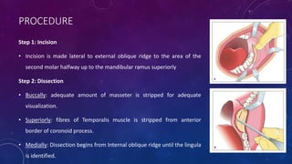
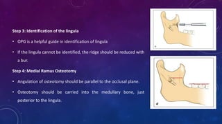
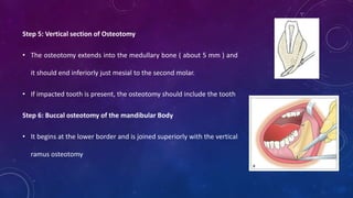
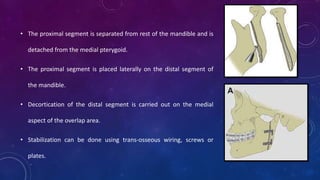
RAMUS OSTEOTOMY
DR. JEFF ZACHARIA
POST GRADUATE STUDENT
DEPT. OF ORAL & MAXILLOFACIAL SURGERY
AJ INSTITUTE OF DENTAL SCIENCES
CONTENTS
a) Introduction
b) Surgical anatomy of mandible
c) Deformities of mandible
d) Classification of deformities of mandible
e) Goals of orthognathic surgery
f) History
g) Classification of mandibular osteotomies
h) Investigations
i) Sagittal Split Osteotomy
j) Vertical Ramus Osteotomy
 “L” Osteotomy
 “C” Osteotomy
k) BSSO vs VRO
l) Conclusion
INTRODUCTION
INTRODUCTION
The development of mandibular osteotomies for correction of dentofacial deformities closely
parallels the advancement of oral and maxillofacial surgery as a specialty more than any
other group of surgical techniques
SURGICAL ANATOMY OF MANDIBLE
• Mandible is the sturdiest bone of the face with strong basal bone
and an alveolar part housing the dentition.
• The mandibular condyles articulate with the temporal bone and
help in rotatory and translatory movements.
• On the medial aspect of the ramus is the mandibular foramen just
posterior to halfway between the anterio-posterior width of the
vertical ramus almost in line with the most concave part of the
anterior border
• Just above is the lingula, a triangular prominence, the
sphenomandibular ligament is attached.
• Mandibular neurovascular bundle enters the mandibular
foramen and runs below the tooth roots in the body of the
jaw, in the inferior alveolar canal.
• It curves upward and backward by about 2 mm and gives out
the mental nerve below the second premolar area and gives
sensory supply to the lower lip and chin
• Orthognathic surgery affects the mandible primarily in two
ways:
• It changes the length of a muscle or it
• changes the direction of muscle function.
• The muscles commonly discussed in orthognathic surgery are
the muscles of mastication and the suprahyoid group of
muscles.
DEFORMITIES OF THE MANDIBLE
CLASSIFICATION OF DEFORMITIES OF THE MANDIBLE
• Deformities of the jaws can be associated with dentoalveolar complex, the skeletal base or both.
• They may be either in excess or a deficiency.
• These problems can occur
in three different vectors
• Antero-posterior
• Transverse
• Vertical
MANDIBULAR EXCESS
Common cause for mandibular excess is either development or genetic.
Clinical features associated with mandibular excess are the following:
• Prognathic mandible
• Anterior cross bite
• Elongated face
• Relatively long lower third of the face
• Concave facial profile
• Lower lip & chin are more anteriorly placed than normal
• Class III relationship of occlusion
• Angle SNB more than 82°
MANDIBULAR DEFICIENCY
Clinical features associated with deficiency are:
• Bird face appearance
• Severe over jet
• Class II relationship of dentition
• Crowding of lower anterior teeth
• Flaring compensation of lower anterior teeth.
• Face appears small.
• Lower third of the face is short
• Labiomental fold is usually short
• Chin neck angle is obtuse
• SNB angle < 78
It is often due to genetical or developmental reasons.
Ankylosis of TMJ, trauma to condyle and aplasia of condyle can cause deficiency
GOALS OF ORTHOGNATHIC SURGERY
FUNCTION
Normal mastication
Speech
Ocular function
Respiratory function
AESTHETICS
Establishment of facial harmony and
balance
STABILITY
Prevention of short & long term
relapse
MINIMIZING TREATMENT
TIME
Provision of efficient & effective
treatment
HISTORY
• 1849: Hullihen performed first anterior mandibular osteotomy for a patient with facial distortion due
to severe burns.
• 1897: Blair and Angle performed a whole mandibular body osteotomy for the correction of
prognathism.
• 1949: Sagittal split was introduced by Schindart
• 1954: Caldwell – Letterman performed vertical sub-sigmoid osteotomy of ramus.
• 1961: Dal Pont changed the lower horizontal cut to a vertical cut on the buccal cortex between
the first and second molars thereby obtaining broader contact surfaces and requiring minimal
muscular displacement.
• 1968: Epker suggested several modifications. These included minimal masseter stripping and
limited medial dissection.
• 1974: Spiessel advocated the rigid internal fixation of the BSSO to promote healing, restore
early function and attenuate relapse.
• 1979: Luhr introduced miniplate fixation in Orthognathic surgery
BODY
OSTEOTOMIES
SUB APICAL
OSTEOTOMIES
RAMUS
OSTEOTOMIES
HORIZONTAL
OSTEOTOMY OF
CHIN
Sagital Split Osteotomy
Vertical Ramus
Osteotomy
Inverted “L” & “C”
Osteotomy
Anterior Sub Apical
Osteotomies
Posterior Sub Apical
Osteotomies
Total Sub Apical
Osteotomies
Condylotomy/
Condylectomy
Anterior To Mental Foramen
• Step osteotomy/ostectomy
Posterior ToMental Foramen
• Y Ostectomy
• Rectangular ostectomy
• Trapezoid ostectomy
• Inverted V ostectomy
CLASSIFICATION OF MANDIBULAR OSTEOTOMIES
INVESTIGATIONS
INVESTIGATIONS IN ORTHOGNATHIC SURGERY
• Facial photographs
• Study models with wax bite recorded in centric occlusion and centric relation
• Lateral cephalogram & PA cephalogram for class III malocclusion and vertical
excess patients to assess transverse problems.
• OPG and intraoral periapical radiographs to assess intra-septal bone available
for surgical cuts.
• Additional records include a CBCT scan.
• 3D facial photos such as 3dMD are optional however a great tool for 3D
virtual planning and prediction for post treatment profile
VARIOUS METHODS TO MEASURE BONE DENSITY
• Digital panoramic radiographs
• Lateral cephalogram & PA cephalogram
• Quantitative ultrasound (QUS)
• Quantitative computerized tomography
• Quantitative CBCT
• Dual energy X ray absorptiometry (DEXA)
• MRI
SAGITTAL SPLIT OSTEOTOMY
REVIEW OF LITERATURE
Published the horizontal sub-condylar osteotomy of the mandible to correct
class II dysgnathias.
• The technique required prolonged IMF which was regarded as an
inconvenience due to lack of bone contact between the osteotomized
segments.
Blair 1907
SCHUCHARDT 1942
Schuchardt modified the horizontal osteotomy by introducing a technique in
which a cortical osteotomy was performed in an oblique way starting just
above the lingula & reaching the buccal cortex 1 cm caudally without
touching the IAN.
• It could be performed intraorally
• More sufficient medullary bone attachment
• Trauner & Obwegeser modified Schuchardt’s technique by increasing
the gap between the horizontal cuts to 25 mm, requiring the surgeon
to address the IAN.
• This innovation resulted from connecting two horizontal cortical cuts
along the lateral oblique ridge and leaving the posterior border of the
ramus untouched.
Trauner & Obwegeser 1955
• The fracture of the ramus was achieved by the chiseling along the lateral cortex. This was called the
sagittal splitting procedure.
• Wider distance between the lingual and buccal cuts increased the overlapping bony amount of the
segments which rendered better stability and better results at a lower risk of pseudoarthosis.
DAL PONT 1961
• Dal Pont’s modification advances & rotates the lower horizontal
cut even further to the buccal cortex of the mandibular body as a
vertical cut between the first and second molars.
• The angle created was approximately 90° leading to an extension
of the connecting cut along the oblique line on the lateral
mandibular aspect through the mylohyoid groove on the lingual
side.
HUNSUCK 1968
• Hunsuck believed that the lingual split of Dal Pont’s osteotomy
would naturally occur given that chisels were used to split the
mandible.
• The buccal vertical cut by Hunsuck was located at the union of
the ascending ramus and the body of the mandible in the tooth
bearing region.
• This area was just distal of the second molar running down to
the mandibular notch anterior of the insertion point of the
massetric muscle.
BELL & SCHENDEL (1977) EPKER (1978)
• They made an anterior vertical cut, through which whole of the
lower border is sectioned through and through.
• The split is kept more laterally by directing the fine osteotomes
down the inner surface of the lateral cortex to produce easier
splitting and greater protection for the inferior alveolar nerve.
• Blood supply to the ramus is preserved as the need to strip the
pterygomassetric sling is eliminated.
SAGITTAL SPLIT OSTEOTOMY PROCEDURE
PROCEDURE
Step 1: Incision
• Incision is made lateral to external oblique ridge to the area of the
second molar halfway up to the mandibular ramus superiorly
Step 2: Dissection
• Buccally: adequate amount of masseter is stripped for adequate
visualization.
• Superiorly: fibres of Temporalis muscle is stripped from anterior
border of coronoid process.
• Medially: Dissection begins from Internal oblique ridge until the lingula
is identified.
Step 3: Identification of the lingula
• OPG is a helpful guide in identification of lingula
• If the lingula cannot be identified, the ridge should be reduced with
a bur.
Step 4: Medial Ramus Osteotomy
• Angulation of osteotomy should be parallel to the occlusal plane.
• Osteotomy should be carried into the medullary bone, just
posterior to the lingula.
Step 5: Vertical section of Osteotomy
• The osteotomy extends into the medullary bone ( about 5 mm ) and
it should end inferiorly just mesial to the second molar.
• If impacted tooth is present, the osteotomy should include the tooth
Step 6: Buccal osteotomy of the mandibular Body
• It begins at the lower border and is joined superiorly with the vertical
ramus osteotomy
Step 7: Defining the osteotomy cut with an osteotome
• It is recommended that the surgeon completes the osteotomy on the
contralateral side before splitting the mandible
• A 10 mm wide osteotome is used to tap along the vertical osteotomy
from the medial osteotomy downward to the buccal osteotomy
• Failure to support the mandible during this step may cause
hemarthrosis or disc displacement.
Step 8: Splitting the mandible
• It is divided into two stages
i. initiation of the split
• the lower border of the mandible splits toward the proximal segment
• the neurovascular bundle is intact and separates from the proximal segment
ii. completion of the split
• the lower border continues to split with the proximal segment
• The neurovascular bundle detaches from the proximal segment as the split
continues
• the inferior alveolar foramen and proximal part of the canal detach from the
proximal segment.
Step 9: Completion of the split
• If the NV bundle is present on the proximal segment, the split should be
stopped and the NV bundle must be carefully dissected
• Any resistance in the split could be due
1. A greenstick fracture of the bone in which the lower border starts
fracturing superiorly toward the distal part of the medial ramus
osteotomy, posterior to the lingula.
2. A fracture of the vertical osteotomy of the ramus anterior to the inferior
alveolar foramen due to stopping the osteotomy short of the lingula.
Step 10: Stripping of musculature
• Stripping of pterygomassetric sling: Inadequate stripping can lead to difficulty in
repositioning the distal segment and inaccurate positioning of the condyle.
• Stripping of pterygoid muscle and stylomandibular ligament: Failure to strip them from the
bone may interfere with the repositioning of the distal segment and may lead to unfavorable
rotation of the proximal segment causing inaccurate positioning of the condyle or rotation of
the proximal segment thereby increasing the potential for relapse.
Step 11: Mobilization of the distal segment
• Segment is mobilized anteriorly using the index finger.
Step 11: Selective odontoplasty and IMF
• Fixation of the central incisors is done first to establish the planned incisor
relationship following by application of IMF to the posterior teeth.
• An acrylic splint can be used.
• Selective odontoplasty should be performed prior to IMF if indicated in the
model surgery.
In case of mandibular set back
• When the teeth have been wired into occlusion and the segment pushed back,
there is bony over lap anteriorly.
• This overlapping bone should be removed using rotary instruments.
• The small piece of bone can be used as grafts. (in areas such as intersegmental
bone defects or in Le Fort I osteotomies)
Step 12: Osteosynthesis
• The proximal and distal segment should be aligned passively with little to
no step on the inferior border.
• A modified Allis clamp can be applied on the ramus to engage both
segments of the bone while fixation.
• Fixation can be achieved using a 2.0 mm 4 holed plate with gap and a
second plate is recommended to attain additional stability.
Drawbacks of presence of third molar
• It weakens the retromolar part of the distal segment;
• increases involvement of the inferior alveolar nerve and increases morbidity of
sensation;
• increases the prevalence of postoperative infection;
• if the tooth is not removed during surgery may interfere with optimal bone contact.
ASYMMETRY OF MANDIBLE
• Corrections include rotation, bodily movement and lateralization of the dentate segment.
• The side that receives the rotated segment, bony contact may occur at the anterior aspect potentially leaving a gap
posteriorly.
• The side from which the distal segment is pivoted away, is left with anterior gap and contact is present only at the
most posterior aspect of the proximal segment.
lateral
displacement of
the condyle
Relocation of
condyle
shift in occlusion
Mandible rotates
towards the
original side.
Relapse
Fixation of two
segments &
closure of
anterior gap
ADVANTAGES OF BSSO
• Broad bony overlap between segments which allows excellent healing.
• Minimal alteration of the natural position of muscles of mastication which prevents relapse
from muscular traction.
• Minimal alteration of the original position of the TMJ which minimizes the risk of athropathy
• Shorter operating time and low complication rate
COMPLICATIONS
• Bleeding can be from IAN bundle, medullary bed, facial vessels and rarely from RMV.
• Bleeding from IAN bundle and medullary bed can be controlled by local measures.
• Bleeding from facial vessels will have to be clamped and tied.
• Bleeding from RMV can be controlled by a gelatin sponge, if oozing is still present, a drain may be kept.
• Lee et al reported severe swelling, ecchymosis and irregular menstrual cycles among female patients who
underwent orthognathic surgery. Physical stress following surgery was attributed to early menstruation
and bleeding tendency in these patients.
INTRA-OP COMPLICATIONS
Excessive bleeding
• Basu claimed that fibrinolysis increases during menstruation which reflects the increased risk of
bleeding during this period.
• Lin et al reported that hypotensive anesthesia using sevoflurane can effectively reduce bleeding
and secure excellent vision during surgery.
UNFAVORABLE SPLITS
• Incidence: 3-20%
 Proximal segment fractures
• Failure to cut the inferior border before applying chisels
• Impacted third molars
• Incomplete transection of the inferior border.
 Distal segment fractures
• Splits short of lingula
• Retromolar fracture
• Inferior border remains attached to the distal segment.
Comminuted fractures
• Occurs most commonly on the buccal cortex.
• Rescue involves completion of the originally planned osteotomy,
positioning of the mandible into the preplanned occlusion and fixation
of the segments with internal fixation devices.
• Other options include aborting the procedure, establishing IMF,
assessing the complication pattern using high resolution CT scans and
allowing the unfavorable fracture segments to heal before reattempting
the treatment.
• Another option is the use of an extra oral approach to permit
visualization, reduction and fixation with reconstruction plates.
RELAPSE
Factors associated with relapse include muscle related physiological effects that are influenced by:
• direction of bone rotation and the amount of bone movement
• asymmetry between left and right side
• changes in teeth position after surgery
• change in condyle position
• change ramus inclination
• change in mandibular plane
• type of fixation
• Unresolved misalignment of upper and lower jaws during orthodontic procedures before surgery
POST-OP COMPLICATIONS
CONDYLAR SAG
• An immediate or late caudal movement of the condyle in the glenoid fossa after surgical
establishment of a pre-planned occlusion and rigid fixation of the bone fragments, leading to a
change in the occlusion.
• Causes occlusal relapse either immediately on release of IMF or in the late post-op phase.
• Classification of condylar sag
 Central condylar sag
 Peripheral condylar sag type 1
 Peripheral condylar sag type 2
Central condylar sag
The condyle is positioned inferiorly in the glenoid fossa
and makes no contact with any part of the fossa.
In the absence of the intracapsular edema or hemarthosis,
the condyle will move superiorly after removal IMF leading
to malocclusion.
Clinical signs of condylar sag
 Bilateral central condylar sag
• Dental midlines correct
• Increased overjet
• Anterior open bite
• Class II malocclusion bilaterally
 Unilateral central condylar sag
• Mandibular dental midline shifted towards the
offending side
• Overjet increased (more towards the offending side
• Class II dental relationship on the offending side
Peripheral condylar sag I
• There is an inferior displacement of the condyle with some contact with
the glenoid fossa with the teeth in temporary IMF and rigid fixation
applied.
• There is no occlusal change in the phase.
• Post-op resorption and subsequent change in condylar shape will lead to
delayed relapse.
Clinical signs
• Difficult to diagnose intra-op because the contact between the condyle &
glenoid fossa supports the occlusion, which may lead to late relapse due to
condylar resorption
Peripheral condylar sag II
• The condyle is positioned correctly in the fossa with
IMF in place.
• However, with the placement of rigid fixation, a
torquing force is applied to the condyle and ramus.
• The tension in the ramus is released when the IMF is
released, and the condyle will move laterally or
medially and slide inferiorly in the fossa.
Clinical signs
 Bilateral condylar sag II
• Dental midlines correct
• Anterior crossbite or edge to edge incisal relationship
• Tendency for class III dental relationship
• Tendency for bilateral posterior open bites
 Unilateral peripheral condylar sag II
• Dental midline shifted towards contralateral side
• Edge to edge incisal relationship with tendency for crossbite on affected side
• Posterior open bite on affected side
• Tendency for class III relationship between canine and molars when mandible is moved to
correct the midline.
NERVE INJURY
• Continuity of the neurovascular bundle must be maintained throughout the process.
• If the nerve is transected, the cut ends should be micro-anastomosed.
• Literature has reported neurological problems persist until 24 weeks after BSSO and recovery
tends to be slower after BSSO than IVRO
• Verweij et al reported that hypoesthesia following BSSO occurred in 4.8 % pf patients less
than 19 years, in 7.9 % of patients aged between 19 – 30 years and in 15.2 % in patients aged
31 years or older. Although the rate of permanent hypoesthesia that persisted until one year
after BSSO was low, the mean recovery time from hypoesthesia was relatively long for older
patients.
NEUROPATHIC PAIN
• Politis et al investigated the types of pain experienced by patients who underwent
orthognathic surgery between 2001 and 2011. Among 982 cases of BSSO, 6 cases of
debilitating chronic neuropathic pain were observed. Direct damage to the inferior alveolar
nerve and its bone environment that had become exposed may have been the cause of
partial axonal injuries.
• Teerijoki-Oksa et al claimed that pain that persists up to one month after surgery indicates
axonal damage, and that pain can continue for even a longer period if axonal damage does
not heal completely. A third of the patients with axonal damage never show complete
recovery.
TMJ DISORDER
• All types of orthognathic surgery can directly or indirectly affect TMJ symptoms
• Orthognathic surgery can improve or worsen the existing symptoms or may not result in any changes
at all.
• The following principles should be followed when performing BSSO:
• All bony interferences that exist between proximal and distal segment should be removed.
• Condylar heads should passively settle into the glenoid fossa during surgery.
• For fixation of bony segments, non rigid fixation using monocortical plates and screws is
recommended. Use of compression plates or lag screws should be avoided.
• Counterclockwise maxillomandibular rotation and a large amount of mandibular advancement can
increase the load and stress on the TMJ. When the load on the TMJ exceeds adaptability, condylar
resorption results.
DELAYED UNION OR NON UNION
• Delayed union or non union of an osteotomy site may occur as a result of poor healing of
hard tissues.
• Risk of non union is high when inadequate fixation is performed after non rigid fixation using
materials such as wires and when anterior displacement of a bony segment is large.
INFECTION
• Post-op infections include cellulitis, abscess and osteomyelitis.
• Davis et al reported that rate of infection is 8% among 2521 patients who underwent
orthognathic surgery and these infections occurred mostly in the mandible.
VERTICAL RAMUS OSTEOTOMY
• First advocated by Robinson & Hinds in 1955
• These osteotomies can be used to advance a retruded mandible as well as to set back a protruded
mandible.
• The development of the vertical osteotomy of the ramus allowed for repositioning of the mandible
under direct vision.
EXTRA ORAL VERTICAL RAMUS OSTEOTOMY
PROCEDURE
• A submandibular incision is placed about 1.5 cm below the angle of the
mandible.
• Dissection is done and the marginal mandibular nerve is identified and
preserved.
• The lower border is identified and the periosteum is reflected superiorly
until the sigmoid notch on the lateral aspect of the mandible.
• The antilingula is identified and the vertical bony cut is performed posterior
to the mandibular foramen from the sigmoid notch to the lower border.
• The proximal segment is separated from rest of the mandible and is
detached from the medial pterygoid.
• The proximal segment is placed laterally on the distal segment of
the mandible.
• Decortication of the distal segment is carried out on the medial
aspect of the overlap area.
• Stabilization can be done using trans-osseous wiring, screws or
plates.
 Advantages
• Adequate access
• Adequate control of bony segments
• Good surface contact between bony fragments
• Minimal relapse tendency
 Disadvantage
• Scar formation
• Potential damage to mandibular branch of facial nerve
• Bleeding due to damage to RMV
INTRAORAL VERTICAL RAMUS OSTEOTOMY (MOOSE 1964)
• It is used when large setback of the mandible is required and for the correction of severe
mandibular asymmetry
Advantages
o Less IAN morbidity
o Less flaring on the proximal segments
o No necessity for rigid fixation
o Existing TMJ dysfunction can be treated.
Disadvantages
o Post op IMF is required
o Strict protocol for active physiotherapy for
two weeks after release of IMF
PROCEDURE
Step 1: Incision and dissection
• Incision is made lateral to external oblique ridge to the area of the
second molar halfway up to the mandibular ramus superiorly
• The dissection is carried out
• Superiorly: till the sigmoid notch
• Inferiorly: to the anti-gonial notch
• Posteriorly: till the angle of the mandible
Step 2: Identification of landmarks
• Various methods to establish the position of the mandibular foramen on the lingual side of
the ramus can be implemented
• The antilingular eminence on the lateral surface of the ramus
• Hall recommends that it is safe to perform the vertical osteotomy 6 mm to 7 mm anterior
to the posterior border of the ramus.
• Fontura et al described that the mandibular foramen is most often located horizontally in
the middle third of the ramus and vertically in the superior third.
• Use of a radioghraph.
Step 3: First vertical Osteotomy
• A guideline can be drawn from the antilingular notch toward the
antigonial notch parallel to the posterior border of the ramus
• A round oscillating saw is used to perform the first osteotomy 2mm
posterior to the guideline (shown at A)
Step 4: Second vertical osteotomy
• The second osteotomy is angulated slightly anteriorly and performed
from the superior end of the reference line into the sigmoid notch.
(shown at B)
Step 5: Mandibular setback
• For setback of 4 mm or less, the inferior cut is carried parallel and about 9
to 10 mm anterior to the posterior border.
• If a setback of >5 mm, the inferior osteotomy is angled anteriorly to
broaden the base of the of the proximal segment.
• This will maintain sufficient residual width of attachment of the masseter
and the medial pterygoid muscle following setback of the distal tooth
bearing segment to prevent condylar sag.
Step 6: Bony interference
1. Occurs between overlapped segments and causes backward rotation of the
proximal segment.
2. Occurs when mandible is setback to 10 mm or more. Contact between condylar
and coronoid processes can block posterior movement.
Step 9: Fixation
• Fixation of the segments is rarely necessary.
• The combined effort of the medial pterygoid and the masseter provides adequate
support and control of the proximal segment. IMF provides adequate control of
distal segment.
• Semi rigid fixation can be done using 2 x 2 mm 4 hole “L” plate over the lateral
surface of the ramus.
• The patient must be maintained on IMF for 7 to 10 days after surgery followed by
light elastic traction to guide the occlusion for the next 4 – 5 weeks while initial
bone healing occurs.
COMPLICATIONS
 Stability
• Goldstein reported incidence of anterior relapse after IVRO in his study.
 Neurologic deficits
• Bell et al preferred the IVRO over SSRO because there was a lower incidence of nerve injury.
• Paulus and Steinhauser found the incidence of mandibular nerve disturbance equal between the
procedures.
• Wang & Waite reported an increased frequency of nerve damage with SSRO
 Avascular necrosis of proximal segment due to excessive soft tissue stripping
 Hemorrhage due to damage to the massetric artery
INVERTED L OSTEOTOMY
• First described by Wassmund and Pichler 1927 and was modified
by Caldwell & Letterman in 1954
• Requires extra oral approach.
• Unpopular due to risk of marginal mandibular nerve injury,
additional time for graft harvest, donor site morbidity.
• Caldwell modified ILO by performing C osteotomy in 1968
• Requires extra oral approach
• Advantage
 greater bony contact and
 Allows rotational advancements.
C OSTEOTOMY
• Indications
 Short ramus height. (conditions like Juvenile idiopathic arthritis , craniofacial microsomia,
craniofacial dysostosis, condylar trauma and mandibular hypoplasia)
 Skeletal anterior open bite
 Mandibular retrognathism.
 In older patients
 Patients with masseteric hypertrophy (less medullary bone)
• Contraindications
 Patients who are prone to hypertrophic scarring or Keloids
 Advantages
• Can correct mandibular prognathism or asymmetries
• Coronoid process and temporalis muscle remain in original position
• Can setback mandible greater distance
• Can lengthen ramus or advance the mandible
Disadvantages
• Bone grafts necessary for ramus lengthening or mandibular advancement
• Healing time may be increased compared to other technique because of poor approximation of
the segments when grafts are not used
BSSO VS VRO
COMPARISON BETWEEN BSSO ANDVRO
BSSO VRO
OSTEOTOMY A-PSaggital split Latero medial cut
Open procedure Blind procedure
Along IAN Rear to IAN
Frequent exposure of IAN No exposure of IAN
BONE HEALING Contact on marrow to
marrow
Contact on cortex to cortex
BONE FIXATION Rigid or semi rigid internal fixation No fixation
CONDYLAR HEAD Original position New equilibrated
position
POST OP IMF
prognosis
None or shorter period Required 7-10 day
CONCLUSION
CONCLUSION
• Orthognathic surgery involves the surgical correction of the facial skeleton to restore the proper
anatomical and functional relationship in patients with dentofacial skeletal abnormalities.
• An important component of orthognathic surgery is BSSO and IVRO which is commonly performed
jaw surgery with or without upper jaw surgery.
• If posterior repositioning is more than 7 to 8 mm is required, BSSO can be difficult and
consideration should be given to IVRO or “L” Osteotomy.
• Cases requiring large advancements, patients with poor soft tissue envelopes and skeletally
immature mandibles are better addressed with Distraction Osteogenesis.
• Undertaking corrections of these deformities requires a thorough knowledge of indications,
complications and techniques.
THANK YOU

Ramus osteotomy

  • 1.
    RAMUS OSTEOTOMY DR. JEFFZACHARIA POST GRADUATE STUDENT DEPT. OF ORAL & MAXILLOFACIAL SURGERY AJ INSTITUTE OF DENTAL SCIENCES
  • 2.
    CONTENTS a) Introduction b) Surgicalanatomy of mandible c) Deformities of mandible d) Classification of deformities of mandible e) Goals of orthognathic surgery f) History g) Classification of mandibular osteotomies
  • 3.
    h) Investigations i) SagittalSplit Osteotomy j) Vertical Ramus Osteotomy  “L” Osteotomy  “C” Osteotomy k) BSSO vs VRO l) Conclusion
  • 4.
  • 5.
    INTRODUCTION The development ofmandibular osteotomies for correction of dentofacial deformities closely parallels the advancement of oral and maxillofacial surgery as a specialty more than any other group of surgical techniques
  • 6.
    SURGICAL ANATOMY OFMANDIBLE • Mandible is the sturdiest bone of the face with strong basal bone and an alveolar part housing the dentition. • The mandibular condyles articulate with the temporal bone and help in rotatory and translatory movements. • On the medial aspect of the ramus is the mandibular foramen just posterior to halfway between the anterio-posterior width of the vertical ramus almost in line with the most concave part of the anterior border
  • 7.
    • Just aboveis the lingula, a triangular prominence, the sphenomandibular ligament is attached. • Mandibular neurovascular bundle enters the mandibular foramen and runs below the tooth roots in the body of the jaw, in the inferior alveolar canal. • It curves upward and backward by about 2 mm and gives out the mental nerve below the second premolar area and gives sensory supply to the lower lip and chin
  • 8.
    • Orthognathic surgeryaffects the mandible primarily in two ways: • It changes the length of a muscle or it • changes the direction of muscle function. • The muscles commonly discussed in orthognathic surgery are the muscles of mastication and the suprahyoid group of muscles.
  • 9.
  • 10.
    CLASSIFICATION OF DEFORMITIESOF THE MANDIBLE • Deformities of the jaws can be associated with dentoalveolar complex, the skeletal base or both. • They may be either in excess or a deficiency. • These problems can occur in three different vectors • Antero-posterior • Transverse • Vertical
  • 11.
    MANDIBULAR EXCESS Common causefor mandibular excess is either development or genetic. Clinical features associated with mandibular excess are the following: • Prognathic mandible • Anterior cross bite • Elongated face • Relatively long lower third of the face • Concave facial profile • Lower lip & chin are more anteriorly placed than normal • Class III relationship of occlusion • Angle SNB more than 82°
  • 12.
    MANDIBULAR DEFICIENCY Clinical featuresassociated with deficiency are: • Bird face appearance • Severe over jet • Class II relationship of dentition • Crowding of lower anterior teeth • Flaring compensation of lower anterior teeth. • Face appears small. • Lower third of the face is short • Labiomental fold is usually short • Chin neck angle is obtuse • SNB angle < 78 It is often due to genetical or developmental reasons. Ankylosis of TMJ, trauma to condyle and aplasia of condyle can cause deficiency
  • 13.
    GOALS OF ORTHOGNATHICSURGERY FUNCTION Normal mastication Speech Ocular function Respiratory function AESTHETICS Establishment of facial harmony and balance STABILITY Prevention of short & long term relapse MINIMIZING TREATMENT TIME Provision of efficient & effective treatment
  • 14.
    HISTORY • 1849: Hullihenperformed first anterior mandibular osteotomy for a patient with facial distortion due to severe burns. • 1897: Blair and Angle performed a whole mandibular body osteotomy for the correction of prognathism. • 1949: Sagittal split was introduced by Schindart • 1954: Caldwell – Letterman performed vertical sub-sigmoid osteotomy of ramus.
  • 15.
    • 1961: DalPont changed the lower horizontal cut to a vertical cut on the buccal cortex between the first and second molars thereby obtaining broader contact surfaces and requiring minimal muscular displacement. • 1968: Epker suggested several modifications. These included minimal masseter stripping and limited medial dissection. • 1974: Spiessel advocated the rigid internal fixation of the BSSO to promote healing, restore early function and attenuate relapse. • 1979: Luhr introduced miniplate fixation in Orthognathic surgery
  • 16.
    BODY OSTEOTOMIES SUB APICAL OSTEOTOMIES RAMUS OSTEOTOMIES HORIZONTAL OSTEOTOMY OF CHIN SagitalSplit Osteotomy Vertical Ramus Osteotomy Inverted “L” & “C” Osteotomy Anterior Sub Apical Osteotomies Posterior Sub Apical Osteotomies Total Sub Apical Osteotomies Condylotomy/ Condylectomy Anterior To Mental Foramen • Step osteotomy/ostectomy Posterior ToMental Foramen • Y Ostectomy • Rectangular ostectomy • Trapezoid ostectomy • Inverted V ostectomy CLASSIFICATION OF MANDIBULAR OSTEOTOMIES
  • 17.
  • 18.
    INVESTIGATIONS IN ORTHOGNATHICSURGERY • Facial photographs • Study models with wax bite recorded in centric occlusion and centric relation • Lateral cephalogram & PA cephalogram for class III malocclusion and vertical excess patients to assess transverse problems. • OPG and intraoral periapical radiographs to assess intra-septal bone available for surgical cuts. • Additional records include a CBCT scan. • 3D facial photos such as 3dMD are optional however a great tool for 3D virtual planning and prediction for post treatment profile
  • 19.
    VARIOUS METHODS TOMEASURE BONE DENSITY • Digital panoramic radiographs • Lateral cephalogram & PA cephalogram • Quantitative ultrasound (QUS) • Quantitative computerized tomography • Quantitative CBCT • Dual energy X ray absorptiometry (DEXA) • MRI
  • 20.
  • 21.
    REVIEW OF LITERATURE Publishedthe horizontal sub-condylar osteotomy of the mandible to correct class II dysgnathias. • The technique required prolonged IMF which was regarded as an inconvenience due to lack of bone contact between the osteotomized segments. Blair 1907
  • 22.
    SCHUCHARDT 1942 Schuchardt modifiedthe horizontal osteotomy by introducing a technique in which a cortical osteotomy was performed in an oblique way starting just above the lingula & reaching the buccal cortex 1 cm caudally without touching the IAN. • It could be performed intraorally • More sufficient medullary bone attachment
  • 23.
    • Trauner &Obwegeser modified Schuchardt’s technique by increasing the gap between the horizontal cuts to 25 mm, requiring the surgeon to address the IAN. • This innovation resulted from connecting two horizontal cortical cuts along the lateral oblique ridge and leaving the posterior border of the ramus untouched. Trauner & Obwegeser 1955
  • 24.
    • The fractureof the ramus was achieved by the chiseling along the lateral cortex. This was called the sagittal splitting procedure. • Wider distance between the lingual and buccal cuts increased the overlapping bony amount of the segments which rendered better stability and better results at a lower risk of pseudoarthosis.
  • 25.
    DAL PONT 1961 •Dal Pont’s modification advances & rotates the lower horizontal cut even further to the buccal cortex of the mandibular body as a vertical cut between the first and second molars. • The angle created was approximately 90° leading to an extension of the connecting cut along the oblique line on the lateral mandibular aspect through the mylohyoid groove on the lingual side.
  • 26.
    HUNSUCK 1968 • Hunsuckbelieved that the lingual split of Dal Pont’s osteotomy would naturally occur given that chisels were used to split the mandible. • The buccal vertical cut by Hunsuck was located at the union of the ascending ramus and the body of the mandible in the tooth bearing region. • This area was just distal of the second molar running down to the mandibular notch anterior of the insertion point of the massetric muscle.
  • 27.
    BELL & SCHENDEL(1977) EPKER (1978) • They made an anterior vertical cut, through which whole of the lower border is sectioned through and through. • The split is kept more laterally by directing the fine osteotomes down the inner surface of the lateral cortex to produce easier splitting and greater protection for the inferior alveolar nerve. • Blood supply to the ramus is preserved as the need to strip the pterygomassetric sling is eliminated.
  • 28.
  • 29.
    PROCEDURE Step 1: Incision •Incision is made lateral to external oblique ridge to the area of the second molar halfway up to the mandibular ramus superiorly Step 2: Dissection • Buccally: adequate amount of masseter is stripped for adequate visualization. • Superiorly: fibres of Temporalis muscle is stripped from anterior border of coronoid process. • Medially: Dissection begins from Internal oblique ridge until the lingula is identified.
  • 30.
    Step 3: Identificationof the lingula • OPG is a helpful guide in identification of lingula • If the lingula cannot be identified, the ridge should be reduced with a bur. Step 4: Medial Ramus Osteotomy • Angulation of osteotomy should be parallel to the occlusal plane. • Osteotomy should be carried into the medullary bone, just posterior to the lingula.
  • 31.
    Step 5: Verticalsection of Osteotomy • The osteotomy extends into the medullary bone ( about 5 mm ) and it should end inferiorly just mesial to the second molar. • If impacted tooth is present, the osteotomy should include the tooth Step 6: Buccal osteotomy of the mandibular Body • It begins at the lower border and is joined superiorly with the vertical ramus osteotomy
  • 32.
    Step 7: Definingthe osteotomy cut with an osteotome • It is recommended that the surgeon completes the osteotomy on the contralateral side before splitting the mandible • A 10 mm wide osteotome is used to tap along the vertical osteotomy from the medial osteotomy downward to the buccal osteotomy • Failure to support the mandible during this step may cause hemarthrosis or disc displacement.
  • 33.
    Step 8: Splittingthe mandible • It is divided into two stages i. initiation of the split • the lower border of the mandible splits toward the proximal segment • the neurovascular bundle is intact and separates from the proximal segment ii. completion of the split • the lower border continues to split with the proximal segment • The neurovascular bundle detaches from the proximal segment as the split continues • the inferior alveolar foramen and proximal part of the canal detach from the proximal segment.
  • 34.
    Step 9: Completionof the split • If the NV bundle is present on the proximal segment, the split should be stopped and the NV bundle must be carefully dissected • Any resistance in the split could be due 1. A greenstick fracture of the bone in which the lower border starts fracturing superiorly toward the distal part of the medial ramus osteotomy, posterior to the lingula. 2. A fracture of the vertical osteotomy of the ramus anterior to the inferior alveolar foramen due to stopping the osteotomy short of the lingula.
  • 35.
    Step 10: Strippingof musculature • Stripping of pterygomassetric sling: Inadequate stripping can lead to difficulty in repositioning the distal segment and inaccurate positioning of the condyle. • Stripping of pterygoid muscle and stylomandibular ligament: Failure to strip them from the bone may interfere with the repositioning of the distal segment and may lead to unfavorable rotation of the proximal segment causing inaccurate positioning of the condyle or rotation of the proximal segment thereby increasing the potential for relapse. Step 11: Mobilization of the distal segment • Segment is mobilized anteriorly using the index finger.
  • 36.
    Step 11: Selectiveodontoplasty and IMF • Fixation of the central incisors is done first to establish the planned incisor relationship following by application of IMF to the posterior teeth. • An acrylic splint can be used. • Selective odontoplasty should be performed prior to IMF if indicated in the model surgery. In case of mandibular set back • When the teeth have been wired into occlusion and the segment pushed back, there is bony over lap anteriorly. • This overlapping bone should be removed using rotary instruments. • The small piece of bone can be used as grafts. (in areas such as intersegmental bone defects or in Le Fort I osteotomies)
  • 37.
    Step 12: Osteosynthesis •The proximal and distal segment should be aligned passively with little to no step on the inferior border. • A modified Allis clamp can be applied on the ramus to engage both segments of the bone while fixation. • Fixation can be achieved using a 2.0 mm 4 holed plate with gap and a second plate is recommended to attain additional stability.
  • 38.
    Drawbacks of presenceof third molar • It weakens the retromolar part of the distal segment; • increases involvement of the inferior alveolar nerve and increases morbidity of sensation; • increases the prevalence of postoperative infection; • if the tooth is not removed during surgery may interfere with optimal bone contact.
  • 39.
    ASYMMETRY OF MANDIBLE •Corrections include rotation, bodily movement and lateralization of the dentate segment. • The side that receives the rotated segment, bony contact may occur at the anterior aspect potentially leaving a gap posteriorly. • The side from which the distal segment is pivoted away, is left with anterior gap and contact is present only at the most posterior aspect of the proximal segment. lateral displacement of the condyle Relocation of condyle shift in occlusion Mandible rotates towards the original side. Relapse Fixation of two segments & closure of anterior gap
  • 42.
    ADVANTAGES OF BSSO •Broad bony overlap between segments which allows excellent healing. • Minimal alteration of the natural position of muscles of mastication which prevents relapse from muscular traction. • Minimal alteration of the original position of the TMJ which minimizes the risk of athropathy • Shorter operating time and low complication rate
  • 43.
  • 44.
    • Bleeding canbe from IAN bundle, medullary bed, facial vessels and rarely from RMV. • Bleeding from IAN bundle and medullary bed can be controlled by local measures. • Bleeding from facial vessels will have to be clamped and tied. • Bleeding from RMV can be controlled by a gelatin sponge, if oozing is still present, a drain may be kept. • Lee et al reported severe swelling, ecchymosis and irregular menstrual cycles among female patients who underwent orthognathic surgery. Physical stress following surgery was attributed to early menstruation and bleeding tendency in these patients. INTRA-OP COMPLICATIONS Excessive bleeding
  • 45.
    • Basu claimedthat fibrinolysis increases during menstruation which reflects the increased risk of bleeding during this period. • Lin et al reported that hypotensive anesthesia using sevoflurane can effectively reduce bleeding and secure excellent vision during surgery.
  • 46.
    UNFAVORABLE SPLITS • Incidence:3-20%  Proximal segment fractures • Failure to cut the inferior border before applying chisels • Impacted third molars • Incomplete transection of the inferior border.  Distal segment fractures • Splits short of lingula • Retromolar fracture • Inferior border remains attached to the distal segment.
  • 47.
    Comminuted fractures • Occursmost commonly on the buccal cortex. • Rescue involves completion of the originally planned osteotomy, positioning of the mandible into the preplanned occlusion and fixation of the segments with internal fixation devices. • Other options include aborting the procedure, establishing IMF, assessing the complication pattern using high resolution CT scans and allowing the unfavorable fracture segments to heal before reattempting the treatment. • Another option is the use of an extra oral approach to permit visualization, reduction and fixation with reconstruction plates.
  • 48.
    RELAPSE Factors associated withrelapse include muscle related physiological effects that are influenced by: • direction of bone rotation and the amount of bone movement • asymmetry between left and right side • changes in teeth position after surgery • change in condyle position • change ramus inclination • change in mandibular plane • type of fixation • Unresolved misalignment of upper and lower jaws during orthodontic procedures before surgery POST-OP COMPLICATIONS
  • 49.
    CONDYLAR SAG • Animmediate or late caudal movement of the condyle in the glenoid fossa after surgical establishment of a pre-planned occlusion and rigid fixation of the bone fragments, leading to a change in the occlusion. • Causes occlusal relapse either immediately on release of IMF or in the late post-op phase. • Classification of condylar sag  Central condylar sag  Peripheral condylar sag type 1  Peripheral condylar sag type 2
  • 50.
    Central condylar sag Thecondyle is positioned inferiorly in the glenoid fossa and makes no contact with any part of the fossa. In the absence of the intracapsular edema or hemarthosis, the condyle will move superiorly after removal IMF leading to malocclusion.
  • 51.
    Clinical signs ofcondylar sag  Bilateral central condylar sag • Dental midlines correct • Increased overjet • Anterior open bite • Class II malocclusion bilaterally  Unilateral central condylar sag • Mandibular dental midline shifted towards the offending side • Overjet increased (more towards the offending side • Class II dental relationship on the offending side
  • 52.
    Peripheral condylar sagI • There is an inferior displacement of the condyle with some contact with the glenoid fossa with the teeth in temporary IMF and rigid fixation applied. • There is no occlusal change in the phase. • Post-op resorption and subsequent change in condylar shape will lead to delayed relapse. Clinical signs • Difficult to diagnose intra-op because the contact between the condyle & glenoid fossa supports the occlusion, which may lead to late relapse due to condylar resorption
  • 53.
    Peripheral condylar sagII • The condyle is positioned correctly in the fossa with IMF in place. • However, with the placement of rigid fixation, a torquing force is applied to the condyle and ramus. • The tension in the ramus is released when the IMF is released, and the condyle will move laterally or medially and slide inferiorly in the fossa.
  • 54.
    Clinical signs  Bilateralcondylar sag II • Dental midlines correct • Anterior crossbite or edge to edge incisal relationship • Tendency for class III dental relationship • Tendency for bilateral posterior open bites  Unilateral peripheral condylar sag II • Dental midline shifted towards contralateral side • Edge to edge incisal relationship with tendency for crossbite on affected side • Posterior open bite on affected side • Tendency for class III relationship between canine and molars when mandible is moved to correct the midline.
  • 55.
    NERVE INJURY • Continuityof the neurovascular bundle must be maintained throughout the process. • If the nerve is transected, the cut ends should be micro-anastomosed. • Literature has reported neurological problems persist until 24 weeks after BSSO and recovery tends to be slower after BSSO than IVRO • Verweij et al reported that hypoesthesia following BSSO occurred in 4.8 % pf patients less than 19 years, in 7.9 % of patients aged between 19 – 30 years and in 15.2 % in patients aged 31 years or older. Although the rate of permanent hypoesthesia that persisted until one year after BSSO was low, the mean recovery time from hypoesthesia was relatively long for older patients.
  • 56.
    NEUROPATHIC PAIN • Politiset al investigated the types of pain experienced by patients who underwent orthognathic surgery between 2001 and 2011. Among 982 cases of BSSO, 6 cases of debilitating chronic neuropathic pain were observed. Direct damage to the inferior alveolar nerve and its bone environment that had become exposed may have been the cause of partial axonal injuries. • Teerijoki-Oksa et al claimed that pain that persists up to one month after surgery indicates axonal damage, and that pain can continue for even a longer period if axonal damage does not heal completely. A third of the patients with axonal damage never show complete recovery.
  • 57.
    TMJ DISORDER • Alltypes of orthognathic surgery can directly or indirectly affect TMJ symptoms • Orthognathic surgery can improve or worsen the existing symptoms or may not result in any changes at all. • The following principles should be followed when performing BSSO: • All bony interferences that exist between proximal and distal segment should be removed. • Condylar heads should passively settle into the glenoid fossa during surgery. • For fixation of bony segments, non rigid fixation using monocortical plates and screws is recommended. Use of compression plates or lag screws should be avoided. • Counterclockwise maxillomandibular rotation and a large amount of mandibular advancement can increase the load and stress on the TMJ. When the load on the TMJ exceeds adaptability, condylar resorption results.
  • 58.
    DELAYED UNION ORNON UNION • Delayed union or non union of an osteotomy site may occur as a result of poor healing of hard tissues. • Risk of non union is high when inadequate fixation is performed after non rigid fixation using materials such as wires and when anterior displacement of a bony segment is large. INFECTION • Post-op infections include cellulitis, abscess and osteomyelitis. • Davis et al reported that rate of infection is 8% among 2521 patients who underwent orthognathic surgery and these infections occurred mostly in the mandible.
  • 59.
  • 60.
    • First advocatedby Robinson & Hinds in 1955 • These osteotomies can be used to advance a retruded mandible as well as to set back a protruded mandible. • The development of the vertical osteotomy of the ramus allowed for repositioning of the mandible under direct vision. EXTRA ORAL VERTICAL RAMUS OSTEOTOMY
  • 61.
    PROCEDURE • A submandibularincision is placed about 1.5 cm below the angle of the mandible. • Dissection is done and the marginal mandibular nerve is identified and preserved. • The lower border is identified and the periosteum is reflected superiorly until the sigmoid notch on the lateral aspect of the mandible. • The antilingula is identified and the vertical bony cut is performed posterior to the mandibular foramen from the sigmoid notch to the lower border.
  • 62.
    • The proximalsegment is separated from rest of the mandible and is detached from the medial pterygoid. • The proximal segment is placed laterally on the distal segment of the mandible. • Decortication of the distal segment is carried out on the medial aspect of the overlap area. • Stabilization can be done using trans-osseous wiring, screws or plates.
  • 63.
     Advantages • Adequateaccess • Adequate control of bony segments • Good surface contact between bony fragments • Minimal relapse tendency  Disadvantage • Scar formation • Potential damage to mandibular branch of facial nerve • Bleeding due to damage to RMV
  • 64.
    INTRAORAL VERTICAL RAMUSOSTEOTOMY (MOOSE 1964) • It is used when large setback of the mandible is required and for the correction of severe mandibular asymmetry Advantages o Less IAN morbidity o Less flaring on the proximal segments o No necessity for rigid fixation o Existing TMJ dysfunction can be treated. Disadvantages o Post op IMF is required o Strict protocol for active physiotherapy for two weeks after release of IMF
  • 65.
    PROCEDURE Step 1: Incisionand dissection • Incision is made lateral to external oblique ridge to the area of the second molar halfway up to the mandibular ramus superiorly • The dissection is carried out • Superiorly: till the sigmoid notch • Inferiorly: to the anti-gonial notch • Posteriorly: till the angle of the mandible
  • 66.
    Step 2: Identificationof landmarks • Various methods to establish the position of the mandibular foramen on the lingual side of the ramus can be implemented • The antilingular eminence on the lateral surface of the ramus • Hall recommends that it is safe to perform the vertical osteotomy 6 mm to 7 mm anterior to the posterior border of the ramus. • Fontura et al described that the mandibular foramen is most often located horizontally in the middle third of the ramus and vertically in the superior third. • Use of a radioghraph.
  • 67.
    Step 3: Firstvertical Osteotomy • A guideline can be drawn from the antilingular notch toward the antigonial notch parallel to the posterior border of the ramus • A round oscillating saw is used to perform the first osteotomy 2mm posterior to the guideline (shown at A) Step 4: Second vertical osteotomy • The second osteotomy is angulated slightly anteriorly and performed from the superior end of the reference line into the sigmoid notch. (shown at B)
  • 68.
    Step 5: Mandibularsetback • For setback of 4 mm or less, the inferior cut is carried parallel and about 9 to 10 mm anterior to the posterior border. • If a setback of >5 mm, the inferior osteotomy is angled anteriorly to broaden the base of the of the proximal segment. • This will maintain sufficient residual width of attachment of the masseter and the medial pterygoid muscle following setback of the distal tooth bearing segment to prevent condylar sag. Step 6: Bony interference 1. Occurs between overlapped segments and causes backward rotation of the proximal segment. 2. Occurs when mandible is setback to 10 mm or more. Contact between condylar and coronoid processes can block posterior movement.
  • 69.
    Step 9: Fixation •Fixation of the segments is rarely necessary. • The combined effort of the medial pterygoid and the masseter provides adequate support and control of the proximal segment. IMF provides adequate control of distal segment. • Semi rigid fixation can be done using 2 x 2 mm 4 hole “L” plate over the lateral surface of the ramus. • The patient must be maintained on IMF for 7 to 10 days after surgery followed by light elastic traction to guide the occlusion for the next 4 – 5 weeks while initial bone healing occurs.
  • 70.
    COMPLICATIONS  Stability • Goldsteinreported incidence of anterior relapse after IVRO in his study.  Neurologic deficits • Bell et al preferred the IVRO over SSRO because there was a lower incidence of nerve injury. • Paulus and Steinhauser found the incidence of mandibular nerve disturbance equal between the procedures. • Wang & Waite reported an increased frequency of nerve damage with SSRO  Avascular necrosis of proximal segment due to excessive soft tissue stripping  Hemorrhage due to damage to the massetric artery
  • 71.
    INVERTED L OSTEOTOMY •First described by Wassmund and Pichler 1927 and was modified by Caldwell & Letterman in 1954 • Requires extra oral approach. • Unpopular due to risk of marginal mandibular nerve injury, additional time for graft harvest, donor site morbidity. • Caldwell modified ILO by performing C osteotomy in 1968 • Requires extra oral approach • Advantage  greater bony contact and  Allows rotational advancements. C OSTEOTOMY
  • 72.
    • Indications  Shortramus height. (conditions like Juvenile idiopathic arthritis , craniofacial microsomia, craniofacial dysostosis, condylar trauma and mandibular hypoplasia)  Skeletal anterior open bite  Mandibular retrognathism.  In older patients  Patients with masseteric hypertrophy (less medullary bone) • Contraindications  Patients who are prone to hypertrophic scarring or Keloids
  • 73.
     Advantages • Cancorrect mandibular prognathism or asymmetries • Coronoid process and temporalis muscle remain in original position • Can setback mandible greater distance • Can lengthen ramus or advance the mandible Disadvantages • Bone grafts necessary for ramus lengthening or mandibular advancement • Healing time may be increased compared to other technique because of poor approximation of the segments when grafts are not used
  • 74.
  • 75.
    COMPARISON BETWEEN BSSOANDVRO BSSO VRO OSTEOTOMY A-PSaggital split Latero medial cut Open procedure Blind procedure Along IAN Rear to IAN Frequent exposure of IAN No exposure of IAN BONE HEALING Contact on marrow to marrow Contact on cortex to cortex BONE FIXATION Rigid or semi rigid internal fixation No fixation CONDYLAR HEAD Original position New equilibrated position POST OP IMF prognosis None or shorter period Required 7-10 day
  • 76.
  • 77.
    CONCLUSION • Orthognathic surgeryinvolves the surgical correction of the facial skeleton to restore the proper anatomical and functional relationship in patients with dentofacial skeletal abnormalities. • An important component of orthognathic surgery is BSSO and IVRO which is commonly performed jaw surgery with or without upper jaw surgery. • If posterior repositioning is more than 7 to 8 mm is required, BSSO can be difficult and consideration should be given to IVRO or “L” Osteotomy. • Cases requiring large advancements, patients with poor soft tissue envelopes and skeletally immature mandibles are better addressed with Distraction Osteogenesis. • Undertaking corrections of these deformities requires a thorough knowledge of indications, complications and techniques.
  • 78.

Editor's Notes

  • #15 Epker: these modications decresed post op swelling, hemorrhage and manipulation of the neuromascular bundle
  • #22 https://www.ncbi.nlm.nih.gov/pmc/articles/PMC4292253/#R7
  • #31 When the medial ramus osteotomy is terminated anterior to the lingula, the bone tends to split anterior to the lingula. The lingula and superior part of the alveolar canal thus remain attached to the proximal segment during the splitting procedure
  • #32 Step 6: Ensure that the cortex of the lower border of the body of the mandible is included in the osteotomy. It is mandatory to include the lingual cortex in the buccal osteotomy to ensure that it forms part of the proximal segment at the start of the split. when there is unilateral condylar hyperplasia
  • #35 The NV bundle is often present on the proximal seg: 1. especially when the mandible is anteroposteriorly excessive or asymmetric, 2. when there is unilateral condylar hyperplasia, 3. when there is an unerupted third molar Sol 1: use a large osteotome to carefully separate the bone attachment between the segments under good visualization. Sol 2: revise the horizontal osteotomy and carefullyseparate the canal and foramen from the distal segment using a small osteotome. Support both segments during detachment to prevent nerve damage.
  • #37 Step 11: Interference at this stage could be due to: an incomplete split a pterygomasseteric sling that has not been completely stripped Sharp bony interference between the proximal and distal segments that prevents free movement between segments
  • #47 Third molars: hence it is advisable to extract the third molars 6 months before the surgery.
  • #54 Both types of peripheral sag are common when bilateral sagittal osteotomies are performed on the mandible and even more so when lag screws are used for fixation. While it is easier to maintain passive position and placement between the proximal and distal segments using mini plates
  • #55 PG 517 Reyneke
  • #67 Antilingular eminence: situated about 10 mm above the level of the occlusal plane and two-thirds posterior to the width of the ramus. The protuberance corresponds to the mandibular foramen on the lingual side of the ramus.
  • #69 Bony interfence 1: bone is reduced from the medial surface of the condylar segment and more rarely from the lateral surface of the distal segment- forward skeletal relapse may result upon release of IMF.
  • #73 Older patients: grater risk of unfavorable split with BSSO because of reduced medullary bone & brittle cortical plates resulting in bad split.
  • #76 McKenna SJ, King EE. Intraoral Vertical Ramus Osteotomy Procedure and Technique. Atlas of the oral and maxillofacial surgery clinics of North America. 2016 Mar 1;24(1):37-43.