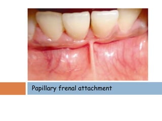
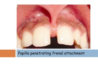
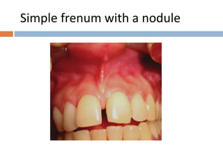
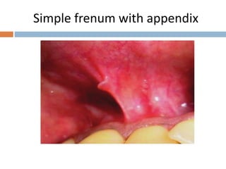
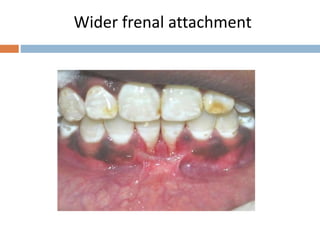
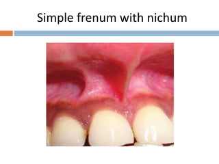
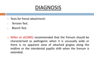
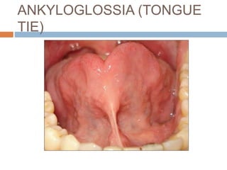
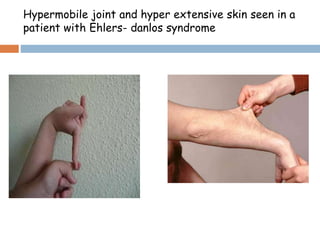
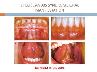
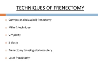
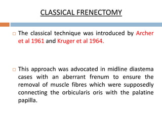
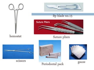
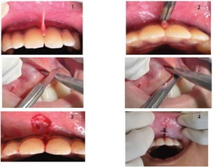
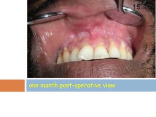
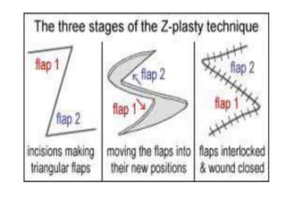
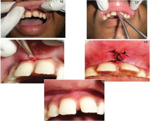
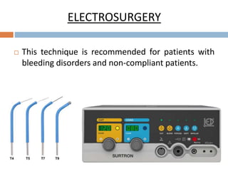
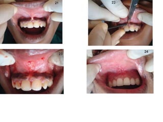
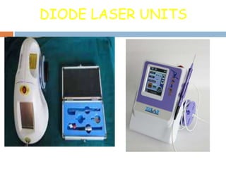
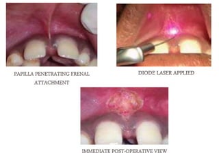
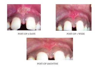
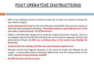
FRENUM ATTACHMENT
AND IT’S MANAGEMENT.
DR BHAUMIK THAKKAR.
PART II P.G. DEPT OF PERIODONTOLOGY.
CONTENTS
 INTRODUCTION
 DEVELOPMENT
 TYPES OF FRENAL ATTACHMENT
 VARIATIONS
 DIAGNOSIS
 ANKYLOGLOSSIA
 COMPLICATIONS OF ANKYLOGLOSSIA
 CLASSIFICATION
 SYNDROMES ASSOCIATED WITH ABNORMAL FRENUM
 COMPLICATIONS OF ABNORMAL FRENUM
 TREATMENT
 CONCLUSION
 REFERENCES
INTRODUCTION
 What is a frenum?
Frenum is a thin fold of mucous membrane with enclosed muscle
fibers that attach the lips to the alveolar mucosa and underlying
periosteum. ( Carranza 10th edition)
A frenulum is a small frenum. There are several frena that are usually
present in a normal oral cavity, most notably the maxillary labial
frenum, the mandibular labial frenum, and the lingual frenum.
Their primary function is to provide stability of the upper and lower lip
and the tongue.
DEVELOPMENT
 The maxillary labial frenum develops as a post eruptive
remnant of the ectolabial bands which connects the tubercle
of the upper lip into the palatine papilla.
 It extends over the alveolar process in infants and forms a
raphe that reaches the palatal papilla.
 Through the growth of alveolar process as the teeth erupt,
this attachment generally changes to assume the adult
configuration.
TYPES OF FRENAL ATTACHMENT
Depending upon the extent of attachment of fibres, frena have been
classified by Placek et al. 1974 as:
 MUCOSAL- The frenal fibres are attached up to the
mucogingival junction.
 GINGIVAL- The fibres are inserted within the attached
gingiva.
 PAPILLARY- The fibres extend into the interdental papilla.
 PAPILLA PENETRATING- The frenal fibres cross the alveolar
process and extend up to palatine papilla.
VARIATIONS
Other variations of normal frenal attachment
Include:
• Simple frenum with a nodule
• Simple frenum with appendix
• Simple frenum with nichum
• Bifid labial frenum
• Persistent tectolabial frenum
• Double frenum
• Wider frenum
Mucosal frenal attachment
Gingival frenal attachment
Papillary frenal attachment
Papilla penetrating frenal attachment
Simple frenum with a nodule
Simple frenum with appendix
Wider frenal attachment
Simple frenum with nichum
DIAGNOSIS
 Tests for frenal attachment:
1. Tension Test.
2. Blanch Test.
 Miller et al(1985) recommended that the frenum should be
characterised as pathogenic when it is unusually wide or
there is no apparent zone of attached gingiva along the
midline or the interdental papilla shift when the frenum is
extended.
ANKYLOGLOSSIA (TONGUE
TIE)
ANKYLOGLOSSIA
 Ankyloglossia or tongue-tie is an uncommon congenital
anomaly that occurs as a result of a short, tight, lingual
frenulum causing difficulty in speech articulation due to
limitation of tongue movement.
WALLACE et al 1963 defined tongue-tie as
“a condition in which the tip of the tongue cannot be protruded
beyond the lower incisor teeth because of a short frenulum
linguae, often containing scar tissue.”
CLINICAL FEATURES OF ANKYLOGLOSSIA
Ankyloglossia leads to :
 Limited mobility of tongue.
 Difficulty in swallowing.
 Difficulty in speech articulation which is evident for consonants like “s, z, t,
d, l, j, zh, ch, th, dg” and it is especially difficult to roll an “r”.
 Notched or “heart-shaped” tongue when it is protruded.
FREE-TONGUE:
The term free-tongue is defined as the length of tongue from the insertion
of lingual frenum from the base of the tongue to the tip of the tongue.
Clinically acceptable, normal range of free-tongue is greater than 16 mm.
(Kotlow et al 1999)
CLASSIFICATION
 Ankyloglossia can be classified into 4 classes based on Kotlow’s
assessment in 1999 (based on length of tongue from insertion of
lingual frenum at base of the tongue to the tip of the tongue) as
follows:
CLASS I: MILD ANKYLOGLOSSIA (12 to 16 mm)
CLASS II: MODERATE ANKYLOGLOSSIA (8 to 11mm)
CLASS III: SEVERE ANKYLOGLOSSIA (3 to 7 mm)
CLASS IV: COMPLETE ANKYLOGLOSSIA (< 3mm)
SYNDROMES ASSOCIATED WITH ABNORMAL
FRENUM
 Ehlers-Danlos syndrome
 Infantile hypertrophic pyloric stenosis
 Holoprosencephaly
 Ellis-van Creveld syndrome
 Oro-facial-digital syndrome
EHLERS-DANLOS SYNDROME
 It is a genetic disorder characterized by hyper
extensive skin and hyper mobile joints with no
gender predilection.
Hypermobile joint and hyper extensive skin seen in a
patient with Ehlers- danlos syndrome
EHLER DANLOS SYNDROME ORAL
MANIFESTATION
DE FELICE ET AL 2001
INFANTILE HYPERTROPHIC PYLORIC STENOSIS
 Occurs commonly in males at a ratio of 4.5 to 1
with an unknown etiology.
 The absence or hypoplasia of mandibular frenum is
seen in patients with this syndrome.
INFANTILE HYPERTROPHIC PYLORIC
STENOSIS
DE FELICE ET AL 2000
HOLOPROSENCEPHALY
 It is an autosomal dominant condition characterized by a
brain malformation due to defects in prosencephalon.
 It is characterized by defects including cyclopia, single
nostril, single central incisor and premaxillary agenesis.
 Absence of labial maxillary frenum is one of the
characteristic features of this condition.
HOLOPROSENCEPHALY
ELLIS-VAN CREVELD SYNDROME
 It is an autosomal recessive disorder mainly affecting enamel, hair and
nails.
 Patients with this syndrome characteristically present with
congenitally missing teeth, abnormal frenal attachment, microdontia
and hexadactyly.
 The most common finding is fusion of the anterior portion of the upper
lip to the maxillary gingival margin, as a result of which no mucobuccal
fold exists, causing the upper lip to present a slight V-shaped notch in
the middle (partial hare lip or lip-tie).
 The anterior portion of the lower ridge is often serrated and presents
with multiple small labial frenula.
Oral manifestations seen in Ellis-van Creveld
syndrome
COMPLICATIONS OF ABNORMAL FRENUM
A frenum becomes a problem if the attachment is too close to the marginal
gingiva.
Tension on the frenum may pull the gingival margin away from the tooth.
This condition may be conducive to plaque accumulation and inhibit proper
tooth brushing.
Abnormal frenum has been found to be associated with:
• Loss of papilla.
• Recession.
• Persistence of midline diastema.
• Difficulty in brushing.
• Malalignment of teeth .
• Compromised denture fit or retention.
TREATMENT
 Techniques for removal of aberrant frenum are :
 Frenotomy
 Frenectomy
Frenectomy : Refers to the complete removal of frenum,
including its attachment to the underlying bone.
It is required in the correction of abnormal diastema
between maxillary central incisors (Friedman 1957).
Frenotomy: Is the incision of the frenum.
It is usually done to relocate the frenal attachment so as to
create a zone of attached gingiva between the gingival
margin and the frenum.
FRENECTOMY
INDICATIONS
1. Gingival or papillary frenal attachment: Where frenal fibres
radiate into marginal gingiva producing gingival retraction and
localized gingival recession.
2. High frenal attachment: Where oral hygiene is hindered by
shallow vestibule caused by high frenal attachment.
3. Ankyloglossia: When lingual frenum interferes with speech.
TECHNIQUES OF FRENECTOMY
 Conventional (classical) frenectomy
 Miller’s technique
 V-Y plasty
 Z plasty
 Frenectomy by using electrocautery
 Laser frenectomy
CLASSICAL FRENECTOMY
 The classical technique was introduced by Archer
et al 1961 and Kruger et al 1964.
 This approach was advocated in midline diastema
cases with an aberrant frenum to ensure the
removal of muscle fibres which were supposedly
connecting the orbicularis oris with the palatine
papilla.
One month post-operative view
DISADVANTAGES
 Causes un-aesthetic labial tissue scarring.
 This may become a matter of concern in case
of high smile line exposing the anterior gingiva.
MILLER’S TECHNIQUE
 This technique was advocated by Miller PD et al in 1985.
 This was proposed for post-orthodontic diastema cases.
 The ideal time for performing this surgery is after the
orthodontic movement is complete and about 6 weeks
before the appliances are removed.
 This allows healing and tissue maturation.
2 weeks post-operative
ADVANTAGES OF MILLER’S TECHNIQUE
 Post-operatively, on healing, there is a continuous band
of gingiva across the midline, that gives a bracing effect
than the scar tissue, thus preventing orthodontic
relapse.
 The transseptal fibres are not disrupted surgically and so,
there is no loss of interdental papilla.
Z- PLASTY TECHNIQUE
 This technique is indicated when:
a) There is hypertrophy of the frenum with a low
insertion, associated with distema.
b) There is a short vestibule.
one month post-operative view
V-Y PLASTY TECHNIQUE
 This technique can be used for lengthening the
localized area, like a broad frena.
This technique is mostly employed in a case of a
papilla type of frenal attachment.
ELECTROSURGERY
 This technique is recommended for patients with
bleeding disorders and non-compliant patients.
ADVANTAGES
This technique offers the advantages of:
 Minimal time consumption.
 Minimal procedural bleeding.
 No need of sutures.
 Healing is by secondary intention as the wound edges are not
approximated with sutures.
LASER FRENECTOMY
The benefits of a laser frenectomy are greater as compared to
traditional techniques .
These include :
 Reduced bleeding during surgery.
 Reduced operating time and rapid postoperative hemostasis,
thus eliminating the need for sutures.
 The lack of need for sutures, as well as improved
postoperative comfort and healing, make this technique
particularly useful for very young patients.
DIODE LASER UNITS
POST OPERATIVE INSTRUCTIONS
 NOT to eat anything until the anesthesia wears off, as there are chances of biting the
lips, cheek or tongue.
 Avoid extremely hot foods for the rest of the day and do NOT rinse out your mouth, as
these will often prolong the bleeding. If bleeding continues, apply light pressure to the
area with a moistened gauze for 20-30 minutes.
 Follow a soft food diet, taking care to avoid the surgical area when chewing. Chew on
the opposite side and do NOT bite into food. Be sure to maintain adequate nutrition and
drink plenty of fluids. Do NOT use a drinking straw, as the suction may dislodge the
blood clot.
 Avoid alcohol and smoking until after your post-operative appointment.
 Maintain normal oral hygiene measures in the areas of mouth not affected by the
surgery. In areas where there is dressing, lightly brush only the biting surfaces of the
teeth. Vigorous rinsing should be avoided!
 Do NOT pull down the lip or cheek.
CONCLUSION
 Frenum may not regularly draw close scrutiny on routine
dental examination.
 While an aberrant frenum can be removed by any of the
modification techniques that have been proposed, a
functional and an aesthetic outcome can be achieved by a
proper technique selection, based on the type of frenal
attachment.
References.
 Carranza 10th and 12th edition.
 Priyanka M, Sruthi R, Ramakrishnan T, Emmadi P, Ambalavanan N.
An overview of frenal attachments. J Indian Soc Periodontol 2013
 Mirko P, Miroslav S, Lubor M. Significance of the labial frenum
attachment in periodontal disease in man. Part I. Classification and
epidemiology of the labial frenum attachment. J Periodontol 1974
 Devishree et al. Journal of Clinical and Diagnostic Research. 2012
November.
 Kotlow LA. Oral diagnosis of abnormal frenum attachments in
neonates and infants: Evaluation and treatment of maxillary
frenum using the Erbium YAG Laser. J Pediatr Dent Care 2004.
 De Felice C, Toti P, Di Maggio G, Parinni S, Bagnoli F. Absence of the
inferior labial and lingual frenula in Ehlers-Danlos syndrome. Lancet
2001
Frenum attachment and it's management.

Frenum attachment and it's management.

  • 1.
    FRENUM ATTACHMENT AND IT’SMANAGEMENT. DR BHAUMIK THAKKAR. PART II P.G. DEPT OF PERIODONTOLOGY.
  • 2.
    CONTENTS  INTRODUCTION  DEVELOPMENT TYPES OF FRENAL ATTACHMENT  VARIATIONS  DIAGNOSIS  ANKYLOGLOSSIA  COMPLICATIONS OF ANKYLOGLOSSIA  CLASSIFICATION  SYNDROMES ASSOCIATED WITH ABNORMAL FRENUM  COMPLICATIONS OF ABNORMAL FRENUM  TREATMENT  CONCLUSION  REFERENCES
  • 3.
    INTRODUCTION  What isa frenum? Frenum is a thin fold of mucous membrane with enclosed muscle fibers that attach the lips to the alveolar mucosa and underlying periosteum. ( Carranza 10th edition) A frenulum is a small frenum. There are several frena that are usually present in a normal oral cavity, most notably the maxillary labial frenum, the mandibular labial frenum, and the lingual frenum. Their primary function is to provide stability of the upper and lower lip and the tongue.
  • 4.
    DEVELOPMENT  The maxillarylabial frenum develops as a post eruptive remnant of the ectolabial bands which connects the tubercle of the upper lip into the palatine papilla.  It extends over the alveolar process in infants and forms a raphe that reaches the palatal papilla.  Through the growth of alveolar process as the teeth erupt, this attachment generally changes to assume the adult configuration.
  • 5.
    TYPES OF FRENALATTACHMENT Depending upon the extent of attachment of fibres, frena have been classified by Placek et al. 1974 as:  MUCOSAL- The frenal fibres are attached up to the mucogingival junction.  GINGIVAL- The fibres are inserted within the attached gingiva.  PAPILLARY- The fibres extend into the interdental papilla.  PAPILLA PENETRATING- The frenal fibres cross the alveolar process and extend up to palatine papilla.
  • 6.
    VARIATIONS Other variations ofnormal frenal attachment Include: • Simple frenum with a nodule • Simple frenum with appendix • Simple frenum with nichum • Bifid labial frenum • Persistent tectolabial frenum • Double frenum • Wider frenum
  • 7.
  • 8.
  • 9.
  • 10.
  • 11.
  • 12.
  • 13.
  • 14.
  • 15.
    DIAGNOSIS  Tests forfrenal attachment: 1. Tension Test. 2. Blanch Test.  Miller et al(1985) recommended that the frenum should be characterised as pathogenic when it is unusually wide or there is no apparent zone of attached gingiva along the midline or the interdental papilla shift when the frenum is extended.
  • 16.
  • 17.
    ANKYLOGLOSSIA  Ankyloglossia ortongue-tie is an uncommon congenital anomaly that occurs as a result of a short, tight, lingual frenulum causing difficulty in speech articulation due to limitation of tongue movement. WALLACE et al 1963 defined tongue-tie as “a condition in which the tip of the tongue cannot be protruded beyond the lower incisor teeth because of a short frenulum linguae, often containing scar tissue.”
  • 18.
    CLINICAL FEATURES OFANKYLOGLOSSIA Ankyloglossia leads to :  Limited mobility of tongue.  Difficulty in swallowing.  Difficulty in speech articulation which is evident for consonants like “s, z, t, d, l, j, zh, ch, th, dg” and it is especially difficult to roll an “r”.  Notched or “heart-shaped” tongue when it is protruded. FREE-TONGUE: The term free-tongue is defined as the length of tongue from the insertion of lingual frenum from the base of the tongue to the tip of the tongue. Clinically acceptable, normal range of free-tongue is greater than 16 mm. (Kotlow et al 1999)
  • 19.
    CLASSIFICATION  Ankyloglossia canbe classified into 4 classes based on Kotlow’s assessment in 1999 (based on length of tongue from insertion of lingual frenum at base of the tongue to the tip of the tongue) as follows: CLASS I: MILD ANKYLOGLOSSIA (12 to 16 mm) CLASS II: MODERATE ANKYLOGLOSSIA (8 to 11mm) CLASS III: SEVERE ANKYLOGLOSSIA (3 to 7 mm) CLASS IV: COMPLETE ANKYLOGLOSSIA (< 3mm)
  • 20.
    SYNDROMES ASSOCIATED WITHABNORMAL FRENUM  Ehlers-Danlos syndrome  Infantile hypertrophic pyloric stenosis  Holoprosencephaly  Ellis-van Creveld syndrome  Oro-facial-digital syndrome
  • 21.
    EHLERS-DANLOS SYNDROME  Itis a genetic disorder characterized by hyper extensive skin and hyper mobile joints with no gender predilection.
  • 22.
    Hypermobile joint andhyper extensive skin seen in a patient with Ehlers- danlos syndrome
  • 23.
    EHLER DANLOS SYNDROMEORAL MANIFESTATION DE FELICE ET AL 2001
  • 24.
    INFANTILE HYPERTROPHIC PYLORICSTENOSIS  Occurs commonly in males at a ratio of 4.5 to 1 with an unknown etiology.  The absence or hypoplasia of mandibular frenum is seen in patients with this syndrome.
  • 25.
  • 26.
    HOLOPROSENCEPHALY  It isan autosomal dominant condition characterized by a brain malformation due to defects in prosencephalon.  It is characterized by defects including cyclopia, single nostril, single central incisor and premaxillary agenesis.  Absence of labial maxillary frenum is one of the characteristic features of this condition.
  • 27.
  • 28.
    ELLIS-VAN CREVELD SYNDROME It is an autosomal recessive disorder mainly affecting enamel, hair and nails.  Patients with this syndrome characteristically present with congenitally missing teeth, abnormal frenal attachment, microdontia and hexadactyly.  The most common finding is fusion of the anterior portion of the upper lip to the maxillary gingival margin, as a result of which no mucobuccal fold exists, causing the upper lip to present a slight V-shaped notch in the middle (partial hare lip or lip-tie).  The anterior portion of the lower ridge is often serrated and presents with multiple small labial frenula.
  • 29.
    Oral manifestations seenin Ellis-van Creveld syndrome
  • 30.
    COMPLICATIONS OF ABNORMALFRENUM A frenum becomes a problem if the attachment is too close to the marginal gingiva. Tension on the frenum may pull the gingival margin away from the tooth. This condition may be conducive to plaque accumulation and inhibit proper tooth brushing. Abnormal frenum has been found to be associated with: • Loss of papilla. • Recession. • Persistence of midline diastema. • Difficulty in brushing. • Malalignment of teeth . • Compromised denture fit or retention.
  • 31.
    TREATMENT  Techniques forremoval of aberrant frenum are :  Frenotomy  Frenectomy Frenectomy : Refers to the complete removal of frenum, including its attachment to the underlying bone. It is required in the correction of abnormal diastema between maxillary central incisors (Friedman 1957). Frenotomy: Is the incision of the frenum. It is usually done to relocate the frenal attachment so as to create a zone of attached gingiva between the gingival margin and the frenum.
  • 32.
    FRENECTOMY INDICATIONS 1. Gingival orpapillary frenal attachment: Where frenal fibres radiate into marginal gingiva producing gingival retraction and localized gingival recession. 2. High frenal attachment: Where oral hygiene is hindered by shallow vestibule caused by high frenal attachment. 3. Ankyloglossia: When lingual frenum interferes with speech.
  • 33.
    TECHNIQUES OF FRENECTOMY Conventional (classical) frenectomy  Miller’s technique  V-Y plasty  Z plasty  Frenectomy by using electrocautery  Laser frenectomy
  • 34.
    CLASSICAL FRENECTOMY  Theclassical technique was introduced by Archer et al 1961 and Kruger et al 1964.  This approach was advocated in midline diastema cases with an aberrant frenum to ensure the removal of muscle fibres which were supposedly connecting the orbicularis oris with the palatine papilla.
  • 37.
  • 38.
    DISADVANTAGES  Causes un-aestheticlabial tissue scarring.  This may become a matter of concern in case of high smile line exposing the anterior gingiva.
  • 39.
    MILLER’S TECHNIQUE  Thistechnique was advocated by Miller PD et al in 1985.  This was proposed for post-orthodontic diastema cases.  The ideal time for performing this surgery is after the orthodontic movement is complete and about 6 weeks before the appliances are removed.  This allows healing and tissue maturation.
  • 41.
  • 42.
    ADVANTAGES OF MILLER’STECHNIQUE  Post-operatively, on healing, there is a continuous band of gingiva across the midline, that gives a bracing effect than the scar tissue, thus preventing orthodontic relapse.  The transseptal fibres are not disrupted surgically and so, there is no loss of interdental papilla.
  • 43.
    Z- PLASTY TECHNIQUE This technique is indicated when: a) There is hypertrophy of the frenum with a low insertion, associated with distema. b) There is a short vestibule.
  • 45.
  • 47.
    V-Y PLASTY TECHNIQUE This technique can be used for lengthening the localized area, like a broad frena. This technique is mostly employed in a case of a papilla type of frenal attachment.
  • 49.
    ELECTROSURGERY  This techniqueis recommended for patients with bleeding disorders and non-compliant patients.
  • 51.
    ADVANTAGES This technique offersthe advantages of:  Minimal time consumption.  Minimal procedural bleeding.  No need of sutures.  Healing is by secondary intention as the wound edges are not approximated with sutures.
  • 52.
    LASER FRENECTOMY The benefitsof a laser frenectomy are greater as compared to traditional techniques . These include :  Reduced bleeding during surgery.  Reduced operating time and rapid postoperative hemostasis, thus eliminating the need for sutures.  The lack of need for sutures, as well as improved postoperative comfort and healing, make this technique particularly useful for very young patients.
  • 53.
  • 56.
    POST OPERATIVE INSTRUCTIONS NOT to eat anything until the anesthesia wears off, as there are chances of biting the lips, cheek or tongue.  Avoid extremely hot foods for the rest of the day and do NOT rinse out your mouth, as these will often prolong the bleeding. If bleeding continues, apply light pressure to the area with a moistened gauze for 20-30 minutes.  Follow a soft food diet, taking care to avoid the surgical area when chewing. Chew on the opposite side and do NOT bite into food. Be sure to maintain adequate nutrition and drink plenty of fluids. Do NOT use a drinking straw, as the suction may dislodge the blood clot.  Avoid alcohol and smoking until after your post-operative appointment.  Maintain normal oral hygiene measures in the areas of mouth not affected by the surgery. In areas where there is dressing, lightly brush only the biting surfaces of the teeth. Vigorous rinsing should be avoided!  Do NOT pull down the lip or cheek.
  • 57.
    CONCLUSION  Frenum maynot regularly draw close scrutiny on routine dental examination.  While an aberrant frenum can be removed by any of the modification techniques that have been proposed, a functional and an aesthetic outcome can be achieved by a proper technique selection, based on the type of frenal attachment.
  • 58.
    References.  Carranza 10thand 12th edition.  Priyanka M, Sruthi R, Ramakrishnan T, Emmadi P, Ambalavanan N. An overview of frenal attachments. J Indian Soc Periodontol 2013  Mirko P, Miroslav S, Lubor M. Significance of the labial frenum attachment in periodontal disease in man. Part I. Classification and epidemiology of the labial frenum attachment. J Periodontol 1974  Devishree et al. Journal of Clinical and Diagnostic Research. 2012 November.  Kotlow LA. Oral diagnosis of abnormal frenum attachments in neonates and infants: Evaluation and treatment of maxillary frenum using the Erbium YAG Laser. J Pediatr Dent Care 2004.  De Felice C, Toti P, Di Maggio G, Parinni S, Bagnoli F. Absence of the inferior labial and lingual frenula in Ehlers-Danlos syndrome. Lancet 2001

Editor's Notes

  • #5 Knox and young in 1962 histologically showed presence of both elastic and muscle fibers in frenulum.
  • #8 The frenal fibres are attached up to the mucogingival junction.
  • #9 The fibres are inserted within the attached gingiva.
  • #10 The fibres extend into the interdental papilla.
  • #11 The frenal fibres cross the alveolar process and extend up to palatine papilla.
  • #16 Tension: lip is move outward n upward for upperlip and downwards for lower lip and sideways. If marginal or interdental pappila moves away from tooth surface then the test is positive. Blanch: to diagnos labial frenum. Done by pulling upper lip outwards and the frenum is diagnosed by blanching of tissue in incisive pappila region.
  • #24 Absence of the inferior labial and lingual frenum.
  • #27 In an autosomal dominant disease, if you inherit the abnormal gene from only one parent, you can get the disease. Often, one of the parents may also have the disease. PROSENCEPHALON:forebrain. CYCLOPIA:fused orbits of eyes into one. PREMAX AGENE: median cleft lip.
  • #37 LA is infiltered. Lip is extended and frenum is gripped with hemostat to the depth of the vestibule. Incisions are made above and below the instrument, the triangular frenum tissue is removed. Underlying fibrous attachment to the bone is exposed by horizontal incision and fibers are seperated. edges of the wound are approximated without creating tension.Only the mucosal extent of incision is sutured.gingival is kept for secondary intention healing.
  • #41 LA is infiltered. A horizontal incision is made to separate the frenulum from the interdental papilla. Care must be taken to extend incisions into the lip as far as necessary, to assure that a remnant of the frenulum is not left on the lip. It was then sutured to obtain primary closure across the midline 
  • #44 Ideal for broad , thick , hypertrophic frenum associated with inter-incisor distema and short vestibule.This technique achieved both removal of fibrous band and vertical lengthening of vestibule.
  • #45 Area is anesthetised with infiltration.frenum held with hemostat. The releasing incision is placed one on the superior border of frenum and other on the inferior border in opposite directions. The Z flaps are raised and then interchanged, so that the length of the frenum is increased. Sutures are placed first through the apices of the flaps and then at the edges of wound to ascertain the adequacy of the flap repositioning.
  • #49 Areais anesthetised with filtration. Frenum held with hemostat. Incision is made in the form of “V” at the undersurface of the frenal attachment. The frenum is relocated at an apical position and the V shape is converted into a Y.wound is then sutured.
  • #51 The area is anesthetized with local infiltration. The frenum is held with a hemostat.By using the loop electrode tip, it is excised.
  • #55 Properly strip, cleave and initiate well the disposable fiber tip. Place topical (small) or a few drops of anesthetic (large) on either side of the frenum attachment. Use 0.8 - 1.4 watts Continous wave ( Less energy without anesthetic). Continue until all vertical fibers are removed and you are at the periosteum. Hydrogen Peroxide or wet cotton pellet to remove tissue tags.