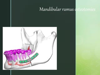
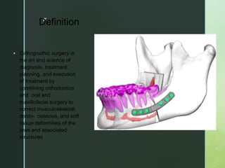
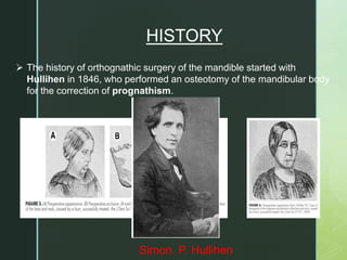
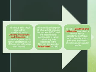
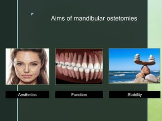
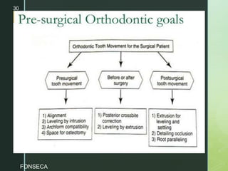
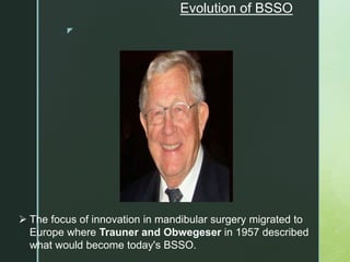
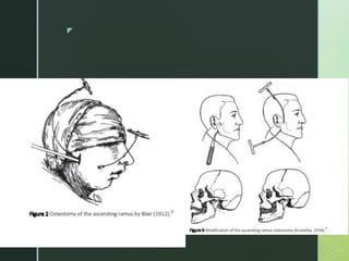
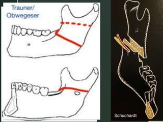
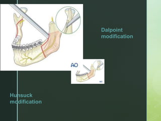
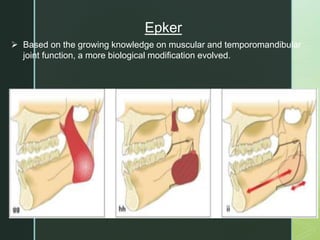
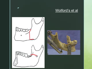
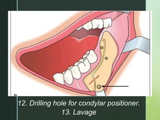
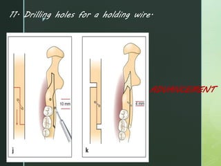
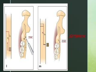
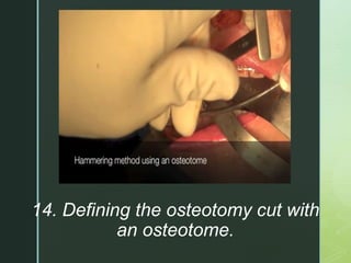
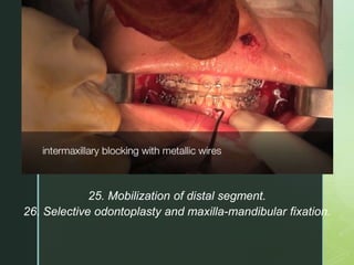
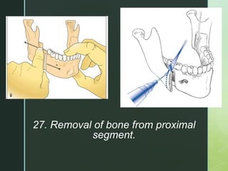
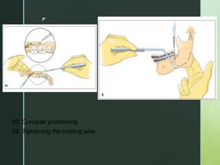
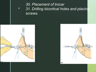
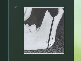
Mandibular ramus osteotomies are performed as part of orthognathic surgery to correct jaw deformities. The history began in the 1840s with body osteotomies and evolved to include ramal osteotomies from the 1920s-1940s. The bilateral sagittal split ramus osteotomy (BSSO) technique described in the 1950s-60s became the standard for mandibular setbacks and advancements. BSSO involves segmental osteotomies, splitting the mandible, repositioning segments, and fixation with plates or screws. Alternative techniques like vertical ramus osteotomy preserve the inferior alveolar nerve but limit distal segment movement. Careful preoperative
This document provides information on the Ramus osteotomy procedure, specifically the sagittal split osteotomy (SSO). It discusses the history and evolution of the SSO technique from its early developments to modern procedures. Key steps of the current SSO procedure are outlined, including incision, dissection, identification of anatomical landmarks, and performing the osteotomies along the medial ramus, vertical body, and buccal cortex before splitting the mandible. The SSO allows correction of mandibular deformities by repositioning the proximal and distal segments.
This document provides an overview of mandibular orthognathic procedures. It begins with an introduction to orthognathic surgery and the history of mandibular osteotomies. It then discusses anatomical and physiological considerations, timing of osteotomies, and various osteotomy techniques including vertical ramus, sagittal split, horizontal ramus, subapical, and total alveolar osteotomies. It also briefly touches on soft tissue changes and complications that can occur with mandibular osteotomies. The document is intended as a reference for various mandibular orthognathic procedures.
This document provides information about mandibular osteotomy and genioplasty procedures. It discusses the goals of mandibular osteotomy which include establishing proper function, aesthetics, stability, and minimizing treatment time. The history of mandibular osteotomy is reviewed dating back to the 1840s. Details are given about the sagittal split ramus osteotomy technique including indications, contraindications, steps, fixation methods, advantages, and complications. Common complications addressed include edema, nerve injury, arthropathy, condylar sag, hemorrhage, infection, and relapse.
This document provides an overview of major surgical procedures, including orthognathic surgeries. It defines orthognathic surgery as combining orthodontics and oral surgery to correct dentofacial deformities. The key steps are described as diagnosis, presurgical orthodontics, surgical treatment planning, mock surgery, the surgery and stabilization, and postsurgical orthodontics. Various surgical methods are outlined for maxillary osteotomies including LeFort I, II, and III, and for mandibular procedures including sagittal split osteotomy and genioplasty. Distraction osteogenesis is also summarized as a technique for gradual bone expansion.
This document provides an overview of orthognathic surgery. It begins with definitions and history, including important developments in mandibular and maxillary osteotomies. It discusses indications, contraindications, diagnosis/assessment, and the pre-surgical orthodontic phase. Surgical procedures covered include bilateral sagittal split osteotomy, LeFort I osteotomy, genioplasty, and implant augmentation. Complications of various procedures are also outlined.
This document provides information about maxillary orthognathic surgery. It discusses the history and types of maxillary osteotomies performed, including Lefort I, II, and III osteotomies. Lefort I osteotomy is described as the workhorse procedure used to correct functional and aesthetic maxillary issues. Complications, patient satisfaction rates, and surgical techniques for performing the various maxillary osteotomies are summarized.
The document discusses maxillary orthognathic procedures, including common dentofacial deformities involving the maxilla, diagnosis and treatment planning, surgical anatomy and approaches, types of mid-face osteotomies, and potential complications. It describes techniques for anterior and posterior segmental maxillary osteotomies, as well as total maxillary osteotomies including LeFort I, II, and III procedures. Complications addressed include vascular compromise, hemorrhage, and infection.
This document provides information on various osteotomies of the mandible including subapical, body, genioplasty, and ramus osteotomies. It discusses the surgical anatomy of the mandible, assessment, techniques, and complications. Key details include the location of the lingula and mandibular foramen, vascular supply, types of mandibular deformities, and techniques for anterior and posterior subapical, body, genioplasty, and sagittal split ramus osteotomies.
Orthognathic surgery involves intentionally sectioning jaw bones to correct dentofacial deformities. This document describes various mandibular and maxillary osteotomy procedures including:
1) Mandibular body osteotomies like anterior, posterior, and mid-symphysis osteotomies.
2) Segmental subapical mandibular surgeries like anterior, posterior, and total subapical osteotomies.
3) Genioplasty procedures to augment, reduce, straighten or lengthen the chin.
4) Mandibular ramus osteotomies including inverted-L, C, and sagittal split osteotomies.
The document discusses pre-prosthetic surgery, which aims to modify the oral environment to better support prosthetic appliances. The goals are to provide a broad, flat ridge with height and a firm mucosal covering. Objectives include eliminating disease, conserving structures, and providing support to withstand forces. The document describes various basic surgical procedures like alveoloplasty, tori removal, and soft tissue procedures to reshape ridges and remove excess tissue in preparation for dentures.
This PowerPoint presentation delivers a technical analysis of the midface orthognathic procedure. Explore surgical techniques, anatomical considerations, and treatment objectives.
The document discusses periodontal flap surgery. It begins by defining a periodontal flap and providing a brief history of flap surgery techniques dating back to the 19th century. It then covers the objectives of periodontal surgery, classifications of flaps, principles of flap design and incision placement. Specific flap techniques are described for pocket elimination, including the Original Widman Flap, Neumann Flap, Modified Flap Operation, Undisplaced Flap, Modified Widman Flap, and Apically Repositioned Flap. The document provides details on incision types, flap elevation, and management. In summary, it provides an overview of periodontal flap surgery, outlining key historical developments, classifications, principles, and specific techniques.
Crown lengthening is a surgical procedure used to expose more of the clinical crown for esthetic or restorative purposes. There are several techniques for crown lengthening including gingivectomy, flap surgery, and lasers. Gingivectomy involves removing gum tissue while flap surgery involves raising a flap and removing underlying bone. The biologic width must be considered to avoid compromising tissue health. Crown lengthening can allow for improved restoration margins or smile appearance.
This document provides an overview of endodontic surgery. It begins with definitions and a brief history of endodontic surgery. It then discusses indications, contraindications, classifications of endodontic surgeries, and recent advances. The document covers various surgical procedures like incision and drainage, flap design, osteotomy, periradicular curettage, root-end resection, root-end preparation, and root-end filling. It provides details on techniques, principles, and advantages/disadvantages of these procedures. Overall, the document serves as a comprehensive guide to endodontic surgery.
This document discusses the management of mandibular angle fractures. It begins by explaining that the mandibular angle is a common site of fracture due to abrupt changes in direction between the body and ramus. It then covers the surgical anatomy of the mandibular angle region and biomechanical considerations for fractures. The document classifies mandibular angle fractures and discusses radiographic examination. It describes surgical approaches like vestibular, submandibular, and retromandibular. Guidelines are provided for managing teeth in the fracture line. Osteosynthesis techniques like Champy's lines and plate placement are outlined. The transbuccal plating system is also summarized.
This document provides an overview of Lefort I osteotomy, including:
- A brief history describing the development of maxillary osteotomy techniques.
- Anatomical considerations and the biologic basis for maxillary osteotomies, which have shown adequate blood supply can be maintained.
- Indications for Lefort I osteotomy include altering vertical dimension, anteroposterior movements, and surgical expansion of the maxilla.
- Types of Lefort I osteotomies are described, including classic, quadrangular, and segmental variations. Postoperative management and potential complications are also outlined.
This document discusses nasal reconstruction using bone grafts. It provides background on the use of autogenous bone grafts for nasal reconstruction since the 1820s. It then discusses alternatives to autogenous grafts due to limitations like limited availability and donor site morbidity. The mechanisms of bone graft healing and factors influencing healing are reviewed. Different types of graft materials including autografts, allografts, alloplasts, and xenografts are described. Common sites for harvesting bone grafts and techniques for nasal reconstruction using various graft materials are summarized.
Maxillary osteotomies are commonly performed to correct dentofacial deformities. The Le Fort I osteotomy involves making cuts in the maxilla to mobilize it for repositioning. It has a high success rate due to the broad soft tissue attachments maintaining the blood supply to the mobilized maxilla. Precise osteotomy cuts, identification of anatomical structures, and accurate repositioning and stabilization are required. Modifications include anterior and posterior subapical osteotomies and quadrangulated osteotomies. Rigid fixation provides stability but requires precise adaptation, while non-rigid fixation offers postoperative flexibility.
TO fraktur mandibula of oral and maxillofasial surgerypptxssuserca681b1
The document discusses mandibular fractures. It begins with an introduction discussing how mandibular fractures commonly involve the jaws and cause malocclusion. It then covers the anatomy, classification, signs and symptoms, clinical examination and diagnosis. It discusses indications for open reduction and internal fixation for mandible fractures. It outlines Champy principles for plate fixation and the surgical approach, preparation, steps and post-operative care for open reduction and internal fixation of a mandibular fracture. It concludes by mentioning potential complications.
endodontic surgery and its current concepts boris saha
This document provides an overview of endodontic surgery and its concepts. It discusses the history and evolution of endodontic surgery techniques. It also covers indications for endodontic surgery, classifications of different surgical procedures, and considerations for pre-surgical treatment planning. Key surgical steps like flap design, osteotomy, and root-end resection are summarized.
The Twin Block Appliance is a functional orthodontic appliance consisting of upper and lower removable bite blocks used to correct Class II malocclusions by repositioning the mandible forward through inclined occlusal planes which provide proprioceptive stimulus for bone growth; it was developed in the 1980s as an improvement on previous functional appliances and provides numerous orthodontic, dental, skeletal, soft tissue and airway benefits when worn as directed.
orthodontic management of Idiopathic condylar resorption part 2MaherFouda2
This document summarizes the management of idiopathic condylar resorption through several case studies and articles. It finds that orthodontic treatment is contraindicated during active idiopathic condylar resorption but can be used once the condition is in remission. For severe cases, orthognathic surgery may be needed but there is a risk of relapse, especially in women. Miniscrew-assisted camouflage treatment helped one patient by retracting teeth and inducing counterclockwise mandibular rotation to improve her lip incompetence and profile.
This document discusses various treatment approaches for idiopathic condylar resorption (ICR), including:
1) Orthodontic treatment is contraindicated during active ICR due to risk of accelerating resorption or litigation.
2) Surgical options like orthognathic surgery place demands on compromised TMJs that may lead to relapse.
3) Combining pre- and post-operative medical management with anti-inflammatory drugs and supplements with orthognathic surgery may help control resorption.
4) Total alloplastic TMJ replacement avoids relying on compromised TMJ tissues, providing an option when other treatments aren't viable.
Orthognathic surgery is used to correct skeletal discrepancies of the jaw bones that cannot be addressed by orthodontics alone. It involves both pre-surgical orthodontics to position the teeth and surgical procedures on the mandible and/or maxilla. Common mandibular procedures include sagittal split osteotomy to move the mandible forwards or backwards and vertical subsigmoid osteotomy to push the mandible back. The main maxillary procedure is a Le Fort I osteotomy where the maxilla is freed and can be repositioned using a buccal incision approach. Both procedures aim to correct functional and aesthetic issues of the jaws and require postoperative orthodontics for detailed occlusion.
This presentation gives information on the pharmacology of Prostaglandins, Thromboxanes and Leukotrienes i.e. Eicosanoids. Eicosanoids are signaling molecules derived from polyunsaturated fatty acids like arachidonic acid. They are involved in complex control over inflammation, immunity, and the central nervous system. Eicosanoids are synthesized through the enzymatic oxidation of fatty acids by cyclooxygenase and lipoxygenase enzymes. They have short half-lives and act locally through autocrine and paracrine signaling.
Applications of NMR in Protein Structure Prediction.pptxAnagha R Anil
This presentation explores the pivotal role of Nuclear Magnetic Resonance (NMR) spectroscopy in predicting protein structures. It delves into the methodologies, advancements, and applications of NMR in determining the three-dimensional configurations of proteins, which is crucial for understanding their function and interactions.
Orthognathic surgery involves intentionally sectioning jaw bones to correct dentofacial deformities. This document describes various mandibular and maxillary osteotomy procedures including:
1) Mandibular body osteotomies like anterior, posterior, and mid-symphysis osteotomies.
2) Segmental subapical mandibular surgeries like anterior, posterior, and total subapical osteotomies.
3) Genioplasty procedures to augment, reduce, straighten or lengthen the chin.
4) Mandibular ramus osteotomies including inverted-L, C, and sagittal split osteotomies.
The document discusses pre-prosthetic surgery, which aims to modify the oral environment to better support prosthetic appliances. The goals are to provide a broad, flat ridge with height and a firm mucosal covering. Objectives include eliminating disease, conserving structures, and providing support to withstand forces. The document describes various basic surgical procedures like alveoloplasty, tori removal, and soft tissue procedures to reshape ridges and remove excess tissue in preparation for dentures.
This PowerPoint presentation delivers a technical analysis of the midface orthognathic procedure. Explore surgical techniques, anatomical considerations, and treatment objectives.
The document discusses periodontal flap surgery. It begins by defining a periodontal flap and providing a brief history of flap surgery techniques dating back to the 19th century. It then covers the objectives of periodontal surgery, classifications of flaps, principles of flap design and incision placement. Specific flap techniques are described for pocket elimination, including the Original Widman Flap, Neumann Flap, Modified Flap Operation, Undisplaced Flap, Modified Widman Flap, and Apically Repositioned Flap. The document provides details on incision types, flap elevation, and management. In summary, it provides an overview of periodontal flap surgery, outlining key historical developments, classifications, principles, and specific techniques.
Crown lengthening is a surgical procedure used to expose more of the clinical crown for esthetic or restorative purposes. There are several techniques for crown lengthening including gingivectomy, flap surgery, and lasers. Gingivectomy involves removing gum tissue while flap surgery involves raising a flap and removing underlying bone. The biologic width must be considered to avoid compromising tissue health. Crown lengthening can allow for improved restoration margins or smile appearance.
This document provides an overview of endodontic surgery. It begins with definitions and a brief history of endodontic surgery. It then discusses indications, contraindications, classifications of endodontic surgeries, and recent advances. The document covers various surgical procedures like incision and drainage, flap design, osteotomy, periradicular curettage, root-end resection, root-end preparation, and root-end filling. It provides details on techniques, principles, and advantages/disadvantages of these procedures. Overall, the document serves as a comprehensive guide to endodontic surgery.
This document discusses the management of mandibular angle fractures. It begins by explaining that the mandibular angle is a common site of fracture due to abrupt changes in direction between the body and ramus. It then covers the surgical anatomy of the mandibular angle region and biomechanical considerations for fractures. The document classifies mandibular angle fractures and discusses radiographic examination. It describes surgical approaches like vestibular, submandibular, and retromandibular. Guidelines are provided for managing teeth in the fracture line. Osteosynthesis techniques like Champy's lines and plate placement are outlined. The transbuccal plating system is also summarized.
This document provides an overview of Lefort I osteotomy, including:
- A brief history describing the development of maxillary osteotomy techniques.
- Anatomical considerations and the biologic basis for maxillary osteotomies, which have shown adequate blood supply can be maintained.
- Indications for Lefort I osteotomy include altering vertical dimension, anteroposterior movements, and surgical expansion of the maxilla.
- Types of Lefort I osteotomies are described, including classic, quadrangular, and segmental variations. Postoperative management and potential complications are also outlined.
This document discusses nasal reconstruction using bone grafts. It provides background on the use of autogenous bone grafts for nasal reconstruction since the 1820s. It then discusses alternatives to autogenous grafts due to limitations like limited availability and donor site morbidity. The mechanisms of bone graft healing and factors influencing healing are reviewed. Different types of graft materials including autografts, allografts, alloplasts, and xenografts are described. Common sites for harvesting bone grafts and techniques for nasal reconstruction using various graft materials are summarized.
Maxillary osteotomies are commonly performed to correct dentofacial deformities. The Le Fort I osteotomy involves making cuts in the maxilla to mobilize it for repositioning. It has a high success rate due to the broad soft tissue attachments maintaining the blood supply to the mobilized maxilla. Precise osteotomy cuts, identification of anatomical structures, and accurate repositioning and stabilization are required. Modifications include anterior and posterior subapical osteotomies and quadrangulated osteotomies. Rigid fixation provides stability but requires precise adaptation, while non-rigid fixation offers postoperative flexibility.
TO fraktur mandibula of oral and maxillofasial surgerypptxssuserca681b1
The document discusses mandibular fractures. It begins with an introduction discussing how mandibular fractures commonly involve the jaws and cause malocclusion. It then covers the anatomy, classification, signs and symptoms, clinical examination and diagnosis. It discusses indications for open reduction and internal fixation for mandible fractures. It outlines Champy principles for plate fixation and the surgical approach, preparation, steps and post-operative care for open reduction and internal fixation of a mandibular fracture. It concludes by mentioning potential complications.
endodontic surgery and its current concepts boris saha
This document provides an overview of endodontic surgery and its concepts. It discusses the history and evolution of endodontic surgery techniques. It also covers indications for endodontic surgery, classifications of different surgical procedures, and considerations for pre-surgical treatment planning. Key surgical steps like flap design, osteotomy, and root-end resection are summarized.
The Twin Block Appliance is a functional orthodontic appliance consisting of upper and lower removable bite blocks used to correct Class II malocclusions by repositioning the mandible forward through inclined occlusal planes which provide proprioceptive stimulus for bone growth; it was developed in the 1980s as an improvement on previous functional appliances and provides numerous orthodontic, dental, skeletal, soft tissue and airway benefits when worn as directed.
orthodontic management of Idiopathic condylar resorption part 2MaherFouda2
This document summarizes the management of idiopathic condylar resorption through several case studies and articles. It finds that orthodontic treatment is contraindicated during active idiopathic condylar resorption but can be used once the condition is in remission. For severe cases, orthognathic surgery may be needed but there is a risk of relapse, especially in women. Miniscrew-assisted camouflage treatment helped one patient by retracting teeth and inducing counterclockwise mandibular rotation to improve her lip incompetence and profile.
This document discusses various treatment approaches for idiopathic condylar resorption (ICR), including:
1) Orthodontic treatment is contraindicated during active ICR due to risk of accelerating resorption or litigation.
2) Surgical options like orthognathic surgery place demands on compromised TMJs that may lead to relapse.
3) Combining pre- and post-operative medical management with anti-inflammatory drugs and supplements with orthognathic surgery may help control resorption.
4) Total alloplastic TMJ replacement avoids relying on compromised TMJ tissues, providing an option when other treatments aren't viable.
Orthognathic surgery is used to correct skeletal discrepancies of the jaw bones that cannot be addressed by orthodontics alone. It involves both pre-surgical orthodontics to position the teeth and surgical procedures on the mandible and/or maxilla. Common mandibular procedures include sagittal split osteotomy to move the mandible forwards or backwards and vertical subsigmoid osteotomy to push the mandible back. The main maxillary procedure is a Le Fort I osteotomy where the maxilla is freed and can be repositioned using a buccal incision approach. Both procedures aim to correct functional and aesthetic issues of the jaws and require postoperative orthodontics for detailed occlusion.
Similar to 11. Mandibular osteotomies psk.pptx (20)
This presentation gives information on the pharmacology of Prostaglandins, Thromboxanes and Leukotrienes i.e. Eicosanoids. Eicosanoids are signaling molecules derived from polyunsaturated fatty acids like arachidonic acid. They are involved in complex control over inflammation, immunity, and the central nervous system. Eicosanoids are synthesized through the enzymatic oxidation of fatty acids by cyclooxygenase and lipoxygenase enzymes. They have short half-lives and act locally through autocrine and paracrine signaling.
Applications of NMR in Protein Structure Prediction.pptxAnagha R Anil
This presentation explores the pivotal role of Nuclear Magnetic Resonance (NMR) spectroscopy in predicting protein structures. It delves into the methodologies, advancements, and applications of NMR in determining the three-dimensional configurations of proteins, which is crucial for understanding their function and interactions.
Receptor Discordance in Breast Carcinoma During the Course of Life
Definition:
Receptor discordance refers to changes in the status of hormone receptors (estrogen receptor ERα, progesterone receptor PgR, and HER2) in breast cancer tumors over time or between primary and metastatic sites.
Causes:
Tumor Evolution:
Genetic and epigenetic changes during tumor progression can lead to alterations in receptor status.
Treatment Effects:
Therapies, especially endocrine and targeted therapies, can selectively pressure tumor cells, causing shifts in receptor expression.
Heterogeneity:
Inherent heterogeneity within the tumor can result in subpopulations of cells with different receptor statuses.
Impact on Treatment:
Therapeutic Resistance:
Loss of ERα or PgR can lead to resistance to endocrine therapies.
HER2 discordance affects the efficacy of HER2-targeted treatments.
Treatment Adjustment:
Regular reassessment of receptor status may be necessary to adjust treatment strategies appropriately.
Clinical Implications:
Prognosis:
Receptor discordance is often associated with a poorer prognosis.
Biopsies:
Obtaining biopsies from metastatic sites is crucial for accurate receptor status assessment and effective treatment planning.
Monitoring:
Continuous monitoring of receptor status throughout the disease course can guide personalized therapy adjustments.
Understanding and managing receptor discordance is essential for optimizing treatment outcomes and improving the prognosis for breast cancer patients.
CLASSIFICATION OF H1 ANTIHISTAMINICS-
FIRST GENERATION ANTIHISTAMINICS-
1)HIGHLY SEDATIVE-DIPHENHYDRAMINE,DIMENHYDRINATE,PROMETHAZINE,HYDROXYZINE 2)MODERATELY SEDATIVE- PHENARIMINE,CYPROHEPTADINE, MECLIZINE,CINNARIZINE
3)MILD SEDATIVE-CHLORPHENIRAMINE,DEXCHLORPHENIRAMINE
TRIPROLIDINE,CLEMASTINE
SECOND GENERATION ANTIHISTAMINICS-FEXOFENADINE,
LORATADINE,DESLORATADINE,CETIRIZINE,LEVOCETIRIZINE,
AZELASTINE,MIZOLASTINE,EBASTINE,RUPATADINE. Mechanism of action of 2nd generation antihistaminics-
These drugs competitively antagonize actions of
histamine at the H1 receptors.
Pharmacological actions-
Antagonism of histamine-The H1 antagonists effectively block histamine induced bronchoconstriction, contraction of intestinal and other smooth muscle and triple response especially wheal, flare and itch. Constriction of larger blood vessel by histamine is also antagonized.
2) Antiallergic actions-Many manifestations of immediate hypersensitivity (type I reactions)are suppressed. Urticaria, itching and angioedema are well controlled.3) CNS action-The older antihistamines produce variable degree of CNS depression.But in case of 2nd gen antihistaminics there is less CNS depressant property as these cross BBB to significantly lesser extent.
4) Anticholinergic action- many H1 blockers
in addition antagonize muscarinic actions of ACh. BUT IN 2ND gen histaminics there is Higher H1 selectivitiy : no anticholinergic side effects
Giloy in Ayurveda - Classical Categorization and SynonymsPlanet Ayurveda
Giloy, also known as Guduchi or Amrita in classical Ayurvedic texts, is a revered herb renowned for its myriad health benefits. It is categorized as a Rasayana, meaning it has rejuvenating properties that enhance vitality and longevity. Giloy is celebrated for its ability to boost the immune system, detoxify the body, and promote overall wellness. Its anti-inflammatory, antipyretic, and antioxidant properties make it a staple in managing conditions like fever, diabetes, and stress. The versatility and efficacy of Giloy in supporting health naturally highlight its importance in Ayurveda. At Planet Ayurveda, we provide a comprehensive range of health services and 100% herbal supplements that harness the power of natural ingredients like Giloy. Our products are globally available and affordable, ensuring that everyone can benefit from the ancient wisdom of Ayurveda. If you or your loved ones are dealing with health issues, contact Planet Ayurveda at 01725214040 to book an online video consultation with our professional doctors. Let us help you achieve optimal health and wellness naturally.
Milan J. Anadkat, MD, and Dale V. Reisner discuss generalized pustular psoriasis in this CME activity titled "Supporting Patient-Centered Care in Generalized Pustular Psoriasis: Communications Strategies to Improve Shared Decision-Making." For the full presentation, please visit us at www.peervoice.com/HUM870.
Congestive Heart failure is caused by low cardiac output and high sympathetic discharge. Diuretics reduce preload, ACE inhibitors lower afterload, beta blockers reduce sympathetic activity, and digitalis has inotropic effects. Newer medications target vasodilation and myosin activation to improve heart efficiency while lowering energy requirements. Combination therapy, following an assessment of cardiac function and volume status, is the most effective strategy to heart failure care.
Spontaneous Bacterial Peritonitis - Pathogenesis , Clinical Features & Manage...Jim Jacob Roy
In this presentation , SBP ( spontaneous bacterial peritonitis ) , which is a common complication in patients with cirrhosis and ascites is described in detail.
The reference for this presentation is Sleisenger and Fordtran's Gastrointestinal and Liver Disease Textbook ( 11th edition ).
TEST BANK For Brunner and Suddarth's Textbook of Medical-Surgical Nursing, 14...Donc Test
TEST BANK For Brunner and Suddarth's Textbook of Medical-Surgical Nursing, 14th Edition (Hinkle, 2017) Verified Chapter's 1 - 73 Complete.pdf
Discover the benefits of homeopathic medicine for irregular periods with our guide on 5 common remedies. Learn how these natural treatments can help regulate menstrual cycles and improve overall menstrual health.
Visit Us: https://drdeepikashomeopathy.com/service/irregular-periods-treatment/
The Children are very vulnerable to get affected with respiratory disease.
In our country, the respiratory Disease conditions are consider as major cause for mortality and Morbidity in Child.
2. z
Definition
Orthognathic surgery is
the art and science of
diagnosis, treatment
planning, and execution
of treatment by
combining orthodontics
and oral and
maxillofacial surgery to
correct musculoskeletal,
dento- osseous, and soft
tissue deformities of the
jaws and associated
structures
3. The history of orthognathic surgery of the mandible started with
Hullihen in 1846, who performed an osteotomy of the mandibular body
for the correction of prognathism.
Simon. P. Hullihen
HISTORY
4. z
• In 1849, Henry Blair developed
osteotomy of mandibular body
for the correction of mandibular
horizontal excess.
Henry Blair
5. The 1920s and 1930s
saw further
modifications by
Limberg, Wassmund,
and Kazanjian of
external approaches to
ramal osteotomies. All
of these had difficulties
with relapse.
The earliest description
of what would become
the modern BSSO and
the first intraoral
approach to a ramal
osteotomy was
described in the
German literature by
Schuchardt in 1942.
In 1954, Caldwell and
Letterman described a
vertical ramus
osteotomy technique,
which was shown to
preserve the inferior
alveolar neurovascular
bundle
13. z
• Medial and forward displacement of the mandibular
disk- by the upper head of the lateral pterygoid
muscle.
• After sectioning - the mandibular condyle is displaced
in the same direction as the disk - by the pull of the
lower head of the lateral pterygoid muscle.
14. z
Muscles
Contribution of suprahyoid muscles in relapse in mandibular
advancement
Ellis and Carlson study (in monkeys) – relieving the suprahyoid
muscles from the symphysis of the mandible decreased the
amount of relapse
Clinical studies – have failed to show a relation between
suprahyoid myotomies and relapse.
17. z
Determination of safe distance away from the apex of
teeth:
• The safer distance is 5 mm but studies have shown that
even 10 mm distance shows pulpal changes.
• Epker BN. Vascular considerations in orthognathic surgery: I. Mandibular osteotomies. Oral surgery, oral medicine, oral
pathology. 1984 May 1;57(5):467-72.
19. z
• The medial horizontal cut be at or just above the tip of the lingula because
a higher cut may be associated with an increased difficulty in splitting or
incidence of unfavorable fracture
7.5 to 13.3 mm above lingula
Buccal & lingual cortex fusion occurs at a rate of
:
• 20% in the anterior ramus
• 39% in the posterior ramus
22. z
Classification of the
topography of the
inferior alveolar nerve.
J Anat 1971;108:433-40
• I = the nerve has a
course near the apices of
the teeth,
• II = the main trunk is
low down in the body
• III = the main trunk is
low down in the body of
the mandible with several
smaller trunks to the
molar teeth
23. z
Revasculirasatio
n and healing
• Intermedullary circulation between the proximal and distal segments
• Margins of osteotomy- avascular
Immediate post-operatively
• Level of hypervascularity around surgical site
• No soft tissue re-attachment
• Isolated areas of sub- periosteal bone formation
One week post-op
• Avascular zone at the proximal osteotomy site
• Necrotic zone at the distal osteotomy site
• No soft tissue attachment at distal necrotic zone
2weeks post-op
• Soft tissue re-attachment
• Vascular anastamoses between proximal and distal segments
• Osteoid formation through out marrow formation
3 weeks post-op
• Circulation reconstituted across the osteotomy site
• Soft tissue re- attachment established
6 weeks post-op
• Circulation between the segments is continuous
12 weeks post- op
25. z
Timing of surgery
As a rule of thumb it is better to wait till the skeletal growth is
completed before doing orthognathic surgery.
Corrective surgical measure even during the growth period, specially if
there is compelling psychological need for such intervention in the
patient.
28
26. z
Role of pre-surgical orthodontics
To eliminate dental mal-relationships
which
prevent surgical repositioning of
fragments
To achieve decompensation by undoing
the natural compensating tooth
alignments.
To create interdental spacing to facilitate
segmental osteotomy and
To perform those tooth movements
which, if done post-surgically, will spoil the
result of surgery.
29
29. z
Evolution of BSSO
The focus of innovation in mandibular surgery migrated to
Europe where Trauner and Obwegeser in 1957 described
what would become today's BSSO.
35. z
BILATERAL SAGITTAL SPLIT RAMUS
OSTEOTOMY
BSSO is similar for all 3 clinical situations , with subtle variations in the
osteotomy and fixation techniques.
INDICATIONS:
Horizontal
mandibular
excess
Horizontal
mandibular
deficiency
Asymmetry
Mandibular
advancement
Mandibular
setback(small-
moderate)
52. z
• 19. Stripping the medial pterygoid msucle and
stylomandibular ligament.
20. Removal of impacted third molars.
21. Smoothing contact areas of bone segments.
22. Placement of a holding wire.
23. Noting the position of IANB
24. Noting position of 3rd molar.
53. z
• 19. Stripping the medial pterygoid msucle and
stylomandibular ligament.
20. Removal of impacted third molars.
21. Smoothing contact areas of bone segments.
22. Placement of a holding wire.
23. Noting the position of IANB
24. Noting position of 3rd molar.
54. z
25. Mobilization of distal segment.
26. Selective odontoplasty and maxilla-mandibular fixation.
59. z
32. Removing MMF ad checking occlusion.
33. Intraop diagnosis of malocclusion.
34. Placement of intraoral and extraoral
sutures.
35. Placement of elastics.
36. Placement of a pressure bandage.
60. z
• With wire at upper and lower border
• Lag screws
• Bicortical screws – 2 or 3 screws are used
• Mini plates
• Bioresorbable plates and screws
Fujioka M, Fujii T,Hirano A. Comparative study of mandibular stability after sagittal split osteotomies:
bicortical versus monocortical osteosynthesis. Cleft palate craniofacial journal 2000; 37:551.
Fixation techniques
61. To define the separation better, a thick, finely
tapered osteotome-10mm wide is driven
between the proximal & distal segments of the
mandible through an anterior corticotomy.It
should not reach IAN
The Dunn dautrey osteotome is driven gently
with only manual force, b/w the buccal cortex &
medulla. The buccal & lingual cortices are
separated with only little resistance as a result
of the complete burring of the post.margin of
the medial osteotomy & inferior margin of
vertical osteotomy
Dunn dautrey osteotome is run carefully down
the inferior border of the mandible.
All contents of IAN is separated from the buccal
attachments , nerve & vessels should be
allowed to fall medial to the osteotome. The
Dunn dautrey osteotome is manually twisted
under visualization of IAN
MANUAL TWIST TECHNIQUE
62. The osteotome should be twisted in the
direction that the distal portion of the
proximal segment is opened & the distal
tips of both proximal segments are
rotated to the buccal side.
This prevents manual twisting force from
being transferred to the TMJ.
Twisting starts from anterior portion of
proximal segments just behind the
vertical buccal osteotome. Splitting by
manual twist force extends from the
mandibular angle to the post. Border of
ascending ramus.
63. z
Vertical ramus
osteotomy
• 1st described by Caldwell and
Letterman in 1954- extra oral
Indications:
Patient with horizontal mand excess.
Mandibular asymmetry.
Contraindications:
Advancement of distal tooth bearing
segment.
Recent condylar #
72. z
COMPARISON BETWEEN SSRO
ANDVRO
McKenna SJ, King EE. Intraoral Vertical Ramus Osteotomy Procedure and Technique. Atlas of the oral and
maxillofacial surgery clinics of North America. 2016 Mar 1;24(1):37-43.
SSRO VRO
OSTEOTOMY PASaggital split Latero medial cut
Open procedure Blind procedure
Along IAN Rear to IAN
Frequent exposure of IAN No exposure of IAN
BONE HEALING Contact on marrow to
marrow
Contact on cortex to
cortex
BONE FIXATION Rigid internal fixation No fixation
CONDYLAR HEAD Original position New equilibrated
position
POST OP IMF
prognosis
None or shorter period
Weakly dependent on pt
Required 7-10 day
Strongly dependent on pt
73. z
REFERENCES
Fonseca- Maxillofacial Surgery Vol. 2
Reyneke Essentials of Orthognathic Surgery Second Edition.
Peterson- Principles of Maxillofacial Surgery.
AOMSI textbook
Bell W, Schendel S: Biological basis for the saggital ramus
split operation J Oral Surg 1977;35;362
Epker BN: Modifications in the saggital split osteotomy of the
mandible. J Oral Surg 1977;35;157.