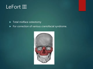
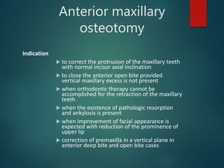
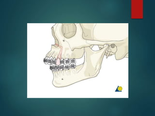
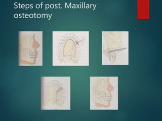
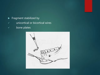
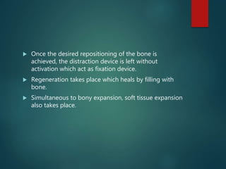
Major surgical
procedures
KIRAN SAJU
IV A
CONTENTS
 Introduction
 Definition
 Steps in orthognathic surgeries
 Envelope of discrepancy
 Pre-operative evaluation
 Presurgical orthodontics
 Surgical treatment objectives
 Mock surgery
 Surgery and stabilization
 Post surgical orthodontics
 Various surgical methods
Introduction
 Definition
 It is the art and science of diagnosis, treatment planning,
execution of treatment by combining orthodontics and oral
and maxillofacial surgery.
Orthognathic surgeries are major surgical procedures
carried out along with orthodontic therapy to correct
dentofacial deformities or orofacial dispropotions
involving maxilla, mandible,or combination.
 Orthognathic surgery basically involves planned fracturing
of the facial skeletal parts and repositioning them as
desired.
 Steps involved are
1. Diagnosis (pre-operative evaluation)
2. Pre-surgical orthodontics
3. Surgical treatment objective
4. Mock surgery
5. Surgery and stabilzation
6. Post surgical orthodontics
Envelope of discrepancy
Transverse envelope of
discrepancy
Envelope of discrepancy
 The inner circle indicates the limits to orthodontic
treatment.
 The middle circle indicates limits to tooth movement
combined with growth modification.
 The outer circle indicates surgical correction.
Timing of surgery
Pre operative evaluation
General medical evaluation
 general medical history should be recorded
 Pulpo-periodontal problems should be relieved prior to
surgery
Socio-psychological evaluation
 Patient is assesed to determine wheather he/she is aware
of existing dentofacial deformity and what they expect out
of surgical therapy.
Preoperative evaluation
Cephalometric evaluation
 It is to determine nature and severity of skeletal problems
Radiographic examination
 IOPA – determine the condition of teeth and alveolar bone
 OPG – evaluation of bony pathologies, TMJ, maxillary sinus
 Submento-vertex view – determine buccolingual thickness of the
mandible and degree of deformity of face
Pre operative evaluation
Study models
 Evaluation of occlusion from all directions. It is used to
assess interarch and intraarch disrepancies.
TMJ evaluation
 Evaluate joint movement and and pathology
Others
 Cone-beam computed tomography
Pre surgical orthodontics
 The objective of pre-surgical orthodontics is to
prepare the patient for intended orthognathic surgery.
Procedures
 Tooth alignment within the arches
Spacing and rotations are eliminated. Space for these is
gained by proximal stripping or even extractions.
 Alteration and co-ordination of the arches
As a general rule orthodontic expansion or contraction
to co-ordinate the upper and lower arches should be
done prior to surgery to provide correct postoperative
occlusal interdigitation.
Procedures
 Decompensation
Most severe skeletal jaw discrepancies are partly compensated by change in
axial inclination of teeth. Eg. Mandibular retrognathism is associated with
proclined lower anteriors and class III patients usually exhibit lingually
tipped lower incisors to partily offset skeletal discrepancy.
Pre surgical orthodontics should correct these positions of compensation to
position the teeth correctly over this supporting bone.
Procedures
 Incisor inclinations
proclined incisors in class II div 1 may need to be retracted
while retroclined incisors in class II div 2 has to be proclined
 Since most patient require post surgical orthodontic
treatment, it is unneccesary to carry out extensive
presurgical treatment.
 Pre surgical preparation should never extend more than
one year.
Surgical treatment objective
(STO)
 Also known as prediction tracing
 Two dimensional visual projection of the changes in the
osseous, dental and soft tissues as a result of orthodontics
and orthognathic surgery.
 Can be drawn manually or using computer software.
Surgical treatment objective
(STO
 Significance
1. Initial STO is prepared before the treatment to
determine orthodontic and surgical goals
2. The final STO is prepared after pre surgical orthodontics
but prior to surgery to determine the exact vertical and
anteroposterior skeletal and soft tissue movements to be
achieved.
Surgical treatment objective
(STO
 Purpose
1.Establish presurgical orthodontic goals
2.Develop an accurate surgical objective that will achieve the
best functional and esthetic result
3.Create a facial profile objective which can be used as a visual
aid in consultation with the patient and family members.
Mock surgery
 Mock surgery is perfomed on the upper and lower models
mounted on articulator.
 Two types of articulator are used
1. arbitrary articulator – when condylar position is not
altered in surgery
2. semiadjustable articulator – when condylar position is
altered in surgery
 Cuts are made in the models and then repositioned in the
desired planned position.
 If surgery is planned in both jaws, maxillary cast is moved first and fixed
on articulator.
 Occusal splint is made in this position for the first stage.
 Then mandibular cast is repositioned to occlude with maxillary cast.
 Second stage occlusal splint is prepared
USES
• To verify whether the planned movements are possible.
• To prepare occlusal splint.
Surgery and stabilisation
 Surgical fracturing and repositioning of bony segments.
 The teeth of upper and lower arches are wired in occlusion
to splints.
 Intermaxillary fixation usually spans for 6-8 weeks
following which splint is removed.
Post surgical orthodontics
 Fine detailing of occlusion and esthetic root paralleling.
 Most cases of post surgical orthodontics are completed by
4-6 months.
 Some skeletal relapse following surgery can be
camouflaged or compensated by appropriate orthodontic
treatment mechanics.
various surgical procedures
1. MAXILLARY SURGERY
i) Total maxillary surgery
 Le Fort I osteotomy
 Le Fort II osteotomy
 Le Fort III ostetomy
ii) Segmental maxillary osteotomy
 Anterior maxillary osteotomy
 Posterior maxillary osteotomy
 Single tooth osteotomy
 Corticotomy
2. Mandibular surgery
i)Ramus osteotomy
-Sagittal split osteotomy
-Extraoral vertical ramus osteotomy
-Intraoral vertical ramus osteotomy
ii) Body osteotomy of mandible
iii) Subapical body osteotomy
-Anterior subapical osteotomy
-Posterior subapical osteotomy
-Total subapical osteotomy
iv) Genioplasty
LeFort I Osteotomy
Indication
 For maxillary advancement.
 Inferior positioning of maxilla.
 Skeletal anterior open bite/levelling of maxilla.
 Superior repositioning of maxilla
Surgical procedure
A horizontal incision is made in the maxillary
vestibule
the mucoperiosteal flap is raised
Entire lateral surface of maxilla is sectioned
The nasoseptal osteotome is used to separate the
nasal septum
A straight osteotome is used to cut the lateral
nasal wall
separate the maxillary tuberosity from the
pterygoid plates.
down fracture the maxilla
Steps of LeFort I osteotomy for maxillary repositioning
Lefort II osteotomy
 It’s a pyramidal naso-orbital maxillary osteotomy.
 For correction of naso-maxillary hypoplasia
LeFort III
 Total midface osteotomy
 For correction of various craniofacial syndrome.
Anterior maxillary
osteotomy
Indication
 to correct the protrusion of the maxillary teeth
with normal incisor axial inclination
 to close the anterior open bite provided
vertical maxillary excess is not present
 when orthodontic therapy cannot be
accomplished for the retraction of the maxillary
teeth
 when the existence of pathologic resorption
and ankylosis is present
 when improvement of facial appearance is
expected with reduction of the prominence of
upper lip
 correction of premaxilla in a vertical plane in
anterior deep bite and open bite cases
Surgical Technique
 a vertical incision is made b/w canine&PM
 soft tissue is raised posteriorly
 A transverse palatal cut
made on buccal aspect
 Osteotomy is performed and the cut can extend upto second
molar
 Anterior segment is mobilized and separated from the nasal
septum
 Its then positioned and fixed using prefabricated occlusal splint
and a soft tissue closure is done.
Posterior Maxillary Osteotomy
Indication
1. to correct posterior cross bite
2. to superiorly reposition a supraerupted posterior segment
3. to close posterior open bite
4. for distal repositioning of maxilla to provide space for proper
eruption of impacted canine or premolars
5. transverse excess or deficiency
6. posterior maxillary hyperplasia
Surgical Technique
 A buccal vestibular incision extending from canine to 2nd molar region.
 Horizontal buccal osteotomy is completed 5mm above the apices of
teeth using rotary instrument or reciprocating saw.
 A vertical osteotomy cut is done either between the adjoining teeth or
by using the extraction site as predetermined.
 Palatal osteotomy is performed with a curved osteotome placed through
the horizontal osteotomy site with operators finger on the palatal tissue
to prevent laceration of palatal mucosa.
 Small curved osteotome is used to separate pterygomaxillary junction.
 the entire posterior segment can be down fractured pedicled on the
palatal mucosa.
 Palatal bone can be removed sufficiently under direct vision
 After completion of osteotomy the posterior segment is
repositioned, the prefabricated occlusal splint is fitted and
wired to teeth.
 Direct wire or bone plate fixation can be done
Steps of post. Maxillary
osteotomy
Sagittal Split Osteotomy
Indication
For correcting mandibular prognathism
For correcting mandibular retrognathism
For correcting crossbites and assymetry
 In this technique the mandibular ramus is split on both sides
in the distal fragment is moved forward and backward.
 For mandibular advancement, the distal segment are moved
forward and teeth are wired into desired occlusion
 For mandibular setback, the distal fragment is pushed back.
The excess bone on the proximal segment is cut off by taking
another vertical incision distal to the previous cut.
GENIOPLASTY
Alteration of chin morphology and movement of chin in
all the three planes is possible to improve the results of
mandibular advancement or reduction or to correct
asymmetry.
Types
 sliding genioplasty
 double horizontal osteotomy
 hinge sliding osteotomy
 propeller genioplasty
Horizontal osteotomy with
advancement
 Incision half way the depth of vestibule and extended to canine
region bilaterally.
 Periosteum left intact on the inferior border
 Line of osteotomy should be 5 mm below canine root & 10 to 15
mm above the inferior border & 5 mm below the lowest mental
foramen
 Fragment stabilized by
 unicortical or bicortical wires
 bone plates
HORIZONTAL OSTEOTOMY
WITH REDUCTION
 Prefabricated chin fixation plate or H shaped plate is
used
 When the chin is set back,postero lingual area has a
palpable step defect.
 To prevent this postero lingual area is contoured
 Labio mental fold is enhanced by contouring the
anterior superior edge.
DOUBLE SLIDING
HORIZONTAL OSTEOTOMY
 In very deficient chin
 Creation of a stepped intermediate wafer of bone between the
inferior fragment and mandible
 This segment is advanced to produce bony contact between upper
and lower fragments
Correction of assymmetry of
chin
 Done in unilateral condylar hyper or hypoplasia where
the chin is deviated.
 Done for the lateral movement of the chin
 Also known as propeller osteotomy
 First osteotomy is performed parallel to the inter
pupillary line
 Second osteotomy is performed parallel to the lower
border of the chin
Altering the width of the chin
 Altering the posterior dimension
 Before the chin is mobilised fix a 4 hole straight plate
at the labial cortex of the chin
 Midline osteotomy is performed both buccal and
lingual cortex
 Chin widened using bone plate as a hinge
 To narrow the chin triangular midline ostectomy is
performed.
Distraction osteogenesis
 It is a surgical procedure or reconstruction of skeletal
deformation.
 It involves gradual, controlled displacement of
surgically created fractures and results in simultaneous
expansion of soft tissue and bone volume.
 This technique of bone formation under the influence
of tensional stress is called distraction osteogenesis.
Distraction osteogenesis
Indications
 combined deficiencies in bone and soft tissue
 expand alveolus for orthodontic tooth movement
 Create site for dental implant
3 step procedure
 Mobilization of bone
 Transport by means of devices
 Fixation of healthy segment
 A mechanical device namely distraction device is used
to produce movement of mobilized bone segment
 Once the desired repositioning of the bone is
achieved, the distraction device is left without
activation which act as fixation device.
 Regeneration takes place which heals by filling with
bone.
 Simultaneous to bony expansion, soft tissue expansion
also takes place.
CONCLUSION
 Only through team effort that the multifaceted problems can be
successfully controlled.
 To accomplish optimal orthodontic treatment a combined effort of
oral and maxillofacial surgeon and orthodontist is essential.
THANK YOU

Major surgical procedures

  • 1.
  • 2.
    CONTENTS  Introduction  Definition Steps in orthognathic surgeries  Envelope of discrepancy  Pre-operative evaluation  Presurgical orthodontics  Surgical treatment objectives  Mock surgery  Surgery and stabilization  Post surgical orthodontics  Various surgical methods
  • 3.
    Introduction  Definition  Itis the art and science of diagnosis, treatment planning, execution of treatment by combining orthodontics and oral and maxillofacial surgery. Orthognathic surgeries are major surgical procedures carried out along with orthodontic therapy to correct dentofacial deformities or orofacial dispropotions involving maxilla, mandible,or combination.
  • 4.
     Orthognathic surgerybasically involves planned fracturing of the facial skeletal parts and repositioning them as desired.  Steps involved are 1. Diagnosis (pre-operative evaluation) 2. Pre-surgical orthodontics 3. Surgical treatment objective 4. Mock surgery 5. Surgery and stabilzation 6. Post surgical orthodontics
  • 5.
  • 6.
  • 7.
    Envelope of discrepancy The inner circle indicates the limits to orthodontic treatment.  The middle circle indicates limits to tooth movement combined with growth modification.  The outer circle indicates surgical correction.
  • 8.
  • 9.
    Pre operative evaluation Generalmedical evaluation  general medical history should be recorded  Pulpo-periodontal problems should be relieved prior to surgery Socio-psychological evaluation  Patient is assesed to determine wheather he/she is aware of existing dentofacial deformity and what they expect out of surgical therapy.
  • 10.
    Preoperative evaluation Cephalometric evaluation It is to determine nature and severity of skeletal problems Radiographic examination  IOPA – determine the condition of teeth and alveolar bone  OPG – evaluation of bony pathologies, TMJ, maxillary sinus  Submento-vertex view – determine buccolingual thickness of the mandible and degree of deformity of face
  • 11.
    Pre operative evaluation Studymodels  Evaluation of occlusion from all directions. It is used to assess interarch and intraarch disrepancies. TMJ evaluation  Evaluate joint movement and and pathology Others  Cone-beam computed tomography
  • 12.
    Pre surgical orthodontics The objective of pre-surgical orthodontics is to prepare the patient for intended orthognathic surgery.
  • 13.
    Procedures  Tooth alignmentwithin the arches Spacing and rotations are eliminated. Space for these is gained by proximal stripping or even extractions.  Alteration and co-ordination of the arches As a general rule orthodontic expansion or contraction to co-ordinate the upper and lower arches should be done prior to surgery to provide correct postoperative occlusal interdigitation.
  • 14.
    Procedures  Decompensation Most severeskeletal jaw discrepancies are partly compensated by change in axial inclination of teeth. Eg. Mandibular retrognathism is associated with proclined lower anteriors and class III patients usually exhibit lingually tipped lower incisors to partily offset skeletal discrepancy. Pre surgical orthodontics should correct these positions of compensation to position the teeth correctly over this supporting bone.
  • 15.
    Procedures  Incisor inclinations proclinedincisors in class II div 1 may need to be retracted while retroclined incisors in class II div 2 has to be proclined
  • 16.
     Since mostpatient require post surgical orthodontic treatment, it is unneccesary to carry out extensive presurgical treatment.  Pre surgical preparation should never extend more than one year.
  • 17.
    Surgical treatment objective (STO) Also known as prediction tracing  Two dimensional visual projection of the changes in the osseous, dental and soft tissues as a result of orthodontics and orthognathic surgery.  Can be drawn manually or using computer software.
  • 18.
    Surgical treatment objective (STO Significance 1. Initial STO is prepared before the treatment to determine orthodontic and surgical goals 2. The final STO is prepared after pre surgical orthodontics but prior to surgery to determine the exact vertical and anteroposterior skeletal and soft tissue movements to be achieved.
  • 19.
    Surgical treatment objective (STO Purpose 1.Establish presurgical orthodontic goals 2.Develop an accurate surgical objective that will achieve the best functional and esthetic result 3.Create a facial profile objective which can be used as a visual aid in consultation with the patient and family members.
  • 20.
    Mock surgery  Mocksurgery is perfomed on the upper and lower models mounted on articulator.  Two types of articulator are used 1. arbitrary articulator – when condylar position is not altered in surgery 2. semiadjustable articulator – when condylar position is altered in surgery  Cuts are made in the models and then repositioned in the desired planned position.
  • 21.
     If surgeryis planned in both jaws, maxillary cast is moved first and fixed on articulator.  Occusal splint is made in this position for the first stage.  Then mandibular cast is repositioned to occlude with maxillary cast.  Second stage occlusal splint is prepared USES • To verify whether the planned movements are possible. • To prepare occlusal splint.
  • 23.
    Surgery and stabilisation Surgical fracturing and repositioning of bony segments.  The teeth of upper and lower arches are wired in occlusion to splints.  Intermaxillary fixation usually spans for 6-8 weeks following which splint is removed.
  • 24.
    Post surgical orthodontics Fine detailing of occlusion and esthetic root paralleling.  Most cases of post surgical orthodontics are completed by 4-6 months.  Some skeletal relapse following surgery can be camouflaged or compensated by appropriate orthodontic treatment mechanics.
  • 25.
    various surgical procedures 1.MAXILLARY SURGERY i) Total maxillary surgery  Le Fort I osteotomy  Le Fort II osteotomy  Le Fort III ostetomy ii) Segmental maxillary osteotomy  Anterior maxillary osteotomy  Posterior maxillary osteotomy  Single tooth osteotomy  Corticotomy
  • 26.
    2. Mandibular surgery i)Ramusosteotomy -Sagittal split osteotomy -Extraoral vertical ramus osteotomy -Intraoral vertical ramus osteotomy ii) Body osteotomy of mandible iii) Subapical body osteotomy -Anterior subapical osteotomy -Posterior subapical osteotomy -Total subapical osteotomy iv) Genioplasty
  • 27.
    LeFort I Osteotomy Indication For maxillary advancement.  Inferior positioning of maxilla.  Skeletal anterior open bite/levelling of maxilla.  Superior repositioning of maxilla
  • 28.
    Surgical procedure A horizontalincision is made in the maxillary vestibule the mucoperiosteal flap is raised Entire lateral surface of maxilla is sectioned The nasoseptal osteotome is used to separate the nasal septum A straight osteotome is used to cut the lateral nasal wall separate the maxillary tuberosity from the pterygoid plates. down fracture the maxilla
  • 29.
    Steps of LeFortI osteotomy for maxillary repositioning
  • 30.
    Lefort II osteotomy It’s a pyramidal naso-orbital maxillary osteotomy.  For correction of naso-maxillary hypoplasia
  • 31.
    LeFort III  Totalmidface osteotomy  For correction of various craniofacial syndrome.
  • 32.
    Anterior maxillary osteotomy Indication  tocorrect the protrusion of the maxillary teeth with normal incisor axial inclination  to close the anterior open bite provided vertical maxillary excess is not present  when orthodontic therapy cannot be accomplished for the retraction of the maxillary teeth  when the existence of pathologic resorption and ankylosis is present  when improvement of facial appearance is expected with reduction of the prominence of upper lip  correction of premaxilla in a vertical plane in anterior deep bite and open bite cases
  • 34.
    Surgical Technique  avertical incision is made b/w canine&PM  soft tissue is raised posteriorly  A transverse palatal cut made on buccal aspect  Osteotomy is performed and the cut can extend upto second molar  Anterior segment is mobilized and separated from the nasal septum  Its then positioned and fixed using prefabricated occlusal splint and a soft tissue closure is done.
  • 35.
    Posterior Maxillary Osteotomy Indication 1.to correct posterior cross bite 2. to superiorly reposition a supraerupted posterior segment 3. to close posterior open bite 4. for distal repositioning of maxilla to provide space for proper eruption of impacted canine or premolars 5. transverse excess or deficiency 6. posterior maxillary hyperplasia
  • 36.
    Surgical Technique  Abuccal vestibular incision extending from canine to 2nd molar region.  Horizontal buccal osteotomy is completed 5mm above the apices of teeth using rotary instrument or reciprocating saw.  A vertical osteotomy cut is done either between the adjoining teeth or by using the extraction site as predetermined.  Palatal osteotomy is performed with a curved osteotome placed through the horizontal osteotomy site with operators finger on the palatal tissue to prevent laceration of palatal mucosa.  Small curved osteotome is used to separate pterygomaxillary junction.  the entire posterior segment can be down fractured pedicled on the palatal mucosa.  Palatal bone can be removed sufficiently under direct vision
  • 37.
     After completionof osteotomy the posterior segment is repositioned, the prefabricated occlusal splint is fitted and wired to teeth.  Direct wire or bone plate fixation can be done
  • 38.
    Steps of post.Maxillary osteotomy
  • 39.
    Sagittal Split Osteotomy Indication Forcorrecting mandibular prognathism For correcting mandibular retrognathism For correcting crossbites and assymetry
  • 40.
     In thistechnique the mandibular ramus is split on both sides in the distal fragment is moved forward and backward.  For mandibular advancement, the distal segment are moved forward and teeth are wired into desired occlusion  For mandibular setback, the distal fragment is pushed back. The excess bone on the proximal segment is cut off by taking another vertical incision distal to the previous cut.
  • 42.
    GENIOPLASTY Alteration of chinmorphology and movement of chin in all the three planes is possible to improve the results of mandibular advancement or reduction or to correct asymmetry. Types  sliding genioplasty  double horizontal osteotomy  hinge sliding osteotomy  propeller genioplasty
  • 43.
    Horizontal osteotomy with advancement Incision half way the depth of vestibule and extended to canine region bilaterally.  Periosteum left intact on the inferior border  Line of osteotomy should be 5 mm below canine root & 10 to 15 mm above the inferior border & 5 mm below the lowest mental foramen
  • 44.
     Fragment stabilizedby  unicortical or bicortical wires  bone plates
  • 45.
    HORIZONTAL OSTEOTOMY WITH REDUCTION Prefabricated chin fixation plate or H shaped plate is used  When the chin is set back,postero lingual area has a palpable step defect.  To prevent this postero lingual area is contoured  Labio mental fold is enhanced by contouring the anterior superior edge.
  • 47.
    DOUBLE SLIDING HORIZONTAL OSTEOTOMY In very deficient chin  Creation of a stepped intermediate wafer of bone between the inferior fragment and mandible  This segment is advanced to produce bony contact between upper and lower fragments
  • 48.
    Correction of assymmetryof chin  Done in unilateral condylar hyper or hypoplasia where the chin is deviated.  Done for the lateral movement of the chin  Also known as propeller osteotomy  First osteotomy is performed parallel to the inter pupillary line  Second osteotomy is performed parallel to the lower border of the chin
  • 50.
    Altering the widthof the chin  Altering the posterior dimension  Before the chin is mobilised fix a 4 hole straight plate at the labial cortex of the chin  Midline osteotomy is performed both buccal and lingual cortex  Chin widened using bone plate as a hinge  To narrow the chin triangular midline ostectomy is performed.
  • 52.
    Distraction osteogenesis  Itis a surgical procedure or reconstruction of skeletal deformation.  It involves gradual, controlled displacement of surgically created fractures and results in simultaneous expansion of soft tissue and bone volume.  This technique of bone formation under the influence of tensional stress is called distraction osteogenesis.
  • 53.
    Distraction osteogenesis Indications  combineddeficiencies in bone and soft tissue  expand alveolus for orthodontic tooth movement  Create site for dental implant
  • 54.
    3 step procedure Mobilization of bone  Transport by means of devices  Fixation of healthy segment  A mechanical device namely distraction device is used to produce movement of mobilized bone segment
  • 55.
     Once thedesired repositioning of the bone is achieved, the distraction device is left without activation which act as fixation device.  Regeneration takes place which heals by filling with bone.  Simultaneous to bony expansion, soft tissue expansion also takes place.
  • 56.
    CONCLUSION  Only throughteam effort that the multifaceted problems can be successfully controlled.  To accomplish optimal orthodontic treatment a combined effort of oral and maxillofacial surgeon and orthodontist is essential.
  • 57.