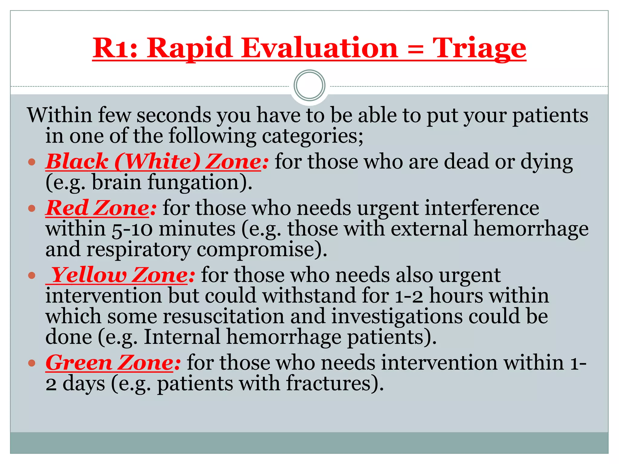
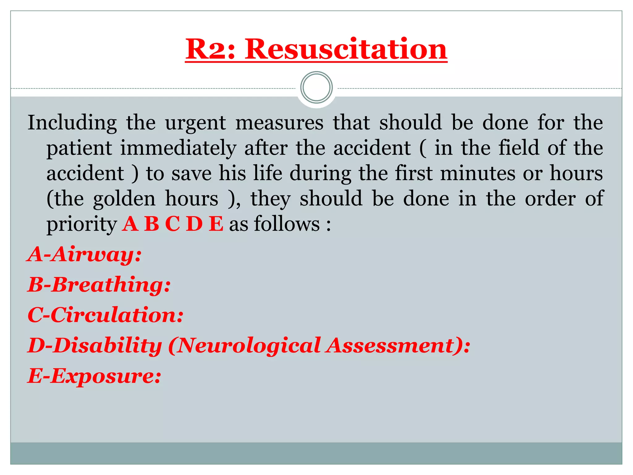
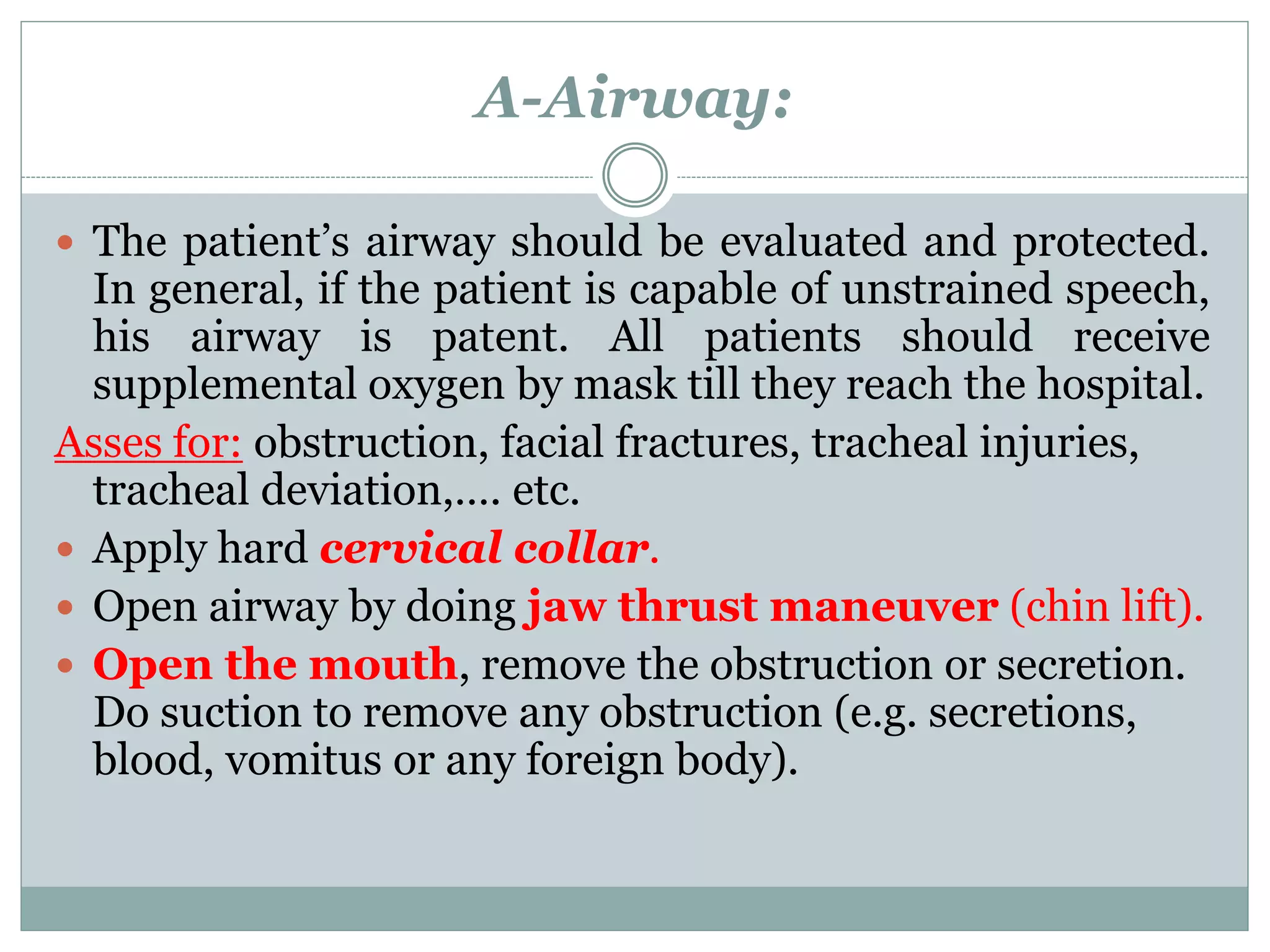
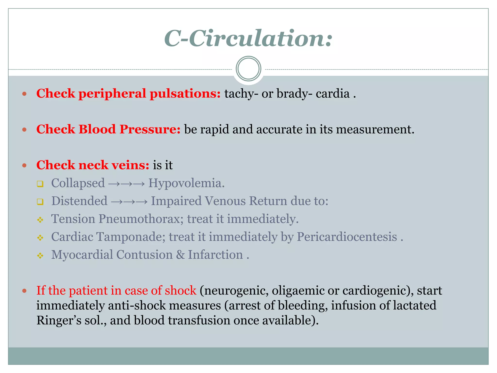
M O H A M E D R I A D
L E C T U R E R O F G E N E R A L S U R G E R Y
Trauma
 Injury to the human body occurs when it is exposed
to sudden transfer of high energy that the body can’t
withstand.
Mechanisms of Injury
 Physical Agents;
○ Kinetic. ○ Electrical.
○ Thermal. ○ Radiation.
 Chemical.
Kinetic Energy Forces:
Blunt Trauma:
 Acceleration/Deceleration: mostly during Motor Vehicle
Accidents (MVA).
 Crushing: In this type of trauma, the priority is to save
patient’s life, then think about patient’s limb.
 Falling from height: Significant trauma occurs if the
victim fall >10 feet or 3 times the person’s height. Type of
trauma depends on the body area striking the ground first.
Penetrating Trauma:
 High velocity → long & short Guns.
 Low velocity → Knives and rifles.
Management of poly-traumatized patient
There are many protocols for management of poly-
trauma, the most universally accepted one is the
protocol of ATLS (ADVANCED TRAUMA LIFE
SUPPORT) which described by the American
College of Surgeons, which consists of 3 steps:
 Primary survey.
 Secondary survey.
 Definitive treatment.
Another protocol is the 5 Rs, as follows
What are the 5 R’s?
 R1: Rapid Evaluation = Triage.
 R2: Resuscitation.
 R3: Radiology and Other Investigations.
 R4: Re-Evaluation.
 R5: Repair and Rehabilitation.
R1: Rapid Evaluation = Triage
Within few seconds you have to be able to put your patients
in one of the following categories;
 Black (White) Zone: for those who are dead or dying
(e.g. brain fungation).
 Red Zone: for those who needs urgent interference
within 5-10 minutes (e.g. those with external hemorrhage
and respiratory compromise).
 Yellow Zone: for those who needs also urgent
intervention but could withstand for 1-2 hours within
which some resuscitation and investigations could be
done (e.g. Internal hemorrhage patients).
 Green Zone: for those who needs intervention within 1-
2 days (e.g. patients with fractures).
R2: Resuscitation
Including the urgent measures that should be done for the
patient immediately after the accident ( in the field of the
accident ) to save his life during the first minutes or hours
(the golden hours ), they should be done in the order of
priority A B C D E as follows :
A-Airway:
B-Breathing:
C-Circulation:
D-Disability (Neurological Assessment):
E-Exposure:
A-Airway:
 The patient’s airway should be evaluated and protected.
In general, if the patient is capable of unstrained speech,
his airway is patent. All patients should receive
supplemental oxygen by mask till they reach the hospital.
Asses for: obstruction, facial fractures, tracheal injuries,
tracheal deviation,…. etc.
 Apply hard cervical collar.
 Open airway by doing jaw thrust maneuver (chin lift).
 Open the mouth, remove the obstruction or secretion.
Do suction to remove any obstruction (e.g. secretions,
blood, vomitus or any foreign body).
 Insert oro-pharyngeal or naso-pharyngeal airway to
maintain patency of airway and to prevent falling back of
the tongue in an unconscious patient. This method is contra-
indicated in conscious patients (stimulates gag reflex and
vomiting)
 Endo-tracheal Intubation (indicated in cases of apnea,
head injuries, air way compromise like maxillofacial injuries,
fracture cervical spine and if there is risk of aspiration).
 Cricothyroidotomy, if there is upper airway obstruction,
and it is impossible to pass an endo-tracheal tube .
B-Breathing:
Check for spontaneous breathing for 10 sec;
 If patient is breathing satisfactorily & PO2 above 90% → just
observe.
 If patient is breathing satisfactorily but PO2 below 90% →
provide 02 therapy via mask 6 L/min, 60% O2 concentration.
 If patient is not breathing or PO2 still declining → manually
ventilate patient with 15L/min, 100% oxygen concentration &
Prepare for intubation and mechanical ventilation.
C-Circulation:
 Check peripheral pulsations: tachy- or brady- cardia .
 Check Blood Pressure: be rapid and accurate in its measurement.
 Check neck veins: is it
 Collapsed →→→ Hypovolemia.
 Distended →→→ Impaired Venous Return due to:
 Tension Pneumothorax; treat it immediately.
 Cardiac Tamponade; treat it immediately by Pericardiocentesis .
 Myocardial Contusion & Infarction .
 If the patient in case of shock (neurogenic, oligaemic or cardiogenic), start
immediately anti-shock measures (arrest of bleeding, infusion of lactated
Ringer’s sol., and blood transfusion once available).
Class I Class II Class III Class IV
Blood Loss mL Up to 750 750-1500 1500-2000 >2000
Blood Loss
%
Up to 15% 15-30% 30-40% >40%
Pulse rate <100 >100 >120 >140
Systolic blood
pressure
Normal Normal Decreased Decreased
Pulse pressure Normal Decreased Decreased Decreased
Respiratory rate 14-20 20-30 30-40 >35
Urine output >30 20-30 5-15 Negligible
Mental status Slightly
anxious
Mildly
anxious
Anxious,
confused
Confused,
lethargic
Fluid
(3:1 rule)
Crystalloid Crystalloid Crystalloid and
blood
Crystalloid
and blood
D-Disability (Neurological Assessment):
Level of consciousness.
 AVPU scale;
 Awake.
 Verbal response.
 Pain response.
 Unresponsive.
 For assessment, apply any scale e.g Glasgow Coma
Scale.
Glascow Coma Scale
 3 – 15 point scale to assess mental status only
 Best observed response
 GCS ≤ 8 is a “coma” and requires intubation for
airway protection
Eye opening
• None = 1
• To painful stimuli only = 2
• To voice only = 3
• Spontaneously open = 4
Verbal response
• None = 1
• Incomprehensible sounds = 2
• Incomprehensible words = 3
• Confused = 4
• Oriented = 5
Motor response
• None = 1
• Decerebrate (extension) posturing = 2
• Decorticate (flexion) posturing = 3
• Withdraws to pain = 4
• Localizes pain = 5
• Follows commands = 6
E-Exposure:
 Remove clothing.
 Observe the chest for bruises, penetrations, and
symmetry.
 Auscultate breath sounds.
 Auscultate heart sounds.
 For total assessment.
After exposure you may find:
 Ecchymosis at site of trauma.
 Grey-Turner sign.
R3: Radiology & Other Investigations.
I- Basic X- Ray Films have to be done for every case
of Polytrauma depends largely on the suspected site
and the doctor who is going to request it. Every
specialty has its own interest;
 General Surgery: erect abdomen
 Cardiothoracic Surgery: chest x-ray
 Neurosurgery: skull and spines
 Orthopedic Surgery: pelvis, spine and fractures
II- Focused Assessment with Sonography for
Trauma (FAST):
Perihepatic
Perisplenic
Pericardium
Pelvis
III-CT Scans:
 Brain and Spine, Abdomen & Pelvis and Chest are
usually needed for assessment of most of cases.
IV - DPL (Diagnostic Peritoneal Lavage):
 This is an invasive diagnostic tool that should be preserved in
cases where diagnosis of intra-abdominal injury is very
doubtful.
 It should be done in the OR (Operating Room) under
complete aseptic precautions and with all facilities ready for
abdominal exploration if needed (even blood).
 Through a stab incision just above the umbilicus, a Ryle tube
is passed to the intra-peritoneal space and one liter of normal
saline infused intra-peritoneal through it. Then we leave the
fluid to come back and we analyze its content.
 Positive result is considered when;
 10cc gross blood.
 RBC >100,000/mm2 . , WBC >500/mm2
 Amylase >175 IU/dL.
 Bile, bacteria, or food.
 This means that this patient is candidate for immediate
exploration.
 Contraindications of DPL:
 Those with clear decision of exploration.
 Prior abdominal surgeries.
 Pregnancy & Obesity.
R4: Re-Evaluation
(Secondary survey)
Now, this is the time of re-evaluation of the patient. It
is done in two steps:
I-History taking: this includes; SAMPLE;
→ Symptoms.
→ Allergies.
→ Medications.
→ Past history.
→ Last meal.
→ Events related to injury.
II-General Examination from Hair to Heal:
1. Head: search for sub-galeal hematoma, sub-
conjunctival hemorrhage, facial fractures,…etc.
2. Neck: pain or tenderness, tracheal deviation, jugular
vein, impaled objects and open wounds, Expanding
neck hematoma.
3. Chest and Heart: rib fractures, pneumo- or hemo-
thorax,…etc.
4. Abdomen & Pelvis: Cullen’s sign, Grey-Turner sign,
Kher’s sign,…etc.
5. Extremities: Fractures, peripheral pulsations, soft
tissue injuries,…etc.
6. Back: bruising, impaled objects, pain and tenderness.
R5: Repair & Rehabilitation.
(Definitive treatment of individual injuries )
Finally, the patient is admitted to the hospital in one of
the following destination sites:
 General Surgery Department.
 Neurosurgery Department.
 Orthopedic Surgery Department.
 Cardiothoracic Surgery Department.
 ICU.

Management of Trauma

  • 1.
    M O HA M E D R I A D L E C T U R E R O F G E N E R A L S U R G E R Y Trauma
  • 2.
     Injury tothe human body occurs when it is exposed to sudden transfer of high energy that the body can’t withstand.
  • 3.
    Mechanisms of Injury Physical Agents; ○ Kinetic. ○ Electrical. ○ Thermal. ○ Radiation.  Chemical.
  • 4.
    Kinetic Energy Forces: BluntTrauma:  Acceleration/Deceleration: mostly during Motor Vehicle Accidents (MVA).  Crushing: In this type of trauma, the priority is to save patient’s life, then think about patient’s limb.  Falling from height: Significant trauma occurs if the victim fall >10 feet or 3 times the person’s height. Type of trauma depends on the body area striking the ground first. Penetrating Trauma:  High velocity → long & short Guns.  Low velocity → Knives and rifles.
  • 5.
    Management of poly-traumatizedpatient There are many protocols for management of poly- trauma, the most universally accepted one is the protocol of ATLS (ADVANCED TRAUMA LIFE SUPPORT) which described by the American College of Surgeons, which consists of 3 steps:  Primary survey.  Secondary survey.  Definitive treatment.
  • 6.
    Another protocol isthe 5 Rs, as follows What are the 5 R’s?  R1: Rapid Evaluation = Triage.  R2: Resuscitation.  R3: Radiology and Other Investigations.  R4: Re-Evaluation.  R5: Repair and Rehabilitation.
  • 7.
    R1: Rapid Evaluation= Triage Within few seconds you have to be able to put your patients in one of the following categories;  Black (White) Zone: for those who are dead or dying (e.g. brain fungation).  Red Zone: for those who needs urgent interference within 5-10 minutes (e.g. those with external hemorrhage and respiratory compromise).  Yellow Zone: for those who needs also urgent intervention but could withstand for 1-2 hours within which some resuscitation and investigations could be done (e.g. Internal hemorrhage patients).  Green Zone: for those who needs intervention within 1- 2 days (e.g. patients with fractures).
  • 8.
    R2: Resuscitation Including theurgent measures that should be done for the patient immediately after the accident ( in the field of the accident ) to save his life during the first minutes or hours (the golden hours ), they should be done in the order of priority A B C D E as follows : A-Airway: B-Breathing: C-Circulation: D-Disability (Neurological Assessment): E-Exposure:
  • 9.
    A-Airway:  The patient’sairway should be evaluated and protected. In general, if the patient is capable of unstrained speech, his airway is patent. All patients should receive supplemental oxygen by mask till they reach the hospital. Asses for: obstruction, facial fractures, tracheal injuries, tracheal deviation,…. etc.  Apply hard cervical collar.  Open airway by doing jaw thrust maneuver (chin lift).  Open the mouth, remove the obstruction or secretion. Do suction to remove any obstruction (e.g. secretions, blood, vomitus or any foreign body).
  • 10.
     Insert oro-pharyngealor naso-pharyngeal airway to maintain patency of airway and to prevent falling back of the tongue in an unconscious patient. This method is contra- indicated in conscious patients (stimulates gag reflex and vomiting)  Endo-tracheal Intubation (indicated in cases of apnea, head injuries, air way compromise like maxillofacial injuries, fracture cervical spine and if there is risk of aspiration).  Cricothyroidotomy, if there is upper airway obstruction, and it is impossible to pass an endo-tracheal tube .
  • 11.
    B-Breathing: Check for spontaneousbreathing for 10 sec;  If patient is breathing satisfactorily & PO2 above 90% → just observe.  If patient is breathing satisfactorily but PO2 below 90% → provide 02 therapy via mask 6 L/min, 60% O2 concentration.  If patient is not breathing or PO2 still declining → manually ventilate patient with 15L/min, 100% oxygen concentration & Prepare for intubation and mechanical ventilation.
  • 12.
    C-Circulation:  Check peripheralpulsations: tachy- or brady- cardia .  Check Blood Pressure: be rapid and accurate in its measurement.  Check neck veins: is it  Collapsed →→→ Hypovolemia.  Distended →→→ Impaired Venous Return due to:  Tension Pneumothorax; treat it immediately.  Cardiac Tamponade; treat it immediately by Pericardiocentesis .  Myocardial Contusion & Infarction .  If the patient in case of shock (neurogenic, oligaemic or cardiogenic), start immediately anti-shock measures (arrest of bleeding, infusion of lactated Ringer’s sol., and blood transfusion once available).
  • 13.
    Class I ClassII Class III Class IV Blood Loss mL Up to 750 750-1500 1500-2000 >2000 Blood Loss % Up to 15% 15-30% 30-40% >40% Pulse rate <100 >100 >120 >140 Systolic blood pressure Normal Normal Decreased Decreased Pulse pressure Normal Decreased Decreased Decreased Respiratory rate 14-20 20-30 30-40 >35 Urine output >30 20-30 5-15 Negligible Mental status Slightly anxious Mildly anxious Anxious, confused Confused, lethargic Fluid (3:1 rule) Crystalloid Crystalloid Crystalloid and blood Crystalloid and blood
  • 14.
    D-Disability (Neurological Assessment): Levelof consciousness.  AVPU scale;  Awake.  Verbal response.  Pain response.  Unresponsive.  For assessment, apply any scale e.g Glasgow Coma Scale.
  • 15.
    Glascow Coma Scale 3 – 15 point scale to assess mental status only  Best observed response  GCS ≤ 8 is a “coma” and requires intubation for airway protection
  • 16.
    Eye opening • None= 1 • To painful stimuli only = 2 • To voice only = 3 • Spontaneously open = 4 Verbal response • None = 1 • Incomprehensible sounds = 2 • Incomprehensible words = 3 • Confused = 4 • Oriented = 5 Motor response • None = 1 • Decerebrate (extension) posturing = 2 • Decorticate (flexion) posturing = 3 • Withdraws to pain = 4 • Localizes pain = 5 • Follows commands = 6
  • 17.
    E-Exposure:  Remove clothing. Observe the chest for bruises, penetrations, and symmetry.  Auscultate breath sounds.  Auscultate heart sounds.  For total assessment. After exposure you may find:  Ecchymosis at site of trauma.  Grey-Turner sign.
  • 18.
    R3: Radiology &Other Investigations. I- Basic X- Ray Films have to be done for every case of Polytrauma depends largely on the suspected site and the doctor who is going to request it. Every specialty has its own interest;  General Surgery: erect abdomen  Cardiothoracic Surgery: chest x-ray  Neurosurgery: skull and spines  Orthopedic Surgery: pelvis, spine and fractures
  • 19.
    II- Focused Assessmentwith Sonography for Trauma (FAST): Perihepatic Perisplenic Pericardium Pelvis
  • 20.
    III-CT Scans:  Brainand Spine, Abdomen & Pelvis and Chest are usually needed for assessment of most of cases.
  • 21.
    IV - DPL(Diagnostic Peritoneal Lavage):  This is an invasive diagnostic tool that should be preserved in cases where diagnosis of intra-abdominal injury is very doubtful.  It should be done in the OR (Operating Room) under complete aseptic precautions and with all facilities ready for abdominal exploration if needed (even blood).  Through a stab incision just above the umbilicus, a Ryle tube is passed to the intra-peritoneal space and one liter of normal saline infused intra-peritoneal through it. Then we leave the fluid to come back and we analyze its content.
  • 22.
     Positive resultis considered when;  10cc gross blood.  RBC >100,000/mm2 . , WBC >500/mm2  Amylase >175 IU/dL.  Bile, bacteria, or food.  This means that this patient is candidate for immediate exploration.  Contraindications of DPL:  Those with clear decision of exploration.  Prior abdominal surgeries.  Pregnancy & Obesity.
  • 23.
    R4: Re-Evaluation (Secondary survey) Now,this is the time of re-evaluation of the patient. It is done in two steps: I-History taking: this includes; SAMPLE; → Symptoms. → Allergies. → Medications. → Past history. → Last meal. → Events related to injury.
  • 24.
    II-General Examination fromHair to Heal: 1. Head: search for sub-galeal hematoma, sub- conjunctival hemorrhage, facial fractures,…etc. 2. Neck: pain or tenderness, tracheal deviation, jugular vein, impaled objects and open wounds, Expanding neck hematoma. 3. Chest and Heart: rib fractures, pneumo- or hemo- thorax,…etc. 4. Abdomen & Pelvis: Cullen’s sign, Grey-Turner sign, Kher’s sign,…etc. 5. Extremities: Fractures, peripheral pulsations, soft tissue injuries,…etc. 6. Back: bruising, impaled objects, pain and tenderness.
  • 25.
    R5: Repair &Rehabilitation. (Definitive treatment of individual injuries ) Finally, the patient is admitted to the hospital in one of the following destination sites:  General Surgery Department.  Neurosurgery Department.  Orthopedic Surgery Department.  Cardiothoracic Surgery Department.  ICU.