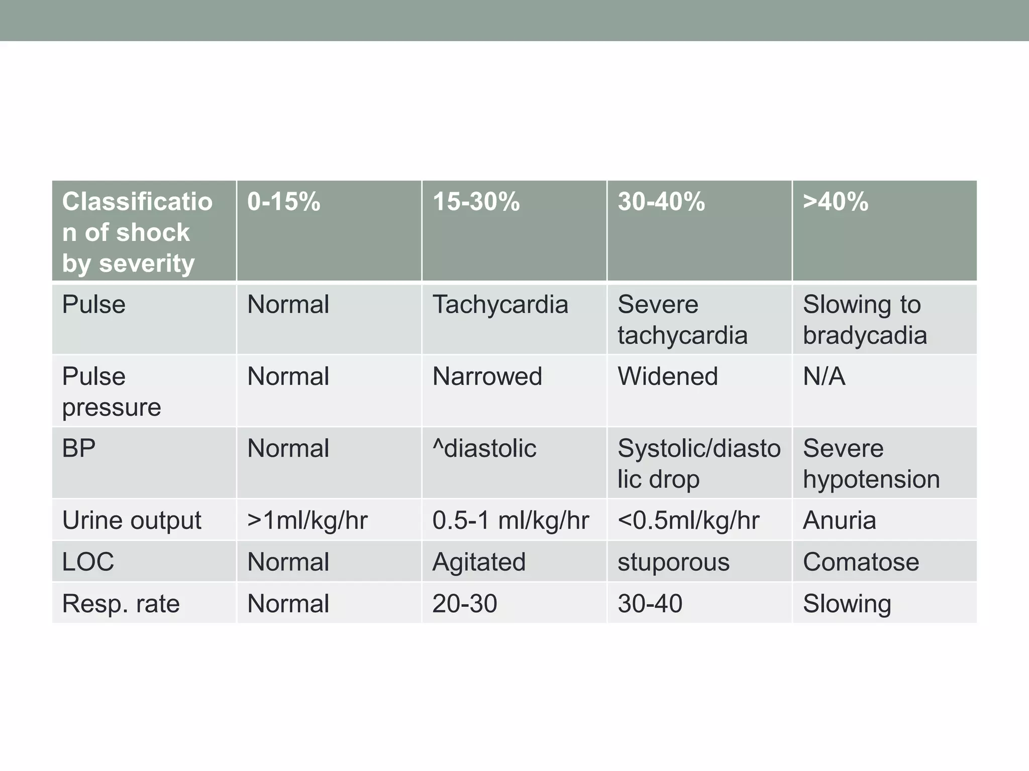
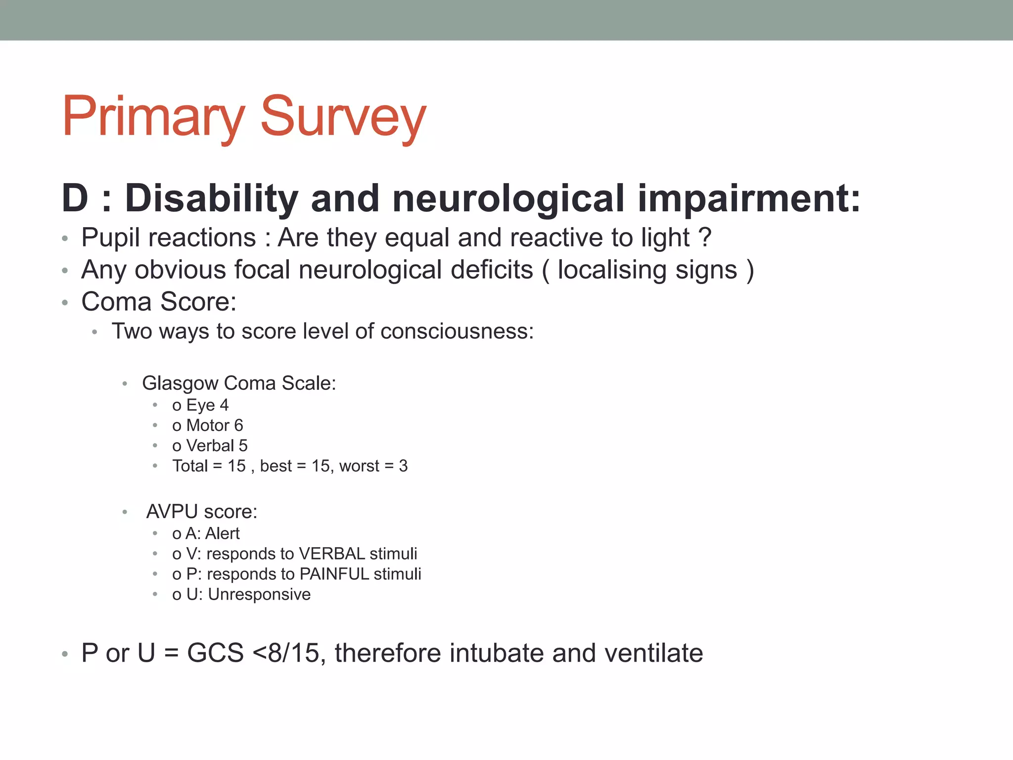
APPROACH TO
POLYTRAUMA PATIENT
DR NE DLAMINI
• The goals of Trauma Resuscitation and Evaluation are to:
1. Identify factors that will save the patient’s life, enable
adequate resuscitation, triage, and
prioritize individual injuries.
2. Discuss mechanism of injury in cases of Blunt and
Penetrating Trauma.
3. Describe the process of evaluation of abdominal
trauma
4. Enable understanding of the principles for the
management of individual intra-abdominal
• Introduction:
Trauma remains the major killer of persons in the age
groups 15 – 40 years of age in South Africa,
and the second biggest killer, after infective diseases, in
children.
The purpose of this chapter is to provide a safe approach
to the polytrauma patient, that identifies
injuries in the order that would kill the patient the fastest if
not adequately addressed.
• Mechanism of injury
There are basically two mechanisms of injury in any trauma
situation ( may co-exist ) :
Blunt Trauma:
Motor vehicle and other transport related incidents
Assault with a blunt object
Fall onto a blunt object
Fall from a height
Penetrating Trauma:
Broken glass penetration
Knife wound
Low-velocity bullet wound
High-velocity bullet wound
Combined:
Bomb blast
Some motor vehicle accidents with impalement
• Injury patterns can sometimes be predicted from knowing
the site of impact and the forces
generated. A good history from EMS staff is helpful here.
Generally though, the incidence of hollow
organ injury is higher in penetrating injury than with blunt
trauma, and solid organs are most often
injured in the patient with a predominantly blunt injury
Approach to the trauma victim
1. Protect yourself
2. Primary Survey
3. History
4. Secondary Survey
5. Definitive treatment
Protect yourself
• Now to the patient:
• We use the Advanced Life support(ATLS) approach as it
is simple and ONE safe way:
• We start with a rapid DeMIST from EMS
Demographics
Mechanism of injury
Injuries identified pre-hospitalario/at referring facility
Signs and symptoms
Treatment given till the patient arrived at you facility
• Then move to the
Primary survey: ABCDE- this is the resuscitation phase
Then history AMPLE
Then secondary survey: the head-to-toe review of all
injuries
Finally: definitive care
Primary Survey
A : Airway
• JAW THRUST
• Assess the airway :
• Is it open?
• Is it maintained, or do I need to intervene & support it?
• Is it potentially threatened?
• Suction away obstructing fluids
• What is a DEFINITIVE airway? (in adults)
• A cuffed ET-tube through the vocal cords with the cuff inflated
• When do I intubate?:
• A: airway not maintained or threatened by swelling or foreign body
• B: need for ventilation
• C: shocked patient, who will need systemic support
• D: Comatose patient
• How do I intubate?
• Pre-oxygenate
• Drug assisted (Rapid Sequence Intubation) if combative
• Collar OFF – manual in-line support of the spine
• Cricoid pressure (BURP)
• Place tube (ETT) through cords – watch the tube go through
• Secure the tube to the mouth (do not tie in the OPA, only the ETT)
Primary Survey
• B: Breathing and Ventilation:
• Is my patient breathing?
• Exclude life-threatening chest injuries:
• Tension pneumothorax
• Open pneumothorax
• Massive haemothorax
• Severe lung contusion underlying rib fractures
• Diaphragm rupture after blunt trauma
• Who needs ventilation? :
• The patient with apnoea
• Adult Respiratory Rate >30 or <10 /min
• Severe flail chest and contusion >25%
• Blood gas: pO2<8 kPa or pCO2>6,5kPa
• GCS <8/15
Technique for placement of a chest drain
• 4th to 5th Intercostal space, anterior
axillary line
• Clean and drape
• Local Anaesthetic
• Incise SKIN and underlying fat
• Blunt dissection of intercostal muscles until
you feel the pleural “pop”
• Widen the aperture
• Make a hole big enough for finger sweep to
check for intra-pleural adhesions
• DO NOT USE ANY SHARP POINTED
TROCHARS
• Place the tip of the drain on the blunt
spreadable forceps and direct it into the
chest aiming always APICO-POSTERIOR,
so that blood can drain from the supine
patient and air will collect at the apex.
• Fix the drain to the chest wall with sutures
and tape, and connect it to a valve-based
or water-seal chest drain set.
Primary survey
C : Circulation and haemorrhage control
The best indicator that a patient is in hypovolaemic shock is
the pulse rate, the ADULT with a
tachycardia >100/min is shocked till proven otherwise.
Importantly it is more about perfusion than
pressure!
Classificatio
n of shock
by severity
0-15% 15-30% 30-40% >40%
Pulse Normal Tachycardia Severe
tachycardia
Slowing to
bradycadia
Pulse
pressure
Normal Narrowed Widened N/A
BP Normal ^diastolic Systolic/diasto
lic drop
Severe
hypotension
Urine output >1ml/kg/hr 0.5-1 ml/kg/hr <0.5ml/kg/hr Anuria
LOC Normal Agitated stuporous Comatose
Resp. rate Normal 20-30 30-40 Slowing
• The treatment of bleeding is to STOP the bleeding. It is
pointless to resuscitate the patient with
fluids and they bleed onto the floor. Control the bleeding
with DIRECT pressure !
• It may be necessary in C to take the patient to theatre and
surgically control the bleeding, especially
if it is in the Chest, Abdomen, Pelvis or Longbones!
Laparotomy is the default operation unless the
chest is the obvious bleeding source.
Initial fluid: Adults 2 litres Modified ringers lactate
children 20ml/kg/hr
• Follow with colloids
• Then blood
• Response to initial fluid challenge:
Immediate and sustained return to vital signs
Transient response with later deterioration
No improvement
Immediate responders: <20%blood loss and bleeding
ceases spontaneously
Transient responders: bleeding within body cavities and
surgical intervention is required
Two life-threatening cardiovascular injuries to detect:
Thoracic aorta rupture with a false aneurysm
NORMAL GREAT VESSEL INJURY
Cardiac Tamponade
Becks triad
Muffled heart sounds
Distended neck veins
Hypotension
Primary Survey
D : Disability and neurological impairment:
• Pupil reactions : Are they equal and reactive to light ?
• Any obvious focal neurological deficits ( localising signs )
• Coma Score:
• Two ways to score level of consciousness:
• Glasgow Coma Scale:
• o Eye 4
• o Motor 6
• o Verbal 5
• Total = 15 , best = 15, worst = 3
• AVPU score:
• o A: Alert
• o V: responds to VERBAL stimuli
• o P: responds to PAINFUL stimuli
• o U: Unresponsive
• P or U = GCS <8/15, therefore intubate and ventilate
Eye opening Verbal response Motor response
Spontaneously 4 Orientated 5 Obeys commands 6
To speech 3 Confused 4 Localizes pain 5
To pain 2 Inappropriate words 3 Flexion (withdrawal) 4
Never 1 Incomprehensible
sounds 2
Flection (decerebrate) 3
Silent 1 Extension 2
No response 1
Primary Survey
E: Exposure and environment
• Undress them completely – ideally cut off the clothing!
• Keep warm with light blanket, warm fluids and warm room
• Log-roll now if you have not done it yet – get them off the
spine-board
History
A = Allergies
M = Medications used
P = Previous medical / surgical history
L = Time of the Last Meal
E = Events surrounding the current admission
Secondary survey
• The secondary survey commences once the primary survey is
complete, and it entails a meticulous head-to-toe evaluation.
Head
Examine the scalp, head, and neck for lacerations, contusions,
and evidence of fractures. Examine the eyes before eyelid
oedema makes this difficult. Look in the ears for cerebrospinal
fluid leaks, tympanic membrane integrity, and to exclude a
haemotympanum.
Thorax
Re-examine the chest for signs of bruising, lacerations,
deformity, and asymmetry. Arrhythmias or acute ischaemic
changes on the ECG may indicate cardiac contusion. A plain
chest x ray is important to exclude pneumothorax,
haemothorax, and diaphragmatic hernia; a widened
mediastinum may indicate aortic injury and requires a chest
computerised tomography, which is also useful in the detection
of rib fractures that may be missed on a plain chest x ray.
Fluid levels in the chest will only be apparent on x ray if the
patient is erect.
• Abdomen
Examine the abdomen for bruising and swelling. Carefully
palpate each of the four quadrants; large volumes of
blood can
be lost into the abdomen, usually from hepatic or splenic
injuries, without gross clinical signs. Diagnostic peritoneal
lavage or ultrasonography can be performed quickly in the
accident and emergency department. Exploratory
laparotomy
must be performed urgently when intra-abdominal
bleeding is
suspected. Women of childbearing age should have a
pregnancy test.
• Limbs
These should be examined for tenderness, bruising, and
deformity. A careful neurological and vascular assessment
must
be made and any fractures reduced and splinted.
Spinal column
The patient should be log rolled to examine the spine for
tenderness and deformity. Sensory and motor deficits,
priapism, and reduced anal tone will indicate the level of
any
cord lesion. Neurogenic shock is manifest by bradycardia
and
hypotension, the severity of which depends on the cord
level of
the lesion.
Baseline Investigations
• ABG
• Baseline FBC/U&E/ X-Match
• X-Rays :
• C – Spine
• Pelvis
• CXR
• AXR
• Skull
• Long bones
• Urine dipstix
Definitive Management :
Blunt Abdominal Trauma
Solid visceral injury
• FAST
• CT Scan if indicated
• Non operative
management if stable and
no evidence of a vascular
injury
Hollow visceral injury
• Laparotomy
Definitive management :
Penetrating abdominal trauma
Operative
• Unstable
• Acute abdomen
• Blood in NGT or on PR
• Free air on X-Ray
Expectant
• Stable
• Soft Undistended
abdomen
• Normal PR
• No free air on X - Ray
Definitive Management :Cardiac
Penetrating
• Thoracotomy
Blunt
• ECG Monitoring
• Exclude Ischaemia
Acknowledgements
• T Hardcastle : An Approach to the polytrauma patient.
UKZN Surgery Notes

Approach_to_the_trauma_patient[1].pptx

  • 1.
  • 2.
    • The goalsof Trauma Resuscitation and Evaluation are to: 1. Identify factors that will save the patient’s life, enable adequate resuscitation, triage, and prioritize individual injuries. 2. Discuss mechanism of injury in cases of Blunt and Penetrating Trauma. 3. Describe the process of evaluation of abdominal trauma 4. Enable understanding of the principles for the management of individual intra-abdominal
  • 3.
    • Introduction: Trauma remainsthe major killer of persons in the age groups 15 – 40 years of age in South Africa, and the second biggest killer, after infective diseases, in children. The purpose of this chapter is to provide a safe approach to the polytrauma patient, that identifies injuries in the order that would kill the patient the fastest if not adequately addressed.
  • 4.
    • Mechanism ofinjury There are basically two mechanisms of injury in any trauma situation ( may co-exist ) : Blunt Trauma: Motor vehicle and other transport related incidents Assault with a blunt object Fall onto a blunt object Fall from a height Penetrating Trauma: Broken glass penetration Knife wound Low-velocity bullet wound High-velocity bullet wound Combined: Bomb blast Some motor vehicle accidents with impalement
  • 5.
    • Injury patternscan sometimes be predicted from knowing the site of impact and the forces generated. A good history from EMS staff is helpful here. Generally though, the incidence of hollow organ injury is higher in penetrating injury than with blunt trauma, and solid organs are most often injured in the patient with a predominantly blunt injury
  • 6.
    Approach to thetrauma victim 1. Protect yourself 2. Primary Survey 3. History 4. Secondary Survey 5. Definitive treatment
  • 7.
  • 8.
    • Now tothe patient: • We use the Advanced Life support(ATLS) approach as it is simple and ONE safe way: • We start with a rapid DeMIST from EMS Demographics Mechanism of injury Injuries identified pre-hospitalario/at referring facility Signs and symptoms Treatment given till the patient arrived at you facility
  • 9.
    • Then moveto the Primary survey: ABCDE- this is the resuscitation phase Then history AMPLE Then secondary survey: the head-to-toe review of all injuries Finally: definitive care
  • 10.
    Primary Survey A :Airway • JAW THRUST • Assess the airway : • Is it open? • Is it maintained, or do I need to intervene & support it? • Is it potentially threatened? • Suction away obstructing fluids • What is a DEFINITIVE airway? (in adults) • A cuffed ET-tube through the vocal cords with the cuff inflated • When do I intubate?: • A: airway not maintained or threatened by swelling or foreign body • B: need for ventilation • C: shocked patient, who will need systemic support • D: Comatose patient • How do I intubate? • Pre-oxygenate • Drug assisted (Rapid Sequence Intubation) if combative • Collar OFF – manual in-line support of the spine • Cricoid pressure (BURP) • Place tube (ETT) through cords – watch the tube go through • Secure the tube to the mouth (do not tie in the OPA, only the ETT)
  • 11.
    Primary Survey • B:Breathing and Ventilation: • Is my patient breathing? • Exclude life-threatening chest injuries: • Tension pneumothorax • Open pneumothorax • Massive haemothorax • Severe lung contusion underlying rib fractures • Diaphragm rupture after blunt trauma • Who needs ventilation? : • The patient with apnoea • Adult Respiratory Rate >30 or <10 /min • Severe flail chest and contusion >25% • Blood gas: pO2<8 kPa or pCO2>6,5kPa • GCS <8/15
  • 12.
    Technique for placementof a chest drain • 4th to 5th Intercostal space, anterior axillary line • Clean and drape • Local Anaesthetic • Incise SKIN and underlying fat • Blunt dissection of intercostal muscles until you feel the pleural “pop” • Widen the aperture • Make a hole big enough for finger sweep to check for intra-pleural adhesions • DO NOT USE ANY SHARP POINTED TROCHARS • Place the tip of the drain on the blunt spreadable forceps and direct it into the chest aiming always APICO-POSTERIOR, so that blood can drain from the supine patient and air will collect at the apex. • Fix the drain to the chest wall with sutures and tape, and connect it to a valve-based or water-seal chest drain set.
  • 13.
    Primary survey C :Circulation and haemorrhage control The best indicator that a patient is in hypovolaemic shock is the pulse rate, the ADULT with a tachycardia >100/min is shocked till proven otherwise. Importantly it is more about perfusion than pressure!
  • 14.
    Classificatio n of shock byseverity 0-15% 15-30% 30-40% >40% Pulse Normal Tachycardia Severe tachycardia Slowing to bradycadia Pulse pressure Normal Narrowed Widened N/A BP Normal ^diastolic Systolic/diasto lic drop Severe hypotension Urine output >1ml/kg/hr 0.5-1 ml/kg/hr <0.5ml/kg/hr Anuria LOC Normal Agitated stuporous Comatose Resp. rate Normal 20-30 30-40 Slowing
  • 15.
    • The treatmentof bleeding is to STOP the bleeding. It is pointless to resuscitate the patient with fluids and they bleed onto the floor. Control the bleeding with DIRECT pressure ! • It may be necessary in C to take the patient to theatre and surgically control the bleeding, especially if it is in the Chest, Abdomen, Pelvis or Longbones! Laparotomy is the default operation unless the chest is the obvious bleeding source. Initial fluid: Adults 2 litres Modified ringers lactate children 20ml/kg/hr • Follow with colloids • Then blood
  • 16.
    • Response toinitial fluid challenge: Immediate and sustained return to vital signs Transient response with later deterioration No improvement Immediate responders: <20%blood loss and bleeding ceases spontaneously Transient responders: bleeding within body cavities and surgical intervention is required Two life-threatening cardiovascular injuries to detect:
  • 17.
    Thoracic aorta rupturewith a false aneurysm NORMAL GREAT VESSEL INJURY
  • 18.
    Cardiac Tamponade Becks triad Muffledheart sounds Distended neck veins Hypotension
  • 19.
    Primary Survey D :Disability and neurological impairment: • Pupil reactions : Are they equal and reactive to light ? • Any obvious focal neurological deficits ( localising signs ) • Coma Score: • Two ways to score level of consciousness: • Glasgow Coma Scale: • o Eye 4 • o Motor 6 • o Verbal 5 • Total = 15 , best = 15, worst = 3 • AVPU score: • o A: Alert • o V: responds to VERBAL stimuli • o P: responds to PAINFUL stimuli • o U: Unresponsive • P or U = GCS <8/15, therefore intubate and ventilate
  • 20.
    Eye opening Verbalresponse Motor response Spontaneously 4 Orientated 5 Obeys commands 6 To speech 3 Confused 4 Localizes pain 5 To pain 2 Inappropriate words 3 Flexion (withdrawal) 4 Never 1 Incomprehensible sounds 2 Flection (decerebrate) 3 Silent 1 Extension 2 No response 1
  • 21.
    Primary Survey E: Exposureand environment • Undress them completely – ideally cut off the clothing! • Keep warm with light blanket, warm fluids and warm room • Log-roll now if you have not done it yet – get them off the spine-board
  • 22.
    History A = Allergies M= Medications used P = Previous medical / surgical history L = Time of the Last Meal E = Events surrounding the current admission
  • 23.
    Secondary survey • Thesecondary survey commences once the primary survey is complete, and it entails a meticulous head-to-toe evaluation. Head Examine the scalp, head, and neck for lacerations, contusions, and evidence of fractures. Examine the eyes before eyelid oedema makes this difficult. Look in the ears for cerebrospinal fluid leaks, tympanic membrane integrity, and to exclude a haemotympanum. Thorax Re-examine the chest for signs of bruising, lacerations, deformity, and asymmetry. Arrhythmias or acute ischaemic changes on the ECG may indicate cardiac contusion. A plain chest x ray is important to exclude pneumothorax, haemothorax, and diaphragmatic hernia; a widened mediastinum may indicate aortic injury and requires a chest computerised tomography, which is also useful in the detection of rib fractures that may be missed on a plain chest x ray. Fluid levels in the chest will only be apparent on x ray if the patient is erect.
  • 24.
    • Abdomen Examine theabdomen for bruising and swelling. Carefully palpate each of the four quadrants; large volumes of blood can be lost into the abdomen, usually from hepatic or splenic injuries, without gross clinical signs. Diagnostic peritoneal lavage or ultrasonography can be performed quickly in the accident and emergency department. Exploratory laparotomy must be performed urgently when intra-abdominal bleeding is suspected. Women of childbearing age should have a pregnancy test.
  • 25.
    • Limbs These shouldbe examined for tenderness, bruising, and deformity. A careful neurological and vascular assessment must be made and any fractures reduced and splinted. Spinal column The patient should be log rolled to examine the spine for tenderness and deformity. Sensory and motor deficits, priapism, and reduced anal tone will indicate the level of any cord lesion. Neurogenic shock is manifest by bradycardia and hypotension, the severity of which depends on the cord level of the lesion.
  • 26.
    Baseline Investigations • ABG •Baseline FBC/U&E/ X-Match • X-Rays : • C – Spine • Pelvis • CXR • AXR • Skull • Long bones • Urine dipstix
  • 27.
    Definitive Management : BluntAbdominal Trauma Solid visceral injury • FAST • CT Scan if indicated • Non operative management if stable and no evidence of a vascular injury Hollow visceral injury • Laparotomy
  • 28.
    Definitive management : Penetratingabdominal trauma Operative • Unstable • Acute abdomen • Blood in NGT or on PR • Free air on X-Ray Expectant • Stable • Soft Undistended abdomen • Normal PR • No free air on X - Ray
  • 29.
    Definitive Management :Cardiac Penetrating •Thoracotomy Blunt • ECG Monitoring • Exclude Ischaemia
  • 30.
    Acknowledgements • T Hardcastle: An Approach to the polytrauma patient. UKZN Surgery Notes