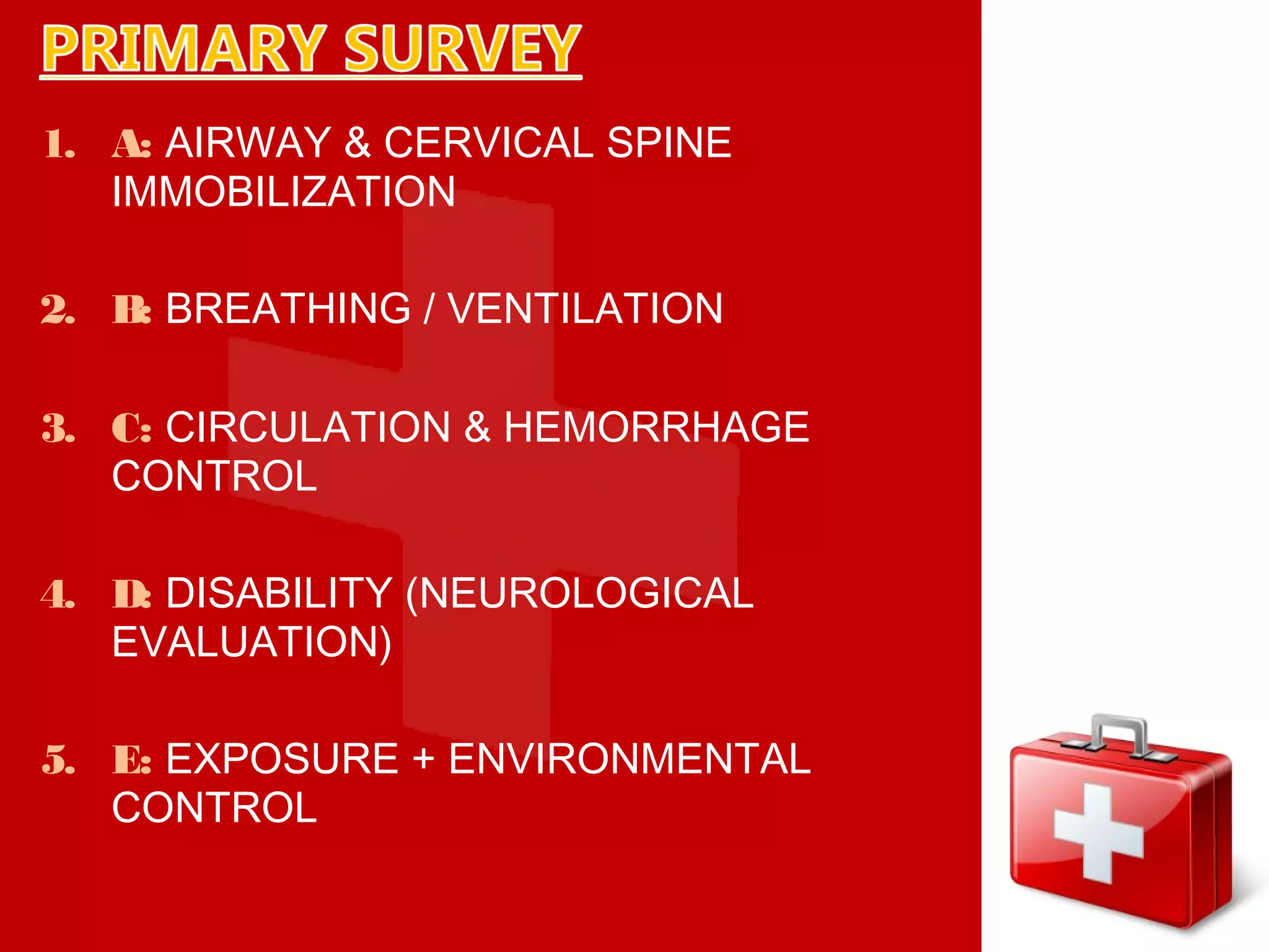
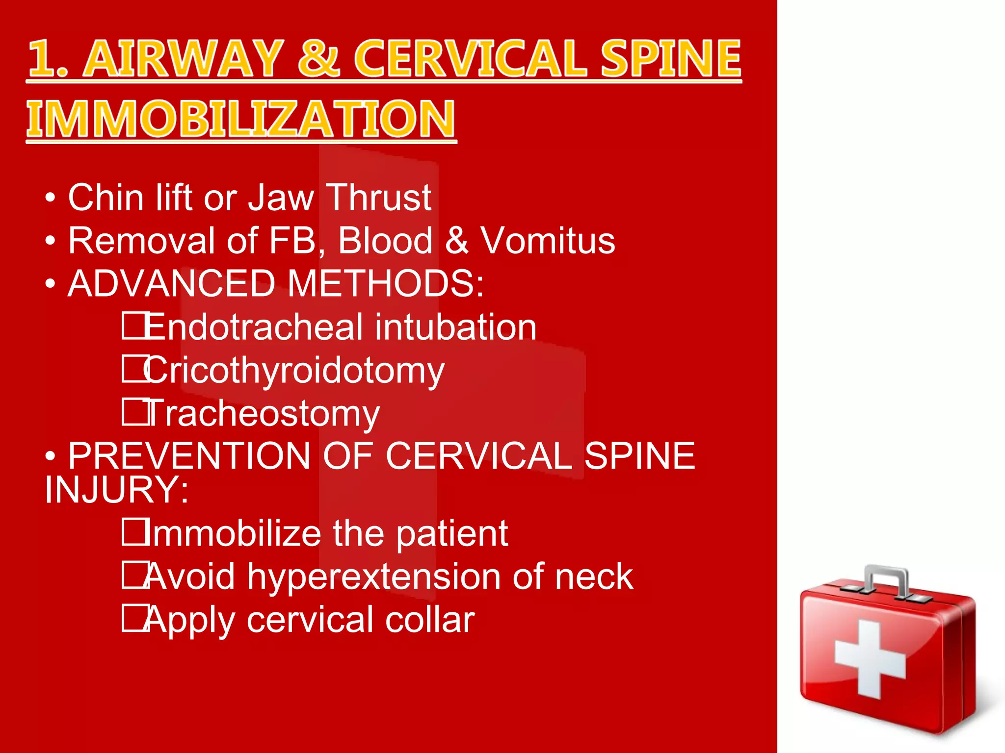
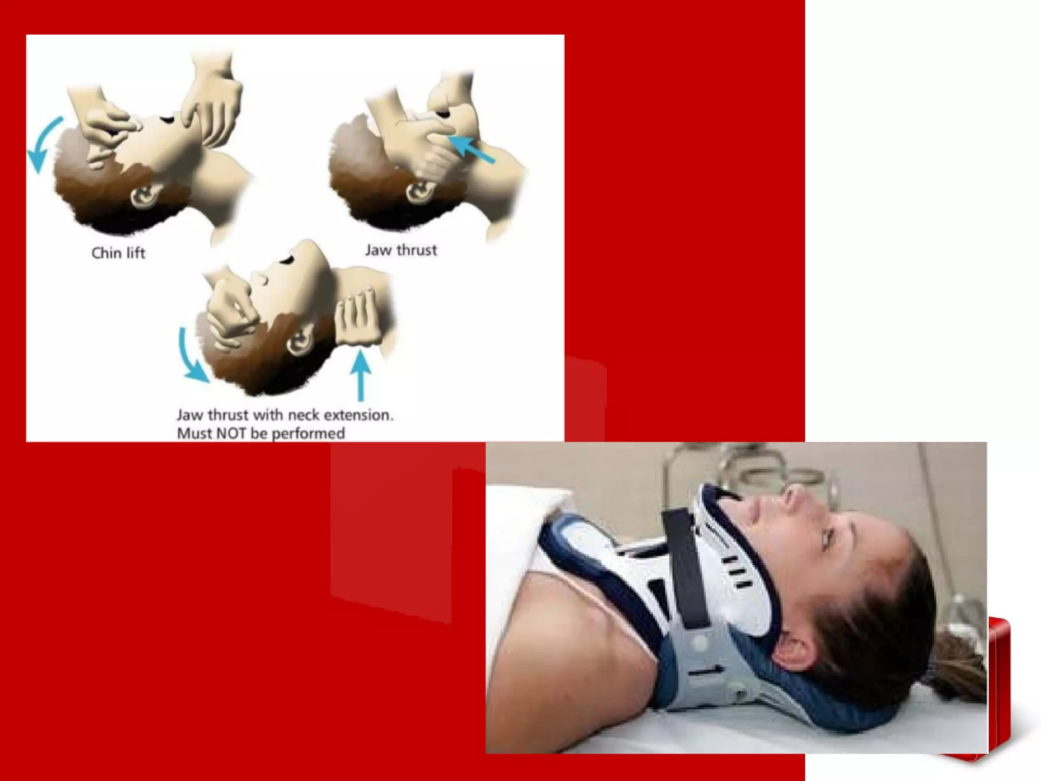
Dr. Saleh Bakry
Associate Professor of Oral and
Maxillofacial Surgery
• A standardized approach to all traumatic
patients.
• A comprehensive assessment and
management of patients in emergency
situation.
• Best utilization of golden hour which lies
between life and death after a traumatic
event.
1. Within Seconds to Minutes
• Brainstem injury
• Aortic rupture
2. Within Minutes to Hours (Golden hours)
• Sub dural Hematoma
• Rupture of Liver & Spleen
3. Within Days to Weeks
• Sepsis & MODS
• Matching the need of your trauma
patient to the facilities (Resources) of
the hospital
ATLS PROTOCOL
1.PRIMARY SURVEY
2.RESUSCITATION
3.SECONDARY SURVEY
4.TERTIARY SURVEY
1. PRIMARY
SURVEY
1. A: AIRWAY & CERVICAL SPINE
IMMOBILIZATION
2. B: BREATHING / VENTILATION
3. C: CIRCULATION & HEMORRHAGE
CONTROL
4. D: DISABILITY (NEUROLOGICAL
EVALUATION)
5. E: EXPOSURE + ENVIRONMENTAL
CONTROL
• Chin lift or Jaw Thrust
• Removal of FB, Blood & Vomitus
• ADVANCED METHODS:
Endotracheal intubation
Cricothyroidotomy
Tracheostomy
• PREVENTION OF CERVICAL SPINE
INJURY:
Immobilize the patient
Avoid hyperextension of neck
Apply cervical collar
1. Noisy breathing.
2. Respiratory distress.
3. Failure to speak, dysphonia.
4. Cyanosis.
5. Confusion, “universal choking
sign”.
1. Hemorrhage accumulation or secretion within the
mouth and oropharynx.
2. Tongue: in an unconscious patient, the tongue may
fall into the pharynx and occlude air way.
3. Foreign bodies: food, chewing gum, dentures, dried
mucous, etc. many fall undetected into oropharynx.
4. Aspiration of vomits in the chest.
5. Edema of the glottis and oropharynx region.
6. Fracture mandible: the tongue may be fall back
and occlude the airway.
7. Fracture maxilla: the soft palate may be fall down on
the posterior part of the tongue and cause
suffocation.
• Airway patency doesn’t assure adequate
ventilation Ô Look for bilateral breath sounds
• Give O2 inhalation
• Check chest wall, lungs & diaphragm by
inspection, palpation, percussion &
auscultation.
• Look for conditions that impair ventilation
 Tension pneumothorax.
 Massive hemothorax.
 Rib fractures.
 Open pneumothorax.
 Pulmonary contusion
Impairment in circulation can lead to
shock, so lookforsigns of shocki.e.
•skin color (pallor)
•narrow pulse pressure
•hypotension
•tachycardia
•level of consciousness
•diminished urine output
Control of hemorrhage:
•Apply direct pressure
•Pneumatic splinting devices
•Access the need for surgical intervention
Fluid replacement therapy
•double i/v lines should be maintained for Fluid
replacement
•adults should be given 2 l bolus fluid (preferred fluid
is ringer lactate better if Warm)
•children should be given @ 20ml/kg bolus Fluid
3 for1 rule:
•A rough guideline for the total Amount of crystalloid
volume acutely is to replace
•Each ml of blood loss with 3 ml of crystalloid fluid.
• Check the level of consciousness
(AVPU/GCS):
 A: alert
 V: responds to vocal stimuli
 P: responds to painful stimuli
 U: unresponsive to all stimuli
• Check pupil size & light reaction.
• Check the level of spinal cord injury level
• Undress completely (use trauma scissors).
• Prevent hypothermia (warm blankets & warm
fluids).
• Early hemorrhage control.
• Warm room temperature should be
maintained.
2. SECONDARY
SURVEY
Does not begin until the primary survey
(ABCDES) is completed, resuscitation
efforts are well established & the patient is
having normalization of vital signs.it
includes:
•Complete history
•Complete head to toe examination
•Reassessment of vital signs
•Complete neurological examination.
•Specific procedures, specific lab.
Investigations
1. A: allergies
2. M: medications
3. P: past illness/ pregnancy
4. L: last meal
5. E: events/ environment/mechanism of
injury:
• Blunt trauma: automobile collisions.
• Penetrating trauma: firearms/stabbing.
• Thermal injuries: burns/explosions.
• Hazardous injuries: chemicals/toxins.
1. HEAD
2. MAXILLOFACIAL STRUCTURES
3. CERVICAL SPINE & NECK
4. CHEST
5. ABDOMEN
6. PERINEUM, RECTUM & VAGINA
7. MUSCULOSKELETAL SYSTEM
8. NEUROLOGICAL SYSTEM
3. TERTIARY
SURVEY
• Comprehensive Treatment of all Injuries.
• Fracture Stabilization.
• Necessary Operative Intervention.
• Appropriate Intensive Care.
• Rehabilitation.
• Stabilization & Appropriate Transfer.
BATTLE SIGN: Bleeding or ecchymosis behind
the ear indicates fracture in the base of the
skull or condylar fracture.
PANDA EYE (RACCOON EYE):
•Indicate fracture of the base of the skull or
zygomatic temporal fracture.
•Appear after 24 hours.
CSF LEAKAGE FROMNOSE (RHINORRHEA)
OREAR(OTORRHEA).
1. Handkerchief test
•The material is collected to dry in a
handkerchief.
•If starch → mucous.
•If not starch → CSF.
2. Test the collected material for glucose: If it is
positive it is C.S.F.
1. Orthopedic.
2. General surgeon.
3. Neurosurgeon.
4. Emergency department physician.
+
5. Maxillofacial surgeon.
6. Ophthalmologist.
7. E.N.T Physician.
8. Anesthetist
THANKYOU

Advanced trauma life support (atls)

  • 1.
    Dr. Saleh Bakry AssociateProfessor of Oral and Maxillofacial Surgery
  • 2.
    • A standardizedapproach to all traumatic patients. • A comprehensive assessment and management of patients in emergency situation. • Best utilization of golden hour which lies between life and death after a traumatic event.
  • 3.
    1. Within Secondsto Minutes • Brainstem injury • Aortic rupture 2. Within Minutes to Hours (Golden hours) • Sub dural Hematoma • Rupture of Liver & Spleen 3. Within Days to Weeks • Sepsis & MODS
  • 4.
    • Matching theneed of your trauma patient to the facilities (Resources) of the hospital ATLS PROTOCOL 1.PRIMARY SURVEY 2.RESUSCITATION 3.SECONDARY SURVEY 4.TERTIARY SURVEY
  • 5.
  • 6.
    1. A: AIRWAY& CERVICAL SPINE IMMOBILIZATION 2. B: BREATHING / VENTILATION 3. C: CIRCULATION & HEMORRHAGE CONTROL 4. D: DISABILITY (NEUROLOGICAL EVALUATION) 5. E: EXPOSURE + ENVIRONMENTAL CONTROL
  • 7.
    • Chin liftor Jaw Thrust • Removal of FB, Blood & Vomitus • ADVANCED METHODS: Endotracheal intubation Cricothyroidotomy Tracheostomy • PREVENTION OF CERVICAL SPINE INJURY: Immobilize the patient Avoid hyperextension of neck Apply cervical collar
  • 10.
    1. Noisy breathing. 2.Respiratory distress. 3. Failure to speak, dysphonia. 4. Cyanosis. 5. Confusion, “universal choking sign”.
  • 11.
    1. Hemorrhage accumulationor secretion within the mouth and oropharynx. 2. Tongue: in an unconscious patient, the tongue may fall into the pharynx and occlude air way. 3. Foreign bodies: food, chewing gum, dentures, dried mucous, etc. many fall undetected into oropharynx. 4. Aspiration of vomits in the chest. 5. Edema of the glottis and oropharynx region. 6. Fracture mandible: the tongue may be fall back and occlude the airway. 7. Fracture maxilla: the soft palate may be fall down on the posterior part of the tongue and cause suffocation.
  • 12.
    • Airway patencydoesn’t assure adequate ventilation Ô Look for bilateral breath sounds • Give O2 inhalation • Check chest wall, lungs & diaphragm by inspection, palpation, percussion & auscultation.
  • 13.
    • Look forconditions that impair ventilation  Tension pneumothorax.  Massive hemothorax.  Rib fractures.  Open pneumothorax.  Pulmonary contusion
  • 14.
    Impairment in circulationcan lead to shock, so lookforsigns of shocki.e. •skin color (pallor) •narrow pulse pressure •hypotension •tachycardia •level of consciousness •diminished urine output
  • 15.
    Control of hemorrhage: •Applydirect pressure •Pneumatic splinting devices •Access the need for surgical intervention Fluid replacement therapy •double i/v lines should be maintained for Fluid replacement •adults should be given 2 l bolus fluid (preferred fluid is ringer lactate better if Warm) •children should be given @ 20ml/kg bolus Fluid 3 for1 rule: •A rough guideline for the total Amount of crystalloid volume acutely is to replace •Each ml of blood loss with 3 ml of crystalloid fluid.
  • 16.
    • Check thelevel of consciousness (AVPU/GCS):  A: alert  V: responds to vocal stimuli  P: responds to painful stimuli  U: unresponsive to all stimuli • Check pupil size & light reaction. • Check the level of spinal cord injury level
  • 17.
    • Undress completely(use trauma scissors). • Prevent hypothermia (warm blankets & warm fluids). • Early hemorrhage control. • Warm room temperature should be maintained.
  • 18.
  • 19.
    Does not beginuntil the primary survey (ABCDES) is completed, resuscitation efforts are well established & the patient is having normalization of vital signs.it includes: •Complete history •Complete head to toe examination •Reassessment of vital signs •Complete neurological examination. •Specific procedures, specific lab. Investigations
  • 20.
    1. A: allergies 2.M: medications 3. P: past illness/ pregnancy 4. L: last meal 5. E: events/ environment/mechanism of injury: • Blunt trauma: automobile collisions. • Penetrating trauma: firearms/stabbing. • Thermal injuries: burns/explosions. • Hazardous injuries: chemicals/toxins.
  • 21.
    1. HEAD 2. MAXILLOFACIALSTRUCTURES 3. CERVICAL SPINE & NECK 4. CHEST 5. ABDOMEN 6. PERINEUM, RECTUM & VAGINA 7. MUSCULOSKELETAL SYSTEM 8. NEUROLOGICAL SYSTEM
  • 22.
  • 23.
    • Comprehensive Treatmentof all Injuries. • Fracture Stabilization. • Necessary Operative Intervention. • Appropriate Intensive Care. • Rehabilitation. • Stabilization & Appropriate Transfer.
  • 24.
    BATTLE SIGN: Bleedingor ecchymosis behind the ear indicates fracture in the base of the skull or condylar fracture.
  • 25.
    PANDA EYE (RACCOONEYE): •Indicate fracture of the base of the skull or zygomatic temporal fracture. •Appear after 24 hours.
  • 26.
    CSF LEAKAGE FROMNOSE(RHINORRHEA) OREAR(OTORRHEA). 1. Handkerchief test •The material is collected to dry in a handkerchief. •If starch → mucous. •If not starch → CSF. 2. Test the collected material for glucose: If it is positive it is C.S.F.
  • 27.
    1. Orthopedic. 2. Generalsurgeon. 3. Neurosurgeon. 4. Emergency department physician. + 5. Maxillofacial surgeon. 6. Ophthalmologist. 7. E.N.T Physician. 8. Anesthetist
  • 28.