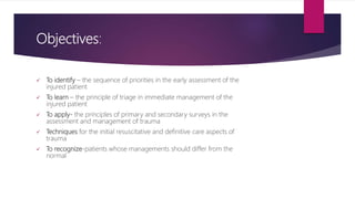
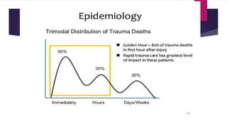
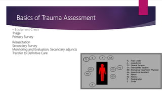
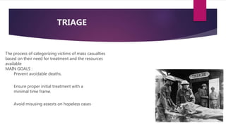
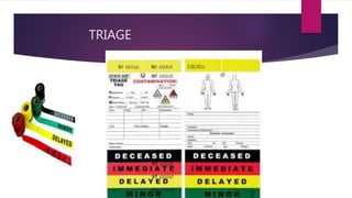
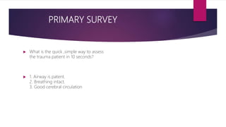
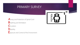
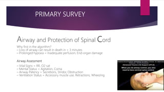
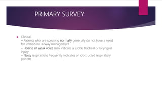
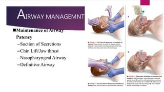
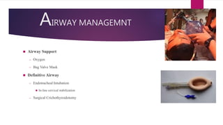
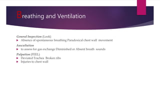
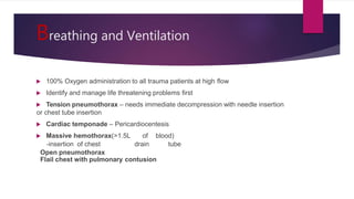
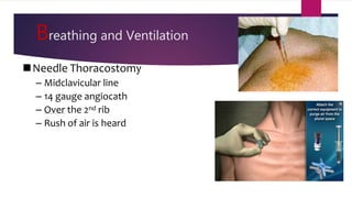
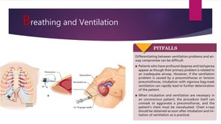
This document provides an overview of Advance Trauma Life Support (ATLS) principles from a presentation. It discusses the importance of the "golden hour" in trauma care. The objectives of ATLS are to prioritize patient assessment and management. It covers the basics of trauma assessment including preparation, triage, primary survey, resuscitation, secondary survey, and transfer to definitive care. The primary survey focuses on addressing life threats in order of airway, breathing, circulation, disability and exposure.
INITIAL ASSESSMENT OF TRAUMA PATIENTS....(INSPIRED FROM CTLS AND ATLS GUIDELI...Prerna Biswal
THIS PRESENTATION WAS MADE AT IMA HOUSE IN BHUBANESWAR,ODISHA, BY DR.NIBEDITA PANI,HOD ,DEPT. OF ANAESTHESIOLOGY AND DR.PRERNA BISWAL,PG,ANAESTHESIOLOGY,SCBMCH,CUTTACK,
INITIAL ASSESSMENT OF TRAUMA PATIENTS....(INSPIRED FROM CTLS AND ATLS GUIDELI...Prerna Biswal
THIS PRESENTATION WAS MADE AT IMA HOUSE IN BHUBANESWAR,ODISHA, BY DR.NIBEDITA PANI,HOD ,DEPT. OF ANAESTHESIOLOGY AND DR.PRERNA BISWAL,PG,ANAESTHESIOLOGY,SCBMCH,CUTTACK,
Approach to a trauma patient - Advanced Trauma Life SupportParthasarathi Ghosh
Approach to a trauma patient from a Critical Care Medicine perspective with basics of Advanced Trauma Life Support.
References - ATLS Manual 10th Edition
This is a presentation which contains basics of polytrauma management,ATLS, triage, critical decision making skills, application of Glasgow coma scale and complications of different management strategies, if not applied properly.
Report Back from SGO 2024: What’s the Latest in Cervical Cancer?bkling
Are you curious about what’s new in cervical cancer research or unsure what the findings mean? Join Dr. Emily Ko, a gynecologic oncologist at Penn Medicine, to learn about the latest updates from the Society of Gynecologic Oncology (SGO) 2024 Annual Meeting on Women’s Cancer. Dr. Ko will discuss what the research presented at the conference means for you and answer your questions about the new developments.
These lecture slides, by Dr Sidra Arshad, offer a quick overview of physiological basis of a normal electrocardiogram.
Learning objectives:
1. Define an electrocardiogram (ECG) and electrocardiography
2. Describe how dipoles generated by the heart produce the waveforms of the ECG
3. Describe the components of a normal electrocardiogram of a typical bipolar leads (limb II)
4. Differentiate between intervals and segments
5. Enlist some common indications for obtaining an ECG
Study Resources:
1. Chapter 11, Guyton and Hall Textbook of Medical Physiology, 14th edition
2. Chapter 9, Human Physiology - From Cells to Systems, Lauralee Sherwood, 9th edition
3. Chapter 29, Ganong’s Review of Medical Physiology, 26th edition
4. Electrocardiogram, StatPearls - https://www.ncbi.nlm.nih.gov/books/NBK549803/
5. ECG in Medical Practice by ABM Abdullah, 4th edition
6. ECG Basics, http://www.nataliescasebook.com/tag/e-c-g-basics
- Video recording of this lecture in English language: https://youtu.be/lK81BzxMqdo
- Video recording of this lecture in Arabic language: https://youtu.be/Ve4P0COk9OI
- Link to download the book free: https://nephrotube.blogspot.com/p/nephrotube-nephrology-books.html
- Link to NephroTube website: www.NephroTube.com
- Link to NephroTube social media accounts: https://nephrotube.blogspot.com/p/join-nephrotube-on-social-media.html
Factory Supply Best Quality Pmk Oil CAS 28578–16–7 PMK Powder in Stockrebeccabio
Factory Supply Best Quality Pmk Oil CAS 28578–16–7 PMK Powder in Stock
Telegram: bmksupplier
signal: +85264872720
threema: TUD4A6YC
You can contact me on Telegram or Threema
Communicate promptly and reply
Free of customs clearance, Double Clearance 100% pass delivery to USA, Canada, Spain, Germany, Netherland, Poland, Italy, Sweden, UK, Czech Republic, Australia, Mexico, Russia, Ukraine, Kazakhstan.Door to door service
Hot Selling Organic intermediates
Lung Cancer: Artificial Intelligence, Synergetics, Complex System Analysis, S...Oleg Kshivets
RESULTS: Overall life span (LS) was 2252.1±1742.5 days and cumulative 5-year survival (5YS) reached 73.2%, 10 years – 64.8%, 20 years – 42.5%. 513 LCP lived more than 5 years (LS=3124.6±1525.6 days), 148 LCP – more than 10 years (LS=5054.4±1504.1 days).199 LCP died because of LC (LS=562.7±374.5 days). 5YS of LCP after bi/lobectomies was significantly superior in comparison with LCP after pneumonectomies (78.1% vs.63.7%, P=0.00001 by log-rank test). AT significantly improved 5YS (66.3% vs. 34.8%) (P=0.00000 by log-rank test) only for LCP with N1-2. Cox modeling displayed that 5YS of LCP significantly depended on: phase transition (PT) early-invasive LC in terms of synergetics, PT N0—N12, cell ratio factors (ratio between cancer cells- CC and blood cells subpopulations), G1-3, histology, glucose, AT, blood cell circuit, prothrombin index, heparin tolerance, recalcification time (P=0.000-0.038). Neural networks, genetic algorithm selection and bootstrap simulation revealed relationships between 5YS and PT early-invasive LC (rank=1), PT N0—N12 (rank=2), thrombocytes/CC (3), erythrocytes/CC (4), eosinophils/CC (5), healthy cells/CC (6), lymphocytes/CC (7), segmented neutrophils/CC (8), stick neutrophils/CC (9), monocytes/CC (10); leucocytes/CC (11). Correct prediction of 5YS was 100% by neural networks computing (area under ROC curve=1.0; error=0.0).
CONCLUSIONS: 5YS of LCP after radical procedures significantly depended on: 1) PT early-invasive cancer; 2) PT N0--N12; 3) cell ratio factors; 4) blood cell circuit; 5) biochemical factors; 6) hemostasis system; 7) AT; 8) LC characteristics; 9) LC cell dynamics; 10) surgery type: lobectomy/pneumonectomy; 11) anthropometric data. Optimal diagnosis and treatment strategies for LC are: 1) screening and early detection of LC; 2) availability of experienced thoracic surgeons because of complexity of radical procedures; 3) aggressive en block surgery and adequate lymph node dissection for completeness; 4) precise prediction; 5) adjuvant chemoimmunoradiotherapy for LCP with unfavorable prognosis.
Title: Sense of Smell
Presenter: Dr. Faiza, Assistant Professor of Physiology
Qualifications:
MBBS (Best Graduate, AIMC Lahore)
FCPS Physiology
ICMT, CHPE, DHPE (STMU)
MPH (GC University, Faisalabad)
MBA (Virtual University of Pakistan)
Learning Objectives:
Describe the primary categories of smells and the concept of odor blindness.
Explain the structure and location of the olfactory membrane and mucosa, including the types and roles of cells involved in olfaction.
Describe the pathway and mechanisms of olfactory signal transmission from the olfactory receptors to the brain.
Illustrate the biochemical cascade triggered by odorant binding to olfactory receptors, including the role of G-proteins and second messengers in generating an action potential.
Identify different types of olfactory disorders such as anosmia, hyposmia, hyperosmia, and dysosmia, including their potential causes.
Key Topics:
Olfactory Genes:
3% of the human genome accounts for olfactory genes.
400 genes for odorant receptors.
Olfactory Membrane:
Located in the superior part of the nasal cavity.
Medially: Folds downward along the superior septum.
Laterally: Folds over the superior turbinate and upper surface of the middle turbinate.
Total surface area: 5-10 square centimeters.
Olfactory Mucosa:
Olfactory Cells: Bipolar nerve cells derived from the CNS (100 million), with 4-25 olfactory cilia per cell.
Sustentacular Cells: Produce mucus and maintain ionic and molecular environment.
Basal Cells: Replace worn-out olfactory cells with an average lifespan of 1-2 months.
Bowman’s Gland: Secretes mucus.
Stimulation of Olfactory Cells:
Odorant dissolves in mucus and attaches to receptors on olfactory cilia.
Involves a cascade effect through G-proteins and second messengers, leading to depolarization and action potential generation in the olfactory nerve.
Quality of a Good Odorant:
Small (3-20 Carbon atoms), volatile, water-soluble, and lipid-soluble.
Facilitated by odorant-binding proteins in mucus.
Membrane Potential and Action Potential:
Resting membrane potential: -55mV.
Action potential frequency in the olfactory nerve increases with odorant strength.
Adaptation Towards the Sense of Smell:
Rapid adaptation within the first second, with further slow adaptation.
Psychological adaptation greater than receptor adaptation, involving feedback inhibition from the central nervous system.
Primary Sensations of Smell:
Camphoraceous, Musky, Floral, Pepperminty, Ethereal, Pungent, Putrid.
Odor Detection Threshold:
Examples: Hydrogen sulfide (0.0005 ppm), Methyl-mercaptan (0.002 ppm).
Some toxic substances are odorless at lethal concentrations.
Characteristics of Smell:
Odor blindness for single substances due to lack of appropriate receptor protein.
Behavioral and emotional influences of smell.
Transmission of Olfactory Signals:
From olfactory cells to glomeruli in the olfactory bulb, involving lateral inhibition.
Primitive, less old, and new olfactory systems with different path
Pulmonary Thromboembolism - etilogy, types, medical- Surgical and nursing man...VarunMahajani
Disruption of blood supply to lung alveoli due to blockage of one or more pulmonary blood vessels is called as Pulmonary thromboembolism. In this presentation we will discuss its causes, types and its management in depth.
2. This focus on urgent problems is first captured by the
“Golden hour” catch phrase and is one of the most
important lessons of ATLS
3. Objectives:
To identify – the sequence of priorities in the early assessment of the
injured patient
To learn – the principle of triage in immediate management of the
injured patient
To apply- the principles of primary and secondary surveys in the
assessment and management of trauma
Techniques for the initial resuscitative and definitive care aspects of
trauma
To recognize-patients whose managements should differ from the
normal
4.
5. Basics of Trauma Assessment
Preparation
– Team Assembly
– Equipment Check
Triage
Primary Survey
Resuscitation
Secondary Survey
Monitoring and Evaluation, Secondary adjuncts
Transfer to Definitive Care
6. The steps in the ATLS principles:
Primary survey with simultaneous resuscitation : identify
and treat what is killing the patient
Secondary survey: proceed to identify all other injuries
Definitive care: develop a definitive management plan
7. TRIAGE
The process of categorizing victims of mass casualties
based on their need for treatment and the resources
available
MAIN GOALS :
Prevent avoidable deaths.
Ensure proper initial treatment with a
minimal time frame.
Avoid misusing assests on hopeless cases
10. PRIMARY SURVEY
What is the quick ,simple way to assess
the trauma patient in 10 seconds?
1. Airway is patent.
2. Breathing intact.
3. Good cerebral circulation
11. PRIMARY SURVEY
Airway and Protection of Spinal Cord
Breathing and Ventilation
Circulation
Disability
Exposure and Control of the Environment
12. PRIMARY SURVEY
Airway and Protection of Spinal Cord
Why first in the algorithm?
– Loss of airway can result in death in < 3 minutes
– Prolonged hypoxia = Inadequate perfusion, End-organ damage
Airway Assessment
– Vital Signs = RR, O2 sat
– Mental Status = Agitation, Coma
– Airway Patency = Secretions, Stridor, Obstruction
– Ventilation Status = Accessory muscle use, Retractions, Wheezing
13. PRIMARY SURVEY
Clinical
– Patients who are speaking normally generally do not have a need
for immediate airway management
– Hoarse or weak voice may indicate a subtle tracheal or laryngeal
injury
– Noisy respirations frequently indicates an obstructed respiratory
pattern
14. AIRWAY MANAGEMNT
Maintenance of Airway
Patency
–Suction of Secretions
–Chin Lift/Jaw thrust
–Nasopharyngeal Airway
–Definitive Airway
16. AIRWAY MANAGEMNT
Needle-
Cricothyroidotomy
• Insertion of a needle
through the
cricothyroid
membrane or Into the
trachea
• provide temporary,
supplemental
oxygenation so that
intubation can be
accomplished on an
urgent rather than an
emergent basis.
17. Surgical-
Cricothyroidotomy
Palpate the thyroid notch, cricothyroid
interval, and the sternal notch for
orientation.
Make a transverse skin incision over
the cricothyroid membrane and
carefully incise through the membrane
transversely.
Insert hemostat or tracheal spreader
into the incision and rotate it 90
degrees to
open the airway.
Insert a proper-size, cuffed
endotracheal tube or tracheostomy
tube into the cricothyroid membrane
incision, directing the tube distally into
the
trachea.
18. Breathing and Ventilation
General Inspection (Look)
Absence of spontaneous breathing Paradoxical chest wall movement
Auscultation
to assess for gas exchange Diminished or Absent breath sounds
Palpation (FEEL)
Deviated Trachea Broken ribs
Injuries to chest wall
19. Breathing and Ventilation
100% Oxygen administration to all trauma patients at high flow
Identify and manage life threatening problems first
Tension pneumothorax – needs immediate decompression with needle insertion
or chest tube insertion
Cardiac temponade – Pericardiocentesis
Massive hemothorax(>1.5L of blood)
-insertion of chest drain tube
Open pneumothorax
Flail chest with pulmonary contusion
22. Chest Tube Insertion
STEP1. Determine the insertion site, usually at the
nipple level (fifth intercostal space), just anterior to the
midaxillary line on the affected
side. A second chest tube may be used for a
hemothorax.
STEP2. Surgically prepare and drape the chest at
the predetermined site of the tube insertion.
STEP3. Locally anesthetize the skin and rib
periosteum.
STEP4. Make a 2- to 3-cm transverse (horizontal)
incision at the predetermined site and
bluntly dissect through the subcutaneous
tissues, just over the top of the rib.
23. Chest Tube Insertion
STEP5. Puncture the parietal pleura with the tip
of a clamp and put a gloved finger into the
incision to avoid injury to other organs and
to clear any adhesions, clots, and so on.
Once the tube in the proper place, remove
the clamp from the tube.
STEP6. Clamp the proximal end of the thoracostomy tube and advance it into the
pleural space to the desired length.
The tube should
be directed posteriorly along the inside of
the chest wall.
24. Chest Tube Insertion
STEP7. Look for “fogging” of the chest tube with
expiration or listen for air movement.
STEP8. Connect the end of the thoracostomy tube
to an underwater-seal apparatus.
STEP9. Suture the tube in place.
STEP10. Apply an occlusive dressing and tape the
tube to the chest.
STEP11. Obtain a chest x-ray film.
STEP12. Obtain arterial blood gas values and/or
institute pulse oximetry monitoring as
necessary.
25. Circulation
Shock
The first step in the initial management of shock in trauma patients is to recognize its
presence
?Is the patient in
?
“Any injured patient who is cool and has tachycardia is considered to be in shock until
proven otherwise”
26. Circulation
Clinical Signs of Shock
Altered mental status
Tachycardia (HR > 100)
Most common sign
Arterial Hypotension (SBP < 90)
Inadequate Tissue Perfusion
Pale skin color , Cool clammy skin ,Delayed cap refill (> 3 seconds) , LOC
, Decreased Urine Output
(UOP < 0.5 ml/kg/hr)
Reliance solely on systolic blood pressure as an indicator of shock can
result in delayed recognition of the shock state.
27. Circulation
The second step in the initial management of shock
is to identify the probable cause of the shock state.
What is the cause of shock?
Hemorrhagic Shock
Nonhemorrhagic Shock
Cardiogenic Shock NeurogenicShock
28. Most injured patients in shock
have hypovolemia, but they may suffer from cardiogenic, obstructive, neurogenic,
and, rarely, septic shock
Hemorrhage is the most common cause of shock in the
injured patient
Multiple: No. of patients and the severity of theirinjuries do not exceed the ability of thefacility to provide care
A copmlete sentence spoken by pt. tells us:
The jet insufflation technique is performed byplacing a large-caliber plastic cannula, 12- to 14-gaugefor adults, and 16- to 18-gauge in children, throughthe cricothyroid membrane into the trachea below thelevel of the obstruction (n FIGURE2-15). The cannula isthen connected to oxygen at 15 L/min (40 to 50 psi)with a Y-connector or a side hole cut in the tubingbetween the oxygen source and the plastic cannula 1 second on and 4 secondsoff, can then be achieved by placing the thumb
Airway patency alone does not ensure adequate ventilation. Adequate gas exchange is required to maximizeoxygenation and carbon dioxide elimination.
Complications of Chest Tube Placement
Injury to intercostal nerve, artery, vein
Injury to lung
Injury to mediastinum
Infection
Allergic reaction to lidocaine
Inappropriate placement of chest tube
Hemorrhage is the most common cause of shock after injury