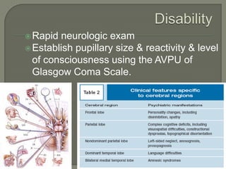
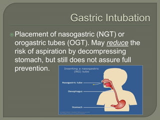
The document provides guidance on the assessment and management of trauma patients. It describes the golden hour period following trauma where rapid assessment is critical. The primary and secondary surveys are outlined, with the primary focusing on stabilizing vital functions like airway, breathing, circulation, disability and exposure. Specific injuries and treatments are discussed for areas like head trauma, spinal trauma, chest trauma, abdominal trauma and genitourinary trauma. Throughout, the emphasis is on stabilizing life-threatening injuries and rapidly diagnosing and treating problems that could impair ventilation or circulation.
INITIAL ASSESSMENT OF TRAUMA PATIENTS....(INSPIRED FROM CTLS AND ATLS GUIDELI...Prerna Biswal
THIS PRESENTATION WAS MADE AT IMA HOUSE IN BHUBANESWAR,ODISHA, BY DR.NIBEDITA PANI,HOD ,DEPT. OF ANAESTHESIOLOGY AND DR.PRERNA BISWAL,PG,ANAESTHESIOLOGY,SCBMCH,CUTTACK,
INITIAL ASSESSMENT OF TRAUMA PATIENTS....(INSPIRED FROM CTLS AND ATLS GUIDELI...Prerna Biswal
THIS PRESENTATION WAS MADE AT IMA HOUSE IN BHUBANESWAR,ODISHA, BY DR.NIBEDITA PANI,HOD ,DEPT. OF ANAESTHESIOLOGY AND DR.PRERNA BISWAL,PG,ANAESTHESIOLOGY,SCBMCH,CUTTACK,
2. The Golden Hour of Trauma
• Period immediately following trauma in which
rapid assessment, diagnosis, and stabilization
must occur.
Primary Survey
• Initial assessment and resuscitation of vital
functions. Prioritization is based on ABC’s of
trauma car.
4. Asses patency of airway
Use jaw thrust or chin lift initially to open airway
Clear foreign bodies
Insert oral or nasal airway when necessary
Obtunded/unconscious patients = intubated
Surgical airway = Cricothyroidotomy used when
unable to intubate.
5. Inspect, Auscultate, & Palpate the chest
Ensure Adequate ventilation & identify &
treat injuries that may immediately impair
ventilation:
• Tension pneumothorax
• Flail chest & Pulmonary Contusion
• Massive Hemothorax
• Open Pneumothorax
6. Place two large-bore peripheral (14- or 16-
gauge) IVs.
Draw blood at time of IV placement
Assess circulatory status (capillary
refill, pulse, skin color)
Control of life-threatening hemorrhage
using direct pressure.
7. Rapid neurologic exam
Establish pupillary size & reactivity & level
of consciousness using the AVPU of
Glasgow Coma Scale.
9. Placement of a urinary catheter is
considered part of the resuscitative phase
that takes place during the primary survey.
Foley is contraindicated when urethral
transection is suspected, such as in the
case of a pelvic fracture. If transection is
suspected, perform retrograde
urethrogram before foley.
10. Signs of Urethral Transection
• Blood at the meatus
• A “high-riding” prostate
• Perineal or scrotal hematoma
• Be suspicious with any pelvic fracture
11. Placement of nasogastric (NGT) or
orogastric tubes (OGT). May reduce the
risk of aspiration by decompressing
stomach, but still does not assure full
prevention.
12. Begins during the primary survey
Life-threatening injuries are tended to as
they are identified.
Fluidtherapy should be initiated with up to
2L of an isotonic (lact. ringer or NSS)
crystalloid solution. Peds Pts should
receive and IV bolus of 20 cc/kg
13. 3-to-1 rule
• Used as a rough estimate for the total amount of
crystalloid volume needed to replace blood loss.
Shock
• Inadequate delivery of oxygen on the cellular level
secondary to tissue hypoperfusion
• In traumatic situations, shock is the result of
hemorrhage until proven otherwise.
14. Shock
• Hypovolemic * Loss of volume
• Hemmorhagic* Blood loss = Loss of volume
• Hypoglycemic
• Septic
• Neurogenic * Sudden loss of ANS control
• Cardiogenic* Failure of the ventricles to function
correctly
15. X-rays of the chest, pelvis, & lat. Cervical Spine
usually occur concurrently during the resuscitation
efforts, but should never interrupt them.
Diagnostic peritoneal lavage & focused abd.
Sonogram for trauma (FAST) are tools used for the
rapid detection of intra-abdominal bleeding that
often occurs early in the resuscitative process.
CT scans should be done only for patients who are
hemodynamically stable.
16. Begins once the primary survey is
complete & resuscitative efforts are well
underway. When possible get an AMPLE
history:
• Allergies
• Medications
• Past medical history/Pregnant?
• Last meal
• Events surrounding the mechanism of Injury
17. Head-to-toe evaluation of the trauma patient;
frequent reassessment is key.
Neurologic examination including glascow
coma scale, procedures, radiologic
examination & laboratory testing occur at this
time if not already accomplished.
Tetanus prophylaxis – immunize as needed
18. ABCs
Nuerologic Exam
Oriented to person, place, time
Pupillary reflex
CT
MRI
Look for sudden changes in level of
consciousness.
Recognize and treat herniation
Assume spinal injury until ruled out!
19. Divided into three zones
• Zone I = lies below the cricoid cartilage.
• Zone II = lies between zones I & III.
• Zone III = lies above the angle of the mandible.
Thesedivisions help drive the diagnostic
and therapeutic management decisions for
penetrating neck injuries
Penetrating Neck Injury: Any injury to the
neck that violates the platysma.
20. Vascular Injuries – Very common and life
threatening. Can lead to
exsanguination, hematoma formation w/
compromise of the airway, & cerebral
vascular accidents (E.g. from transection
of the carotid artery or air embolus.)
21. Nonvascular Injuries
• Injury to the larynx & trachea including fracture of the
thyroid cartilage, dislocation of the tracheal cartilages
& arytenoids leading to airway compromise & often a
difficult intubation
• Esophageal injury does occur & as with penetrating
neck injury, does not often manifest initially. (Very high
morbidity/mortality if missed!)
22. Obtain soft tissue films of the neck for clues to
the presence of soft tissue hematoma &
subcutaneous emphysema & a CXR for possible
pneumothorax.
Surgical Exploration is indicated for
• Expanding hematoma, Subcutaneous
emphysema, Tracheal deviation, Change is voice
quality, Air bubbling through the wound.
• Pulses should be palpated to identify deficits & thrills &
auscultated for bruits.
• A Neurologic exam should be performed to identify brachial
plexus and/or CNS deficits as well as Horner’s Syndrome.
23. ZoneII Injuries with instability or enlarging
hematoma require exploration in the
operating room.
Injuries
to Zones I or III may be taken to
OR or managed conservatively using a
combination of
angiography, bronchoscopy, esophagosco
py, gastrografin or barium studies, & CT
scanning.
24. Primary treatment focus on the ABC’s of resuscitation
General observation: Abrasions, Laceration,
deformities.
Palpation for localization of pain
Neurological examination
Cranial nerves
Motor & Sensory function
Reflexes
Rectal tone
Balbacavernosus Reflex
Incontinence (Loss of control of bladder, bowel)
25. Pericardial Tamponade – Sonogram
• Needle Pericardiocentesis
Blunt Cardiac Trauma – ECG
• MVA, Fall, Crush, Blast, Direct violent trauma
Pneumothorax – Upright CXR
• Chest Tube (thoracostomy) confirmed by x-ray
Tension Pneumothorax – Upright CXR
• Needle decompression then tube thoracostomy
Hemothorax
• > 200cc blood for costophrenic angle to be seen on CXR
26. Gunshot wound creates damage via 3 mech.
• Direct injury from the bullet itself
• Injury from fragmentation of the bullet
• Indirect injury from the resultant shock wave
Stab wound is limited to direct damage of
object of impalement.
Blunt injuries also have three mechanisms
• Injury from the direct blow
• Crush injury
• Deceleration injury
27. Physical Examination
• Seat-Belt Sign – ecchymotic area found in the distribution of the
lower ant. abd. Wall & can be associated with perforation of the
bladder or bowel as well as lumbar distraction fracture.
• Cullen Sign – (Periumbilical ecchymosis) indicative of
intraperitoneal hemorrhage
• Grey Turner’s Sign – (Flank ecchymosis) indicative of
retroperitoneal hemorrhage
• Kehr’s Sign – L. shoulder or neck pain 2° to splenic rupture. It
increases when pt. is in trendelenburg position or with L. upper
quadrant palpatation (Caused by diaphragmatic irritation).
Tests
• Perforation: AXR & CXR to look for free air.
• Diaphragmatic injury: CXR looking for blurring of the diaphragm,
hemothorax, or bowel gas patterns above the diaphragm
28. Other Tests
• Diagnostic Peritoneal Lavage (DPL)
• CT scanning
• Angiography
• Serial Hematocrits
Should be obtained during the observation period of the
hemodynamically stable patient
• Laparoscopy
29. Mechanism
• Largely penetrating (GSW>>Stab wound)
75% of pts. With penetrating injury to the pancreas will
have associated injuries to the aorta, portal vein, or
inferior vena cava.
Diagnosis
• Inspect pancreas during laparotomies for other indications
• Check Amylase
• CT – look for parenchymal fracture, intraparenchymal hematoma,
lesser sac fluid, fluid between splenic vein & pancreatic body,
retroperitoneal hematoma or fluid
• Endoscopic Retrograde Cholangiopancreatography (ERCP) if
stable.
30.
31. Diagnosis done in a retrograde fashion
• Work your way up from the urethra to the kidneys and renal
vasculature.
Signs & Symptoms
• Flank or groin pain, blood @ urethral meatus, ecchymoses on
perineum and/or genitalia, evidence of pelvic fracture, rectal bleeding,
a “high-riding” or superiorly displaced prostate.
U/A
• Gross Hematuria = GU injury & often pelvic fracture as well
• Should be done to check for microscopic hematuria
• Microscopic hematuria is usually self-limited
Retrograde Cystogram & Urethrogram
• Take pre-injection KUB film and take before foley placement.
• Contrast into pouch of douglas = intraperitoneal
• Contrast into behind bladder = extraperitoneal bladder rupture
32. Bladder Rupture
• Intraperitoneal
Usually occurs 2 to blunt trauma to a full bladder.
Tx. Surgical Repair.
• Extraperitoneal
Usually occurs 2 to pelvic fracture
Tx. Nonsurgical management by Foley drainage.
Ureteral injury
• Least common GU injury, surgical repair, dx. IVP or CT during
search for renal injury
Renal Injury
• Commonly diagnosed by CT w/ contrast
• Grade IV & V operative, the rest are non-surgically managed.
33. Grade Injury Description
Renal
Injury Scale
I Contusion Hematuria, urologic studies normal
Hematoma Subcapsular, nonexpanding w/o parenchymal
laceration.
II Hematoma Nonexpanding perirenal hematoma in
retroperitoneum
Laceration <1cm depth of renal cortex w/o urinary extravasation
III Laceration <1cm depth of renal cortex w/o urinary extravasation
and/or collecting system rupture.
IV Laceration Extends through cortex, medulla, & collecting system
Vascular Renal art. or vein injury w/ contained hemmorhage
V Laceration Completely shattered kidney
Vascular Avulsion of renal hilum that devascularizes kidney
34.
35. ABCs
Primary and burn specific secondary survey
As a general rule, burns over less than 15% of the body surface area
are not associated with an extensive capillary leak, and children with
burns of this size can be treated with fluid administered at 150% of a
calculated maintenance rate and close observation of their hydration
status. Those who are able and willing to take fluid by mouth may be
given fluid by mouth, with additional fluid administered intravenously
at a maintenance rate.