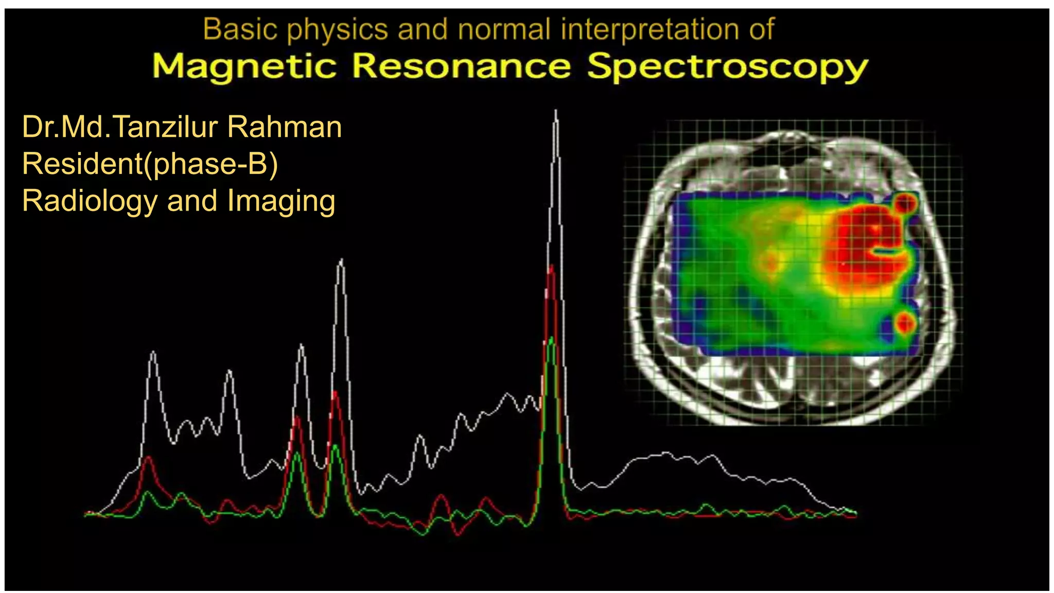
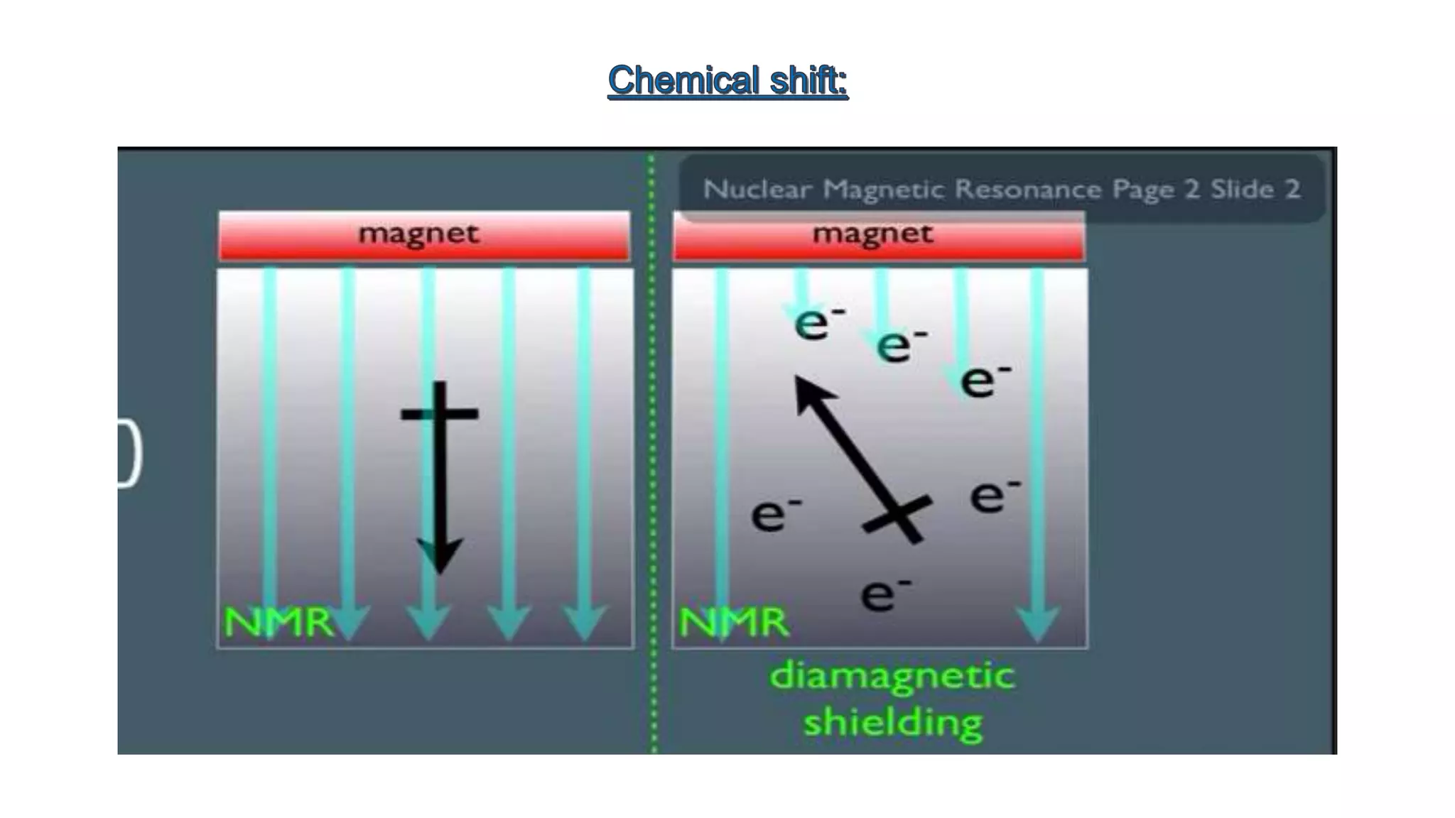
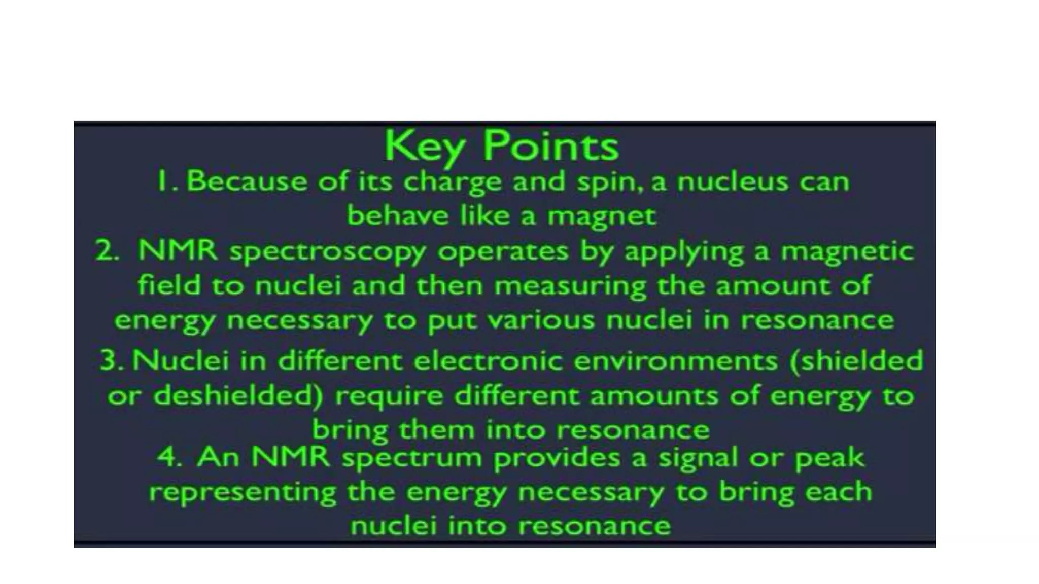
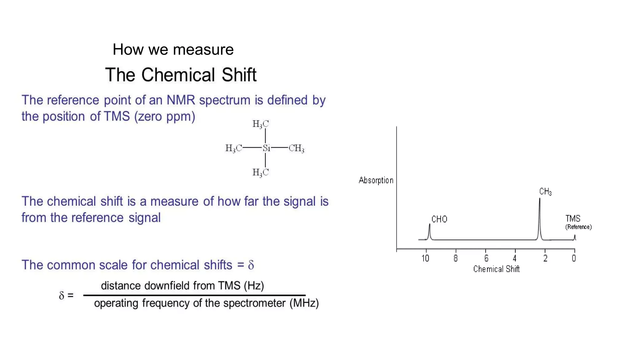
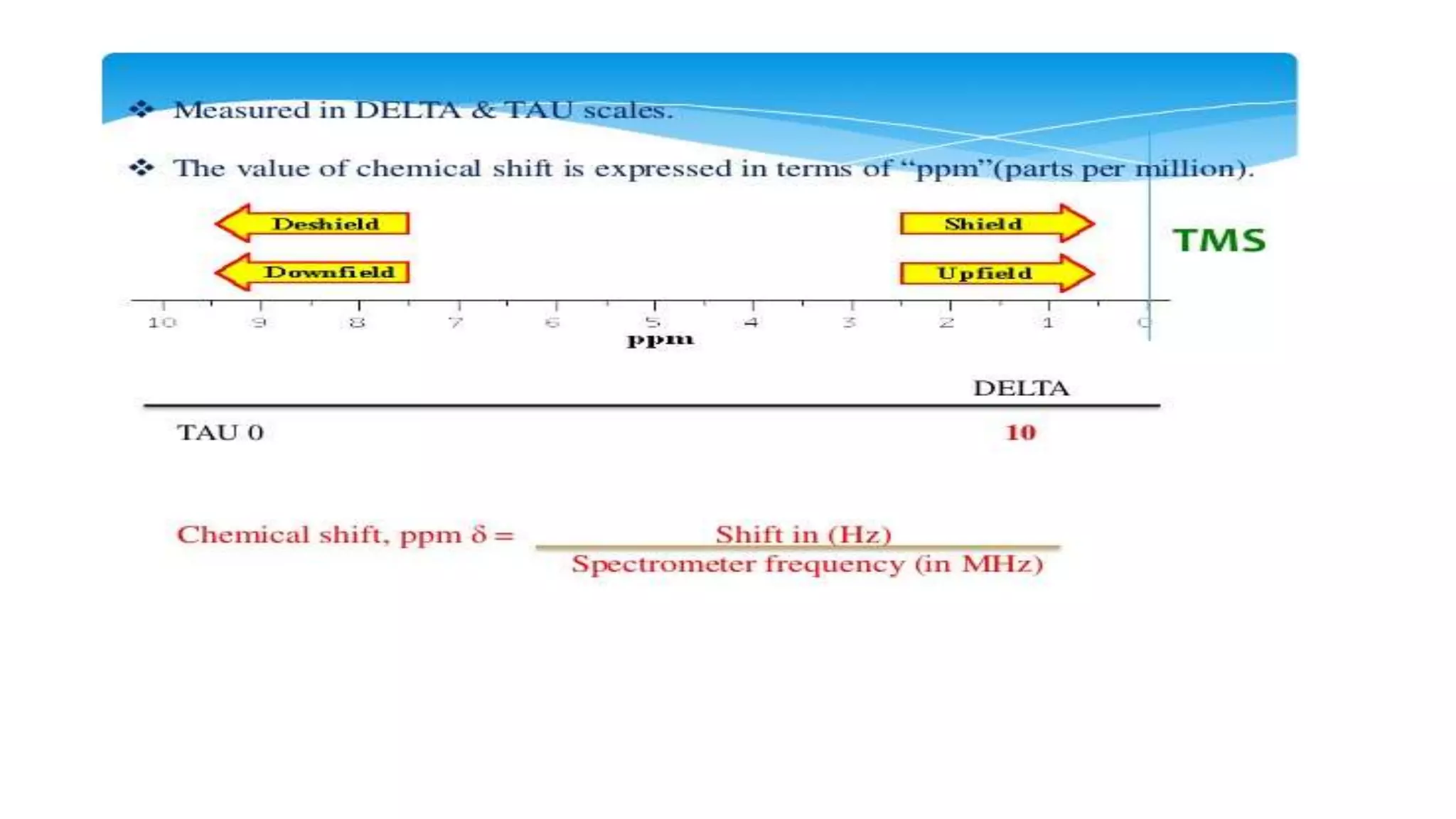
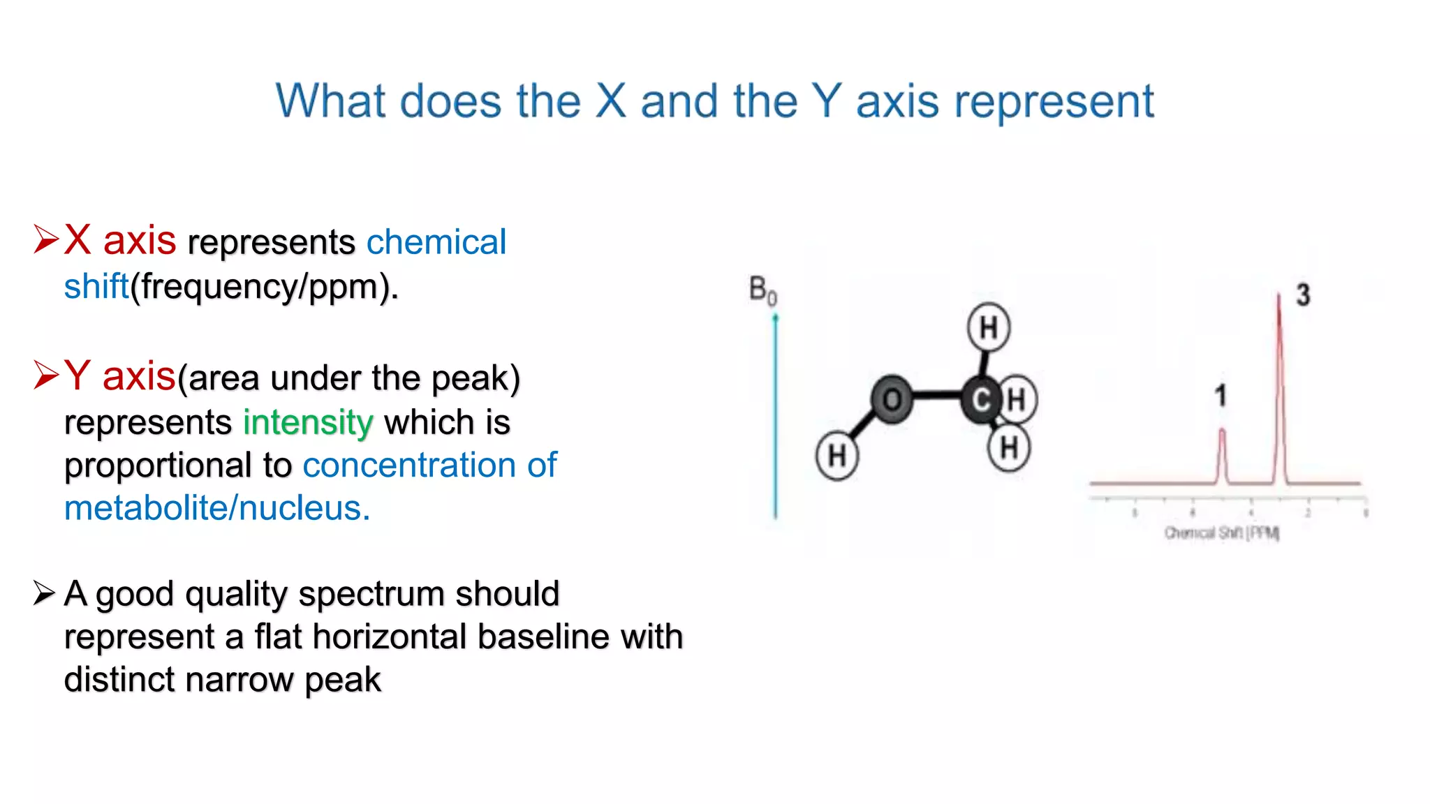
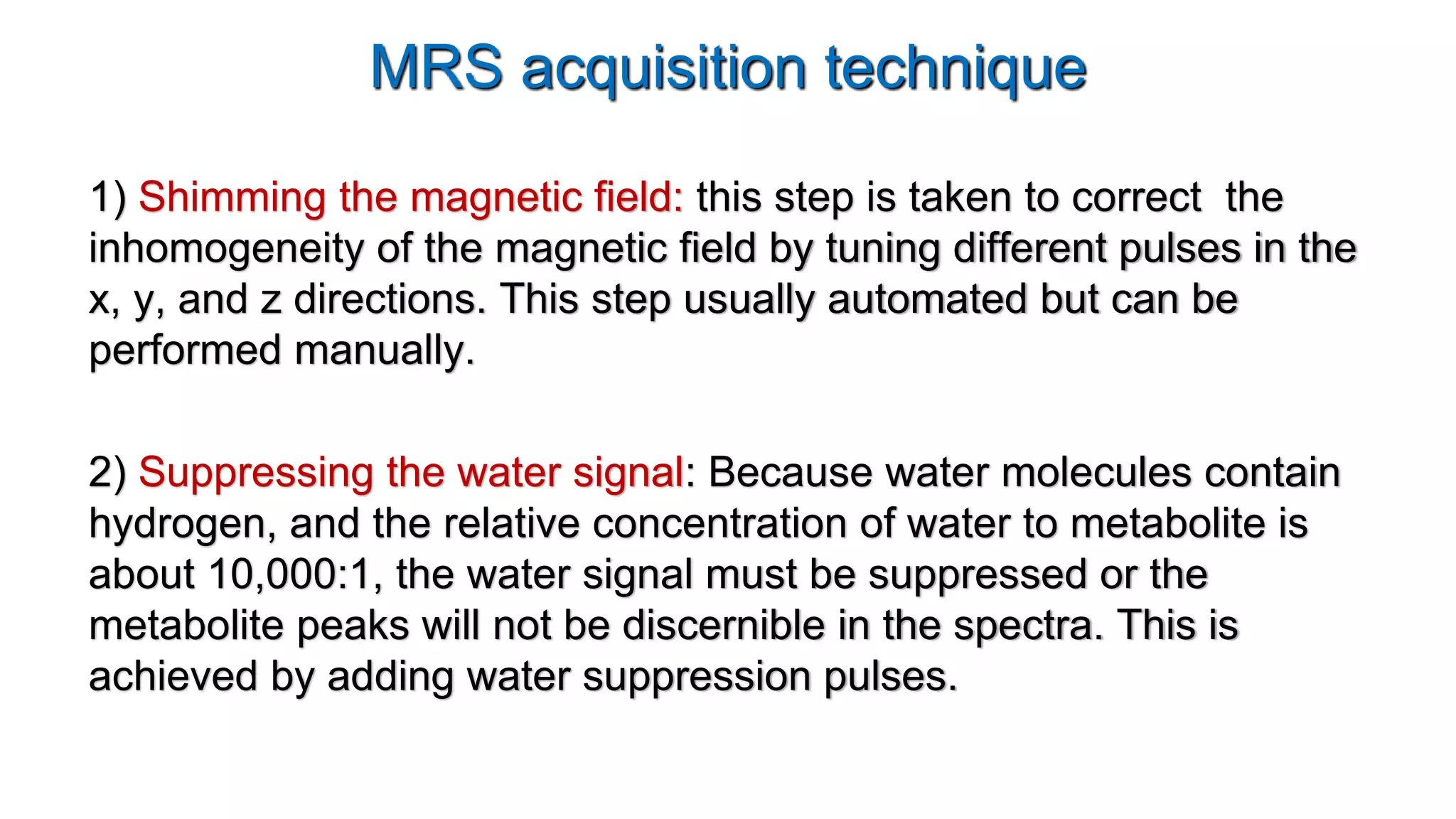
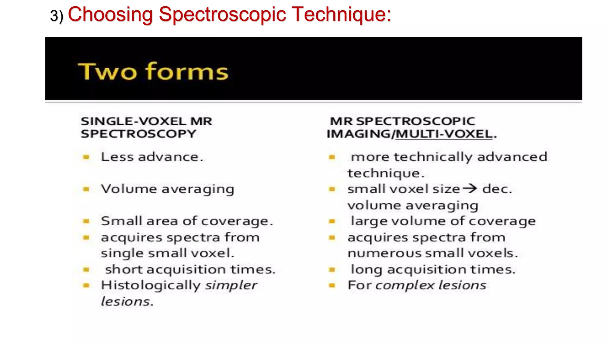
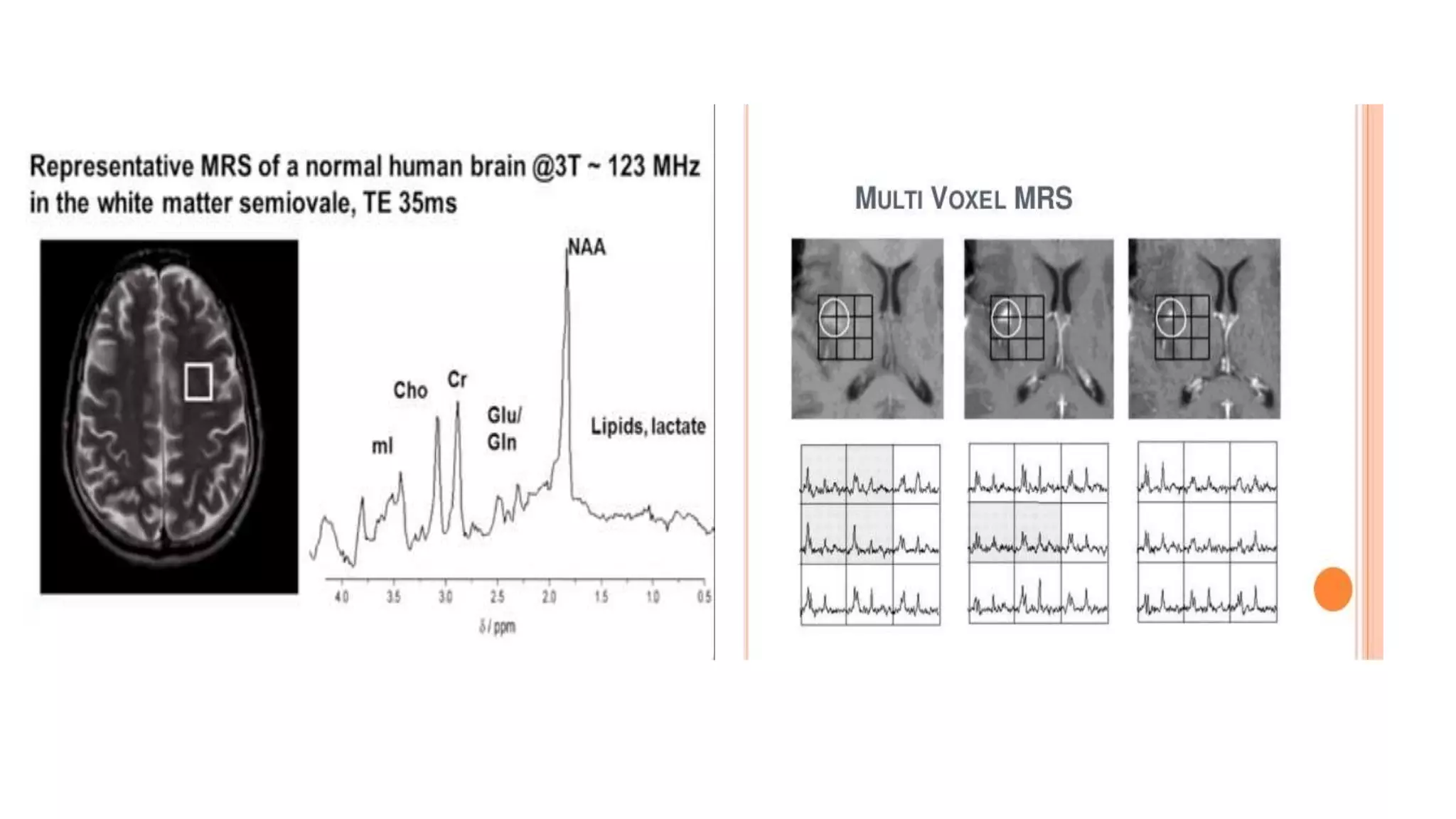
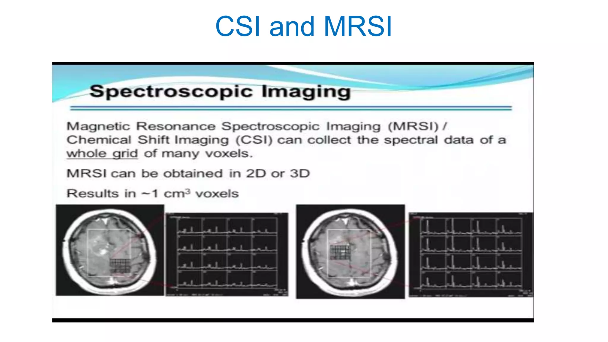
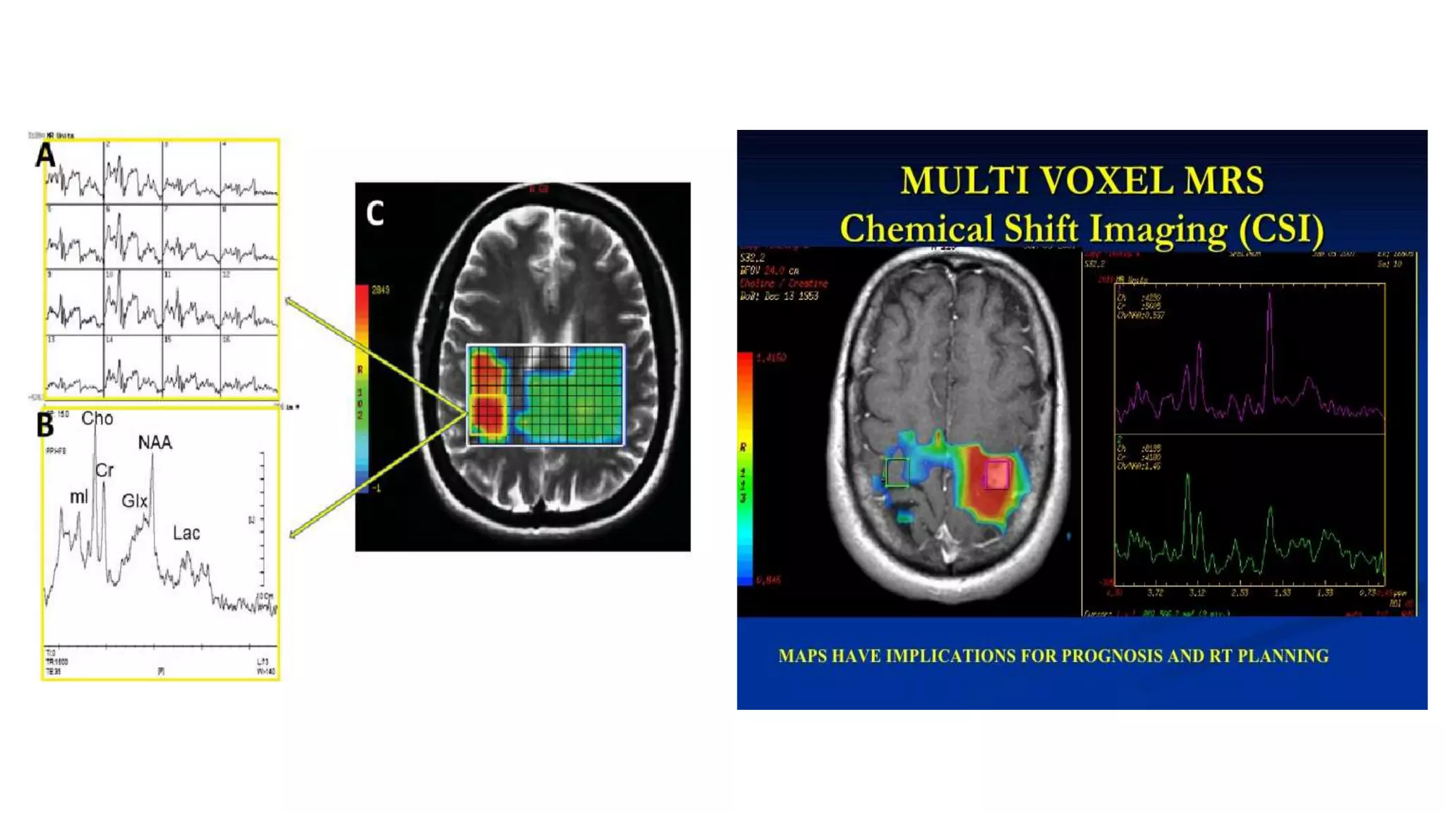
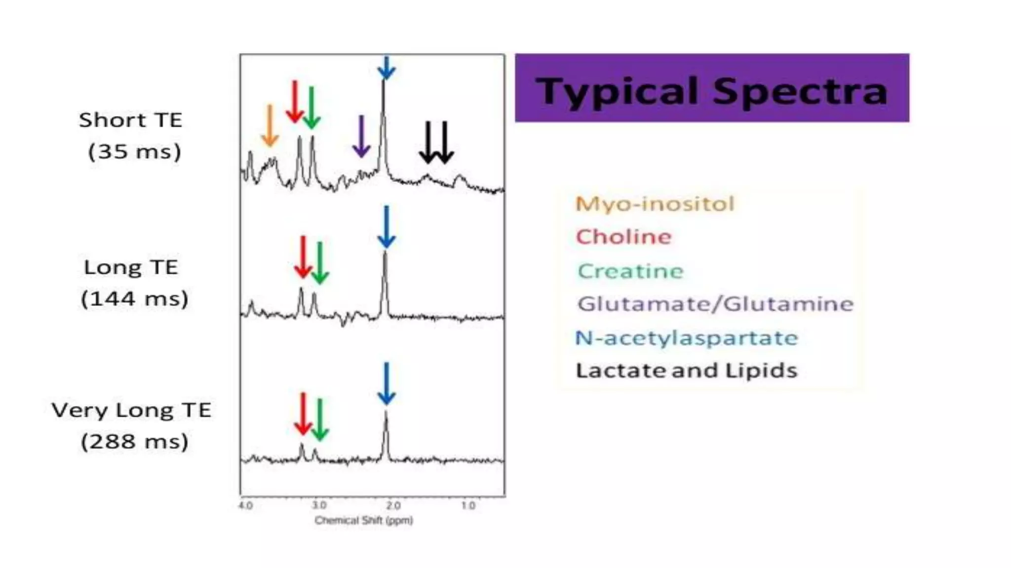
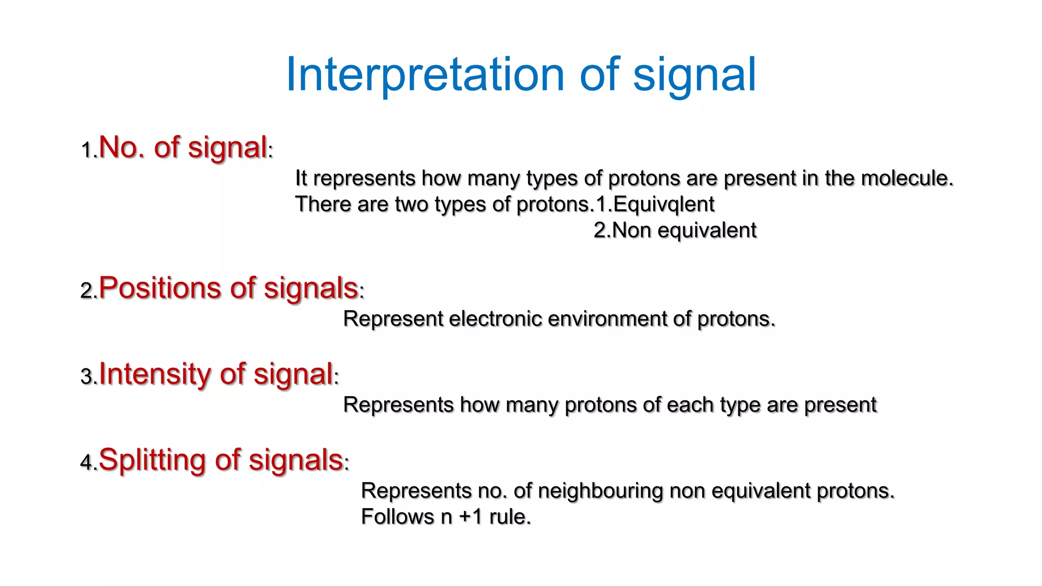
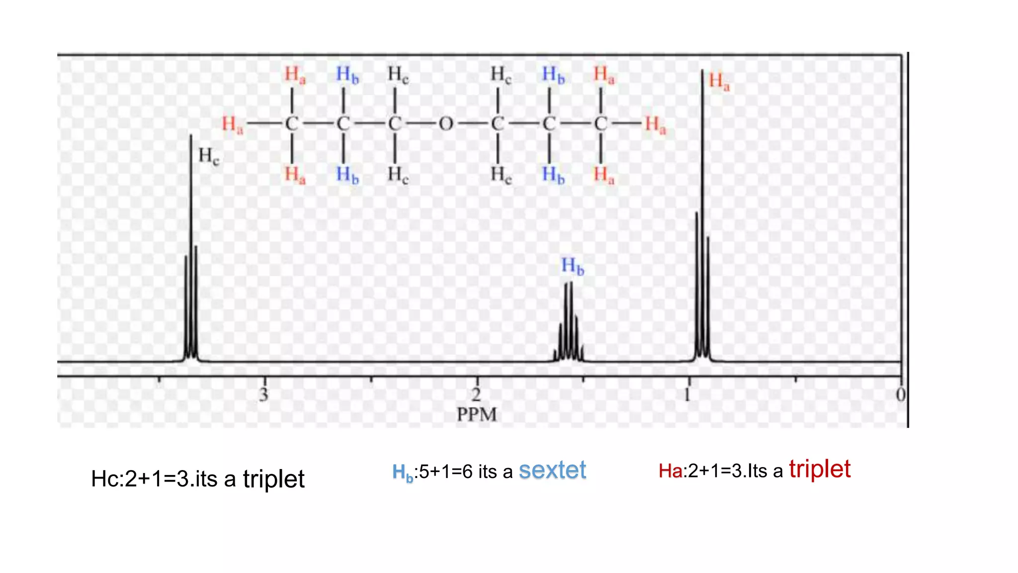
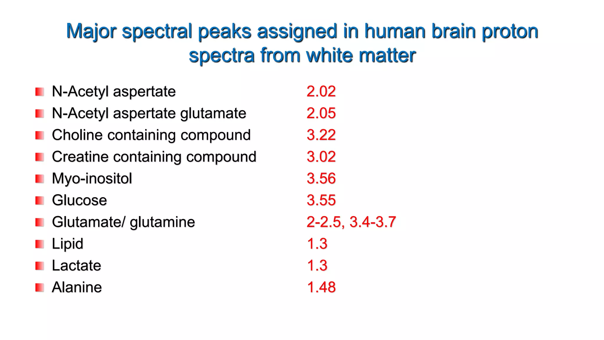
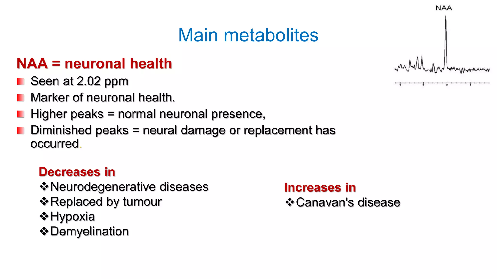
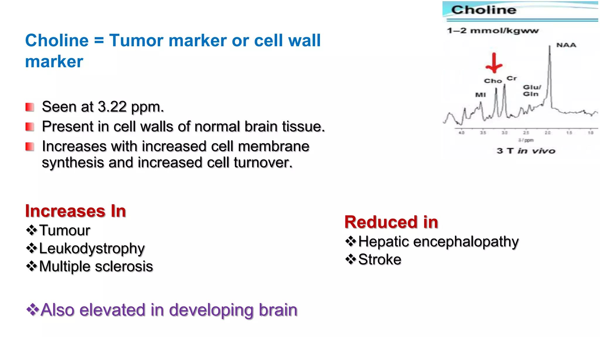
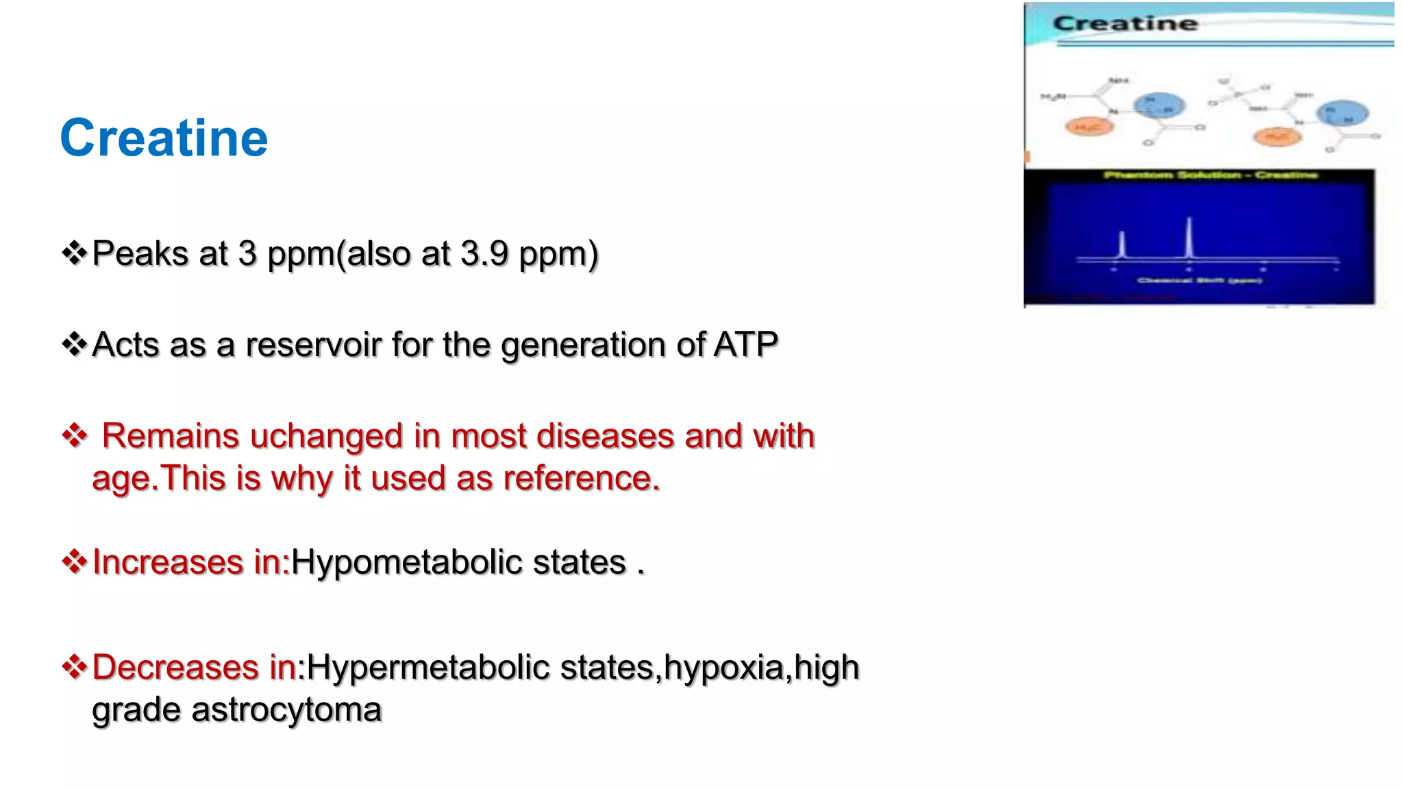
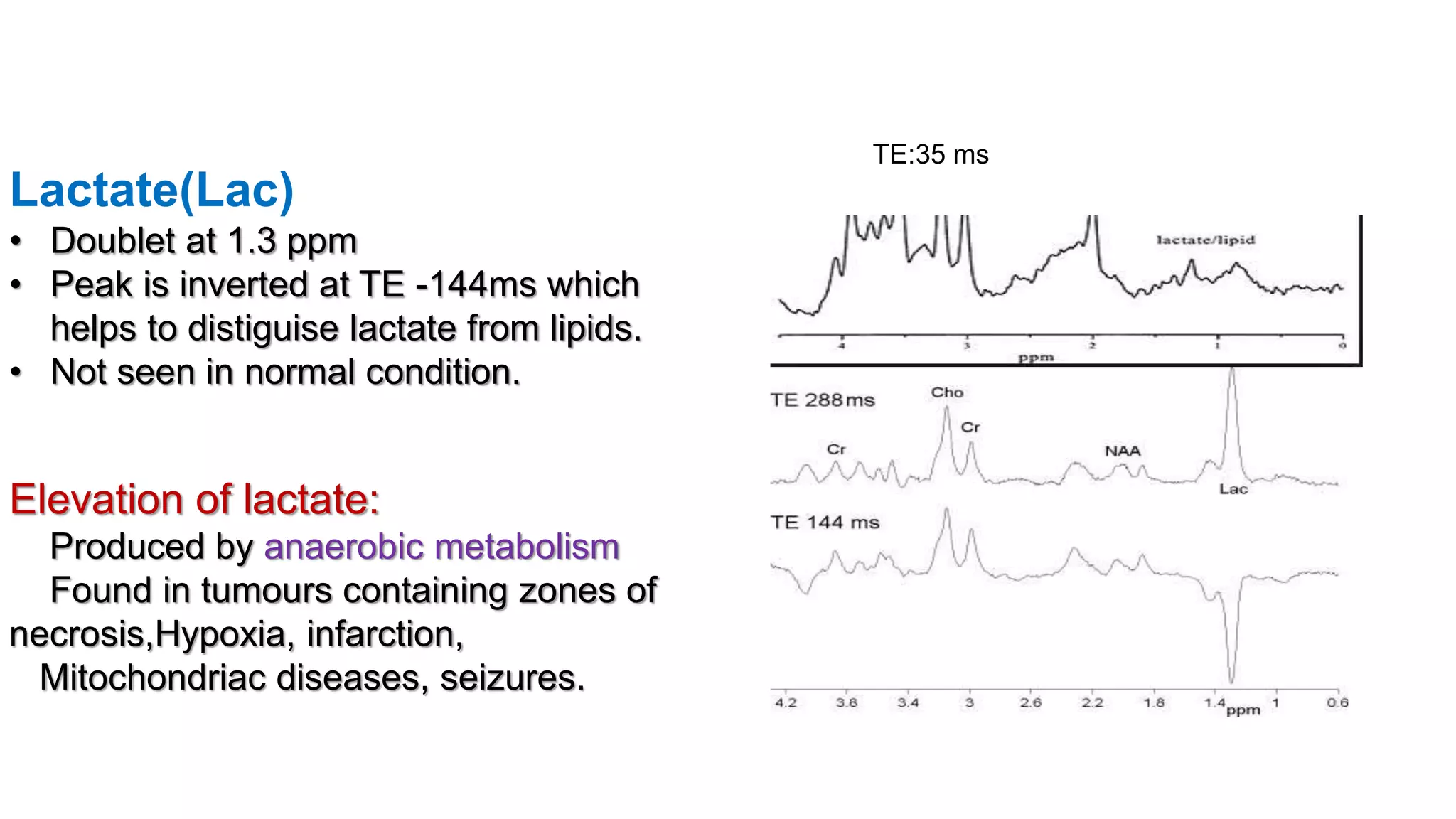
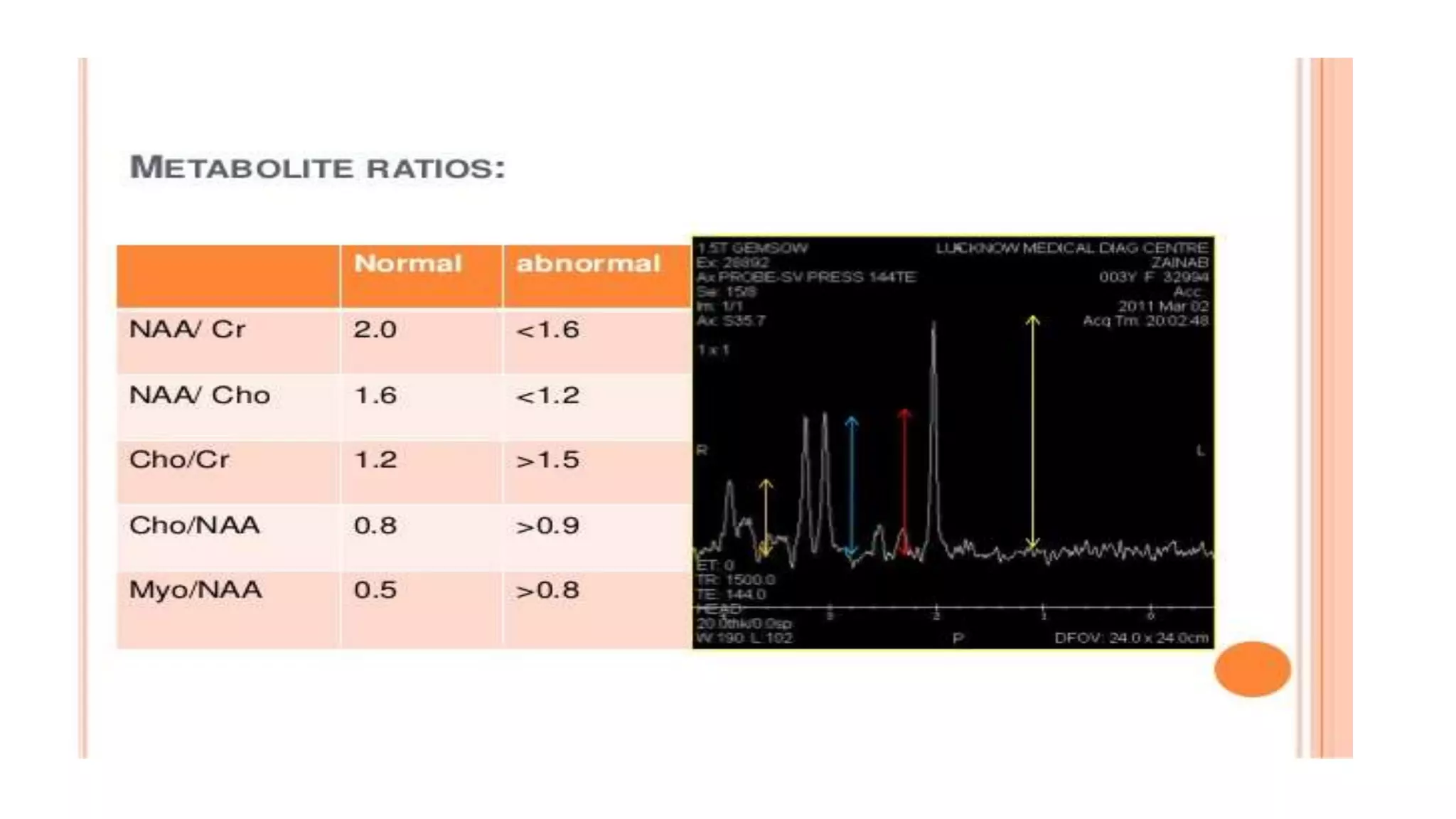
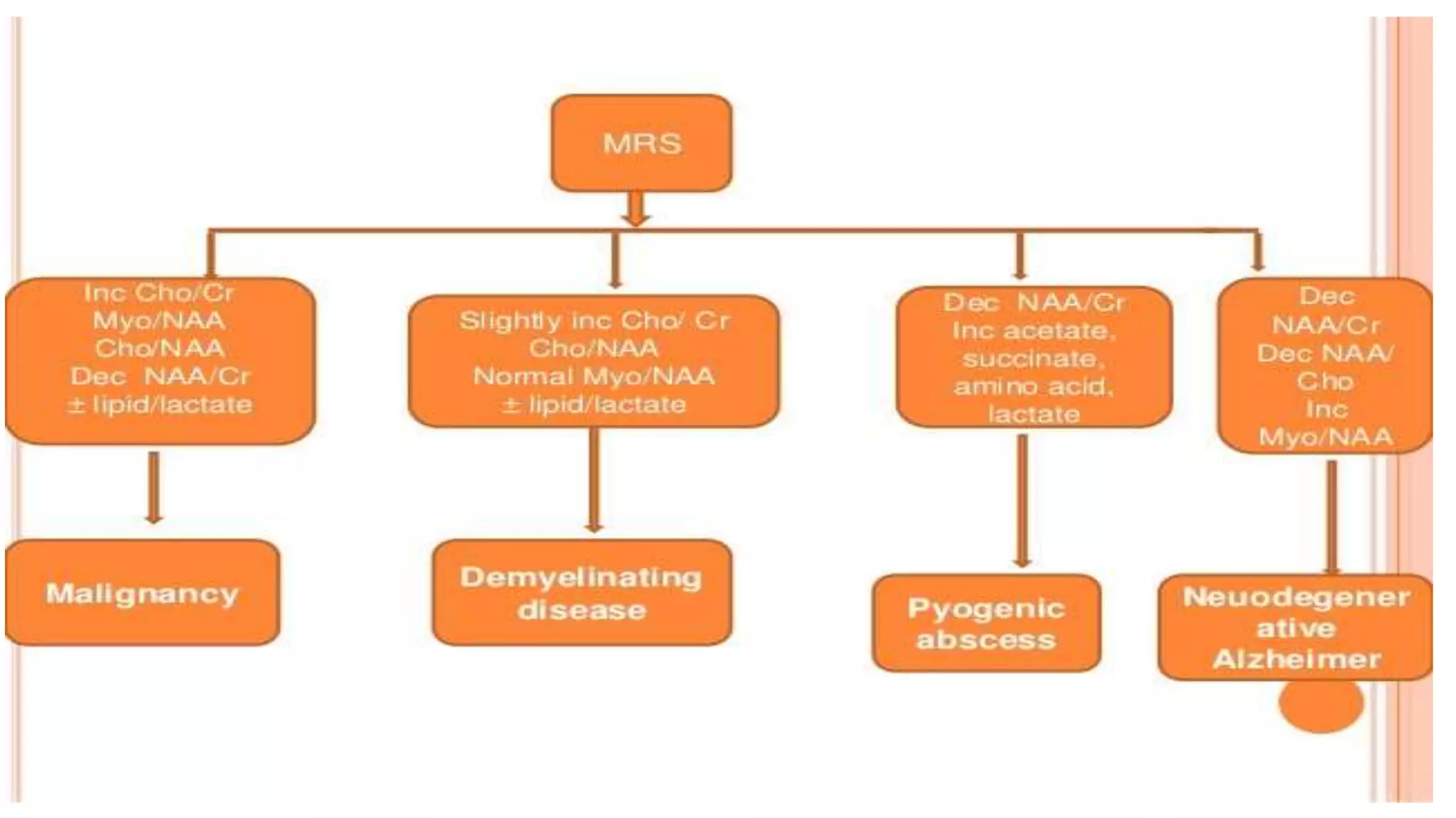
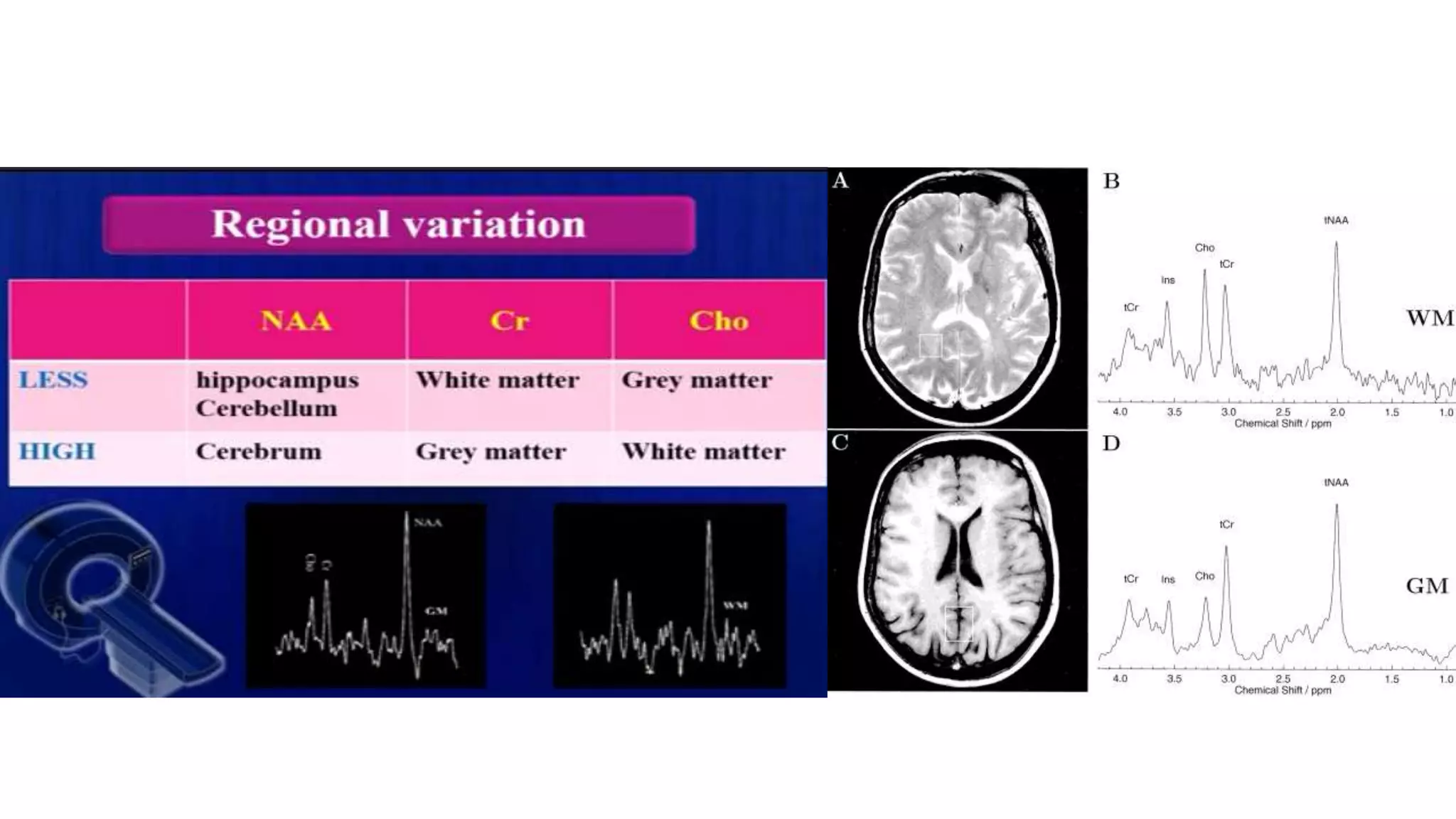
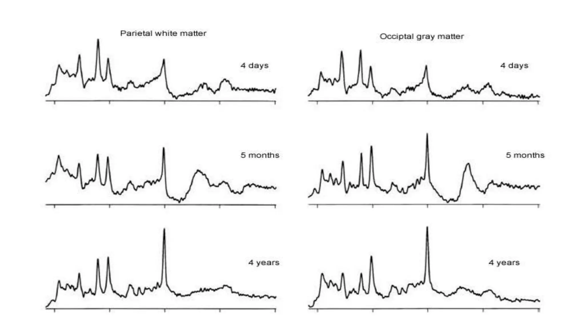
Dr.Md.Tanzilur Rahman
Resident(phase-B)
Radiology and Imaging
What is MRS
MR spectroscopy is the use of magnetic resonance in
quantification of metabolites and the study of their distribution in
different tissues.
Rather than displaying MRI proton signals on a gray scale as an
image, depending on its relative signal strength,MRS displays
the quantities as a spectrum.
The resonance frequency of each metabolites is represented on
a graph and expressed as parts per million(ppm).
History
Got nobel prize in 1952
Basic physics of MRS
Theoretically MRS can be performed with spins or nuclei of
1H,13C,19F,23Na and 31P.
But In present, MRS clinical uses are mainly 1H(Hydrogen) &
31P(Phosphorus) spectroscopy.
Basic physics of MRS
 The aim of MRS itself is to detect small metabolites. To detect
these small metabolites large signal from water protons need
to be suppressed.
Basic physics of MRS
Chemical shift and shielding
It forms the basis of MRS.Protons precess at different frequencies
in different chemical environment.This phenomenon is called
chemical shift.
So,different metabolites give peak at different position because of
chemical shift.
It is expressed as PPM(parts per million).
1 PPM=1millionth of Larmor frequency.
Chemical shift:
→
OR
Resonance
How we measure
X axis represents chemical
shift(frequency/ppm).
Y axis(area under the peak)
represents intensity which is
proportional to concentration of
metabolite/nucleus.
A good quality spectrum should
represent a flat horizontal baseline with
distinct narrow peak
MRS acquisition technique
1) Shimming the magnetic field: this step is taken to correct the
inhomogeneity of the magnetic field by tuning different pulses in the
x, y, and z directions. This step usually automated but can be
performed manually.
2) Suppressing the water signal: Because water molecules contain
hydrogen, and the relative concentration of water to metabolite is
about 10,000:1, the water signal must be suppressed or the
metabolite peaks will not be discernible in the spectra. This is
achieved by adding water suppression pulses.
3) Choosing Spectroscopic Technique:
CSI and MRSI
Spectroscopic technique
Four methods commonly used for localisation in clinical
practice:
STEAM (Stimulated echo acquisition method)- three 90 degree excited
pulse applied along three planes. Short TE (20ms) is used.
PRESS (point resolved spectroscopy)- one 90 degree and two 180
degree pulse are applied along three planes. longer TE (270ms) is used.
ISIS(Image Selective In vivo Spectroscopy):Used in 31P spectroscopy.
CSI(chemical shift Imaging):Used for multi voxel spectrosopy.
Shimming
The process of making magnetic field homogenous is called shimming.
The homogenecity required for MRI is about .5 ppm whereas for MRS it is
about .1 ppm.
Selection of MRS parameters:
TR & TE are important parameters. Improved SNR is obtained at longer TR.
TEs commonly used are 20-30ms,135-145ms & 270ms. At longer TEs
more than 135ms peaks of major brain metabolites are visible.
Long TE- Choline(cho).
Creatine(cr).
N-acetyl aspartate(NAA).
Lactate.
Short TE- Myoinositol.
Glutamate.
Glutamine.
Glycine.
Lipid.
How to obtain quality spectra?
• Appropriate shimming
• Adequate water suppression by CHESS
• Adequate voxel adjustment to avoid
blood, bone, CSF ,cysts and air since susceptibility artifacts
may hamper the expected normal molecular distributions.
Interpretation of signal
1.No. of signal:
It represents how many types of protons are present in the molecule.
There are two types of protons.1.Equivqlent
2.Non equivalent
2.Positions of signals:
Represent electronic environment of protons.
3.Intensity of signal:
Represents how many protons of each type are present
4.Splitting of signals:
Represents no. of neighbouring non equivalent protons.
Follows n +1 rule.
Ha:2+1=3.Its a tripletHb:5+1=6 its a sextetHc:2+1=3.its a triplet
N-Acetyl aspertate 2.02
N-Acetyl aspertate glutamate 2.05
Choline containing compound 3.22
Creatine containing compound 3.02
Myo-inositol 3.56
Glucose 3.55
Glutamate/ glutamine 2-2.5, 3.4-3.7
Lipid 1.3
Lactate 1.3
Alanine 1.48
Major spectral peaks assigned in human brain proton
spectra from white matter
Main metabolites
NAA = neuronal health
Seen at 2.02 ppm
Marker of neuronal health.
Higher peaks = normal neuronal presence,
Diminished peaks = neural damage or replacement has
occurred.
Decreases in
Neurodegenerative diseases
Replaced by tumour
Hypoxia
Demyelination
Increases in
Canavan's disease
Choline = Tumor marker or cell wall
marker
Seen at 3.22 ppm.
Present in cell walls of normal brain tissue.
Increases with increased cell membrane
synthesis and increased cell turnover.
Increases In
Tumour
Leukodystrophy
Multiple sclerosis
Also elevated in developing brain
Reduced in
Hepatic encephalopathy
Stroke
Creatine
Peaks at 3 ppm(also at 3.9 ppm)
Acts as a reservoir for the generation of ATP
 Remains uchanged in most diseases and with
age.This is why it used as reference.
Increases in:Hypometabolic states .
Decreases in:Hypermetabolic states,hypoxia,high
grade astrocytoma
Lactate(Lac)
• Doublet at 1.3 ppm
• Peak is inverted at TE -144ms which
helps to distiguise lactate from lipids.
• Not seen in normal condition.
Elevation of lactate:
Produced by anaerobic metabolism
Found in tumours containing zones of
necrosis,Hypoxia, infarction,
Mitochondriac diseases, seizures.
TE:35 ms
Myo-Inositol(synthesized in glial cells)
 Peaks at 3.5-3.6 ppm.
 Visualised at short TE.
 Considered as glial marker
 Elevation may reflect gliosis.
 Proposed inflammatory marker.
 Important marker in grading gliomas:
Low:in high grade gliomas
High:in low grade gliomas
High in:Gliosis
Alzheimer's disease,
Down syndrome,
DM.
Other metabolites
Neonates Elderly
Cho. and mi is high
Gradual increase in NAA due to
neuronal maturation
After 2 yrs spectrum is identical to
adult
Elevated Cho.
Elevated Cr.
Diminished NAA
Age variation
The three-steps approach to spectral analysis
Step 1: The quality assurance phase. Is it an adequate spectrum?
Step 2: Is Hunter's angle normal?
Step 3: Starting from the right side of the graph, count off the
location and check quantities of The Good (NAA=2.02ppm), The
Bad(Cho=3.22), and The Ugly(LL=0.9-1.33ppm).
Hunter's angle
Neurosurgeon, Hunter Sheldon, at Huntington Medical Research Institutes.
Instead of doing complex ratios and analysis of the spectra, he simply used
his pocket comb. He placed his comb on the spectrum at approximately a 450
angle and connected several of the peaks. If the angle and peaks roughly
corresponded to the 450 angle, the curve was probably normal.
If the peaks strayed off the comb's angle, the curve was abnormal. This is a
quick, useful method to read MRS and determine normal from abnormal.
It is important to remember, however, that this angle was used with STEAM
spectra from the brain.
RECENT ADVANCEMENT
Whole brain MRS analysis Metabolic parametric maps
Breast spectroscopy
GluCEST(Chemical exchange saturaton transfer MRS)
Prostate MRS Liver MRS to see Fat fraction
Reference
 John.R.Haaga:CT and MRI of the whole body
 Jonathan H. Gillard: Clinical MR Neuroimaging
 MRI made easy
 Effect of voxel position on single voxel MRS findings:Peter.E.Ricci at.el
 Radiopaedia.org
Basic physics and normal interpretation of MRS

Basic physics and normal interpretation of MRS

  • 1.
  • 2.
    What is MRS MRspectroscopy is the use of magnetic resonance in quantification of metabolites and the study of their distribution in different tissues. Rather than displaying MRI proton signals on a gray scale as an image, depending on its relative signal strength,MRS displays the quantities as a spectrum. The resonance frequency of each metabolites is represented on a graph and expressed as parts per million(ppm).
  • 3.
  • 4.
  • 7.
    Basic physics ofMRS Theoretically MRS can be performed with spins or nuclei of 1H,13C,19F,23Na and 31P. But In present, MRS clinical uses are mainly 1H(Hydrogen) & 31P(Phosphorus) spectroscopy.
  • 8.
    Basic physics ofMRS  The aim of MRS itself is to detect small metabolites. To detect these small metabolites large signal from water protons need to be suppressed.
  • 10.
  • 11.
    Chemical shift andshielding It forms the basis of MRS.Protons precess at different frequencies in different chemical environment.This phenomenon is called chemical shift. So,different metabolites give peak at different position because of chemical shift. It is expressed as PPM(parts per million). 1 PPM=1millionth of Larmor frequency.
  • 12.
  • 13.
  • 17.
  • 20.
  • 22.
    X axis representschemical shift(frequency/ppm). Y axis(area under the peak) represents intensity which is proportional to concentration of metabolite/nucleus. A good quality spectrum should represent a flat horizontal baseline with distinct narrow peak
  • 23.
    MRS acquisition technique 1)Shimming the magnetic field: this step is taken to correct the inhomogeneity of the magnetic field by tuning different pulses in the x, y, and z directions. This step usually automated but can be performed manually. 2) Suppressing the water signal: Because water molecules contain hydrogen, and the relative concentration of water to metabolite is about 10,000:1, the water signal must be suppressed or the metabolite peaks will not be discernible in the spectra. This is achieved by adding water suppression pulses.
  • 24.
  • 26.
  • 28.
    Spectroscopic technique Four methodscommonly used for localisation in clinical practice: STEAM (Stimulated echo acquisition method)- three 90 degree excited pulse applied along three planes. Short TE (20ms) is used. PRESS (point resolved spectroscopy)- one 90 degree and two 180 degree pulse are applied along three planes. longer TE (270ms) is used. ISIS(Image Selective In vivo Spectroscopy):Used in 31P spectroscopy. CSI(chemical shift Imaging):Used for multi voxel spectrosopy.
  • 29.
    Shimming The process ofmaking magnetic field homogenous is called shimming. The homogenecity required for MRI is about .5 ppm whereas for MRS it is about .1 ppm.
  • 30.
    Selection of MRSparameters: TR & TE are important parameters. Improved SNR is obtained at longer TR. TEs commonly used are 20-30ms,135-145ms & 270ms. At longer TEs more than 135ms peaks of major brain metabolites are visible. Long TE- Choline(cho). Creatine(cr). N-acetyl aspartate(NAA). Lactate. Short TE- Myoinositol. Glutamate. Glutamine. Glycine. Lipid.
  • 32.
    How to obtainquality spectra? • Appropriate shimming • Adequate water suppression by CHESS • Adequate voxel adjustment to avoid blood, bone, CSF ,cysts and air since susceptibility artifacts may hamper the expected normal molecular distributions.
  • 33.
    Interpretation of signal 1.No.of signal: It represents how many types of protons are present in the molecule. There are two types of protons.1.Equivqlent 2.Non equivalent 2.Positions of signals: Represent electronic environment of protons. 3.Intensity of signal: Represents how many protons of each type are present 4.Splitting of signals: Represents no. of neighbouring non equivalent protons. Follows n +1 rule.
  • 34.
    Ha:2+1=3.Its a tripletHb:5+1=6its a sextetHc:2+1=3.its a triplet
  • 35.
    N-Acetyl aspertate 2.02 N-Acetylaspertate glutamate 2.05 Choline containing compound 3.22 Creatine containing compound 3.02 Myo-inositol 3.56 Glucose 3.55 Glutamate/ glutamine 2-2.5, 3.4-3.7 Lipid 1.3 Lactate 1.3 Alanine 1.48 Major spectral peaks assigned in human brain proton spectra from white matter
  • 36.
    Main metabolites NAA =neuronal health Seen at 2.02 ppm Marker of neuronal health. Higher peaks = normal neuronal presence, Diminished peaks = neural damage or replacement has occurred. Decreases in Neurodegenerative diseases Replaced by tumour Hypoxia Demyelination Increases in Canavan's disease
  • 37.
    Choline = Tumormarker or cell wall marker Seen at 3.22 ppm. Present in cell walls of normal brain tissue. Increases with increased cell membrane synthesis and increased cell turnover. Increases In Tumour Leukodystrophy Multiple sclerosis Also elevated in developing brain Reduced in Hepatic encephalopathy Stroke
  • 38.
    Creatine Peaks at 3ppm(also at 3.9 ppm) Acts as a reservoir for the generation of ATP  Remains uchanged in most diseases and with age.This is why it used as reference. Increases in:Hypometabolic states . Decreases in:Hypermetabolic states,hypoxia,high grade astrocytoma
  • 39.
    Lactate(Lac) • Doublet at1.3 ppm • Peak is inverted at TE -144ms which helps to distiguise lactate from lipids. • Not seen in normal condition. Elevation of lactate: Produced by anaerobic metabolism Found in tumours containing zones of necrosis,Hypoxia, infarction, Mitochondriac diseases, seizures. TE:35 ms
  • 41.
    Myo-Inositol(synthesized in glialcells)  Peaks at 3.5-3.6 ppm.  Visualised at short TE.  Considered as glial marker  Elevation may reflect gliosis.  Proposed inflammatory marker.  Important marker in grading gliomas: Low:in high grade gliomas High:in low grade gliomas High in:Gliosis Alzheimer's disease, Down syndrome, DM.
  • 42.
  • 47.
    Neonates Elderly Cho. andmi is high Gradual increase in NAA due to neuronal maturation After 2 yrs spectrum is identical to adult Elevated Cho. Elevated Cr. Diminished NAA Age variation
  • 49.
    The three-steps approachto spectral analysis Step 1: The quality assurance phase. Is it an adequate spectrum? Step 2: Is Hunter's angle normal? Step 3: Starting from the right side of the graph, count off the location and check quantities of The Good (NAA=2.02ppm), The Bad(Cho=3.22), and The Ugly(LL=0.9-1.33ppm).
  • 50.
    Hunter's angle Neurosurgeon, HunterSheldon, at Huntington Medical Research Institutes. Instead of doing complex ratios and analysis of the spectra, he simply used his pocket comb. He placed his comb on the spectrum at approximately a 450 angle and connected several of the peaks. If the angle and peaks roughly corresponded to the 450 angle, the curve was probably normal. If the peaks strayed off the comb's angle, the curve was abnormal. This is a quick, useful method to read MRS and determine normal from abnormal. It is important to remember, however, that this angle was used with STEAM spectra from the brain.
  • 53.
    RECENT ADVANCEMENT Whole brainMRS analysis Metabolic parametric maps
  • 54.
  • 55.
    Prostate MRS LiverMRS to see Fat fraction
  • 58.
    Reference  John.R.Haaga:CT andMRI of the whole body  Jonathan H. Gillard: Clinical MR Neuroimaging  MRI made easy  Effect of voxel position on single voxel MRS findings:Peter.E.Ricci at.el  Radiopaedia.org