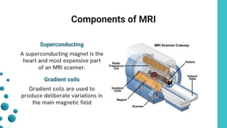
Magnetic Resonance
Imaging ( MRI )
Presented By :
Name : Reduan Ahmad
ID : 232-35-016
Section: A
Batch: 41
Who is the father of MRI ?
Raymond Vahan Damadian
(March 16, 1936 – August 3, 2022) was
an American physician, and inventor of
the first nuclear magnetic resonance
(NMR) scanning machine.
Table of contents
01
04
02
05
03
Introduction How MRI works Components of MRI
Equation Shape of MRI Machine
MRI :
● Big magnet and radio waves
take picture inside our body , no
radiation involved.
● Super clear body pictures, helps
doctors see problems with out
hunting us .
● Secret sauce of MRI , makes
detailed pictures possible .
Introduction
01
How MRI works
• Big magnet
• Radio waves
• Signal
• Picture
A superconducting magnet is the
heart and most expensive part
of an MRI scanner.
Gradient coils are used to
produce deliberate variations in
the main magnetic field
Components of MRI
Superconducting
Gradient coils
Components of MRI
Gradiant coils
• Red coil – Create a varying
magnetic field from left to right .
• Yellow coil – Create a varying
magnetic field from top to bottom.
• Blue coil – Create a varying Magnetic
field from head to toe.
Components of MRI
RF Coils
• RF coils are the “antenna” of the
MRI system
• They are simply a loop of wire
either circular or rectangular .
[Radiofrequency]
Start RF pulses ( Excitation – Protons jump to
higher energy state by absorbing radiation ).
Each proton is rotating
around its axis 63,000,000
rotation per second . The 63
MHz rotation is in the
frequency range called Radio
frequency.
Stop RF pulses (Relaxation – Protons return to their
original state emitting radiation)
Equation
The Larmor equation is given by:
Here,
• ω is how fast spins move (in radians
per second),
• γ is a constant for each atom type
(gyromagnetic ratio),
• B is the magnetic field strength.
ω =γB
Shape of MRI Machine
Closed MRI
Open MRI
Open MRI Closed MRI
Open MRI :
• High field typically 1.5 T – 3
T(Tesla)
• High Image quality
• Fast imaging
• Advance application
Closed MRI :
• Low field typically 0.2 T – 0.4 T (Tesla)
• Low image quality
• Slow imaging
• Limited application
Thank You

Magnetic Resonance Imaging ( MRI ) Presentation

  • 1.
    Magnetic Resonance Imaging (MRI ) Presented By : Name : Reduan Ahmad ID : 232-35-016 Section: A Batch: 41
  • 2.
    Who is thefather of MRI ? Raymond Vahan Damadian (March 16, 1936 – August 3, 2022) was an American physician, and inventor of the first nuclear magnetic resonance (NMR) scanning machine.
  • 3.
    Table of contents 01 04 02 05 03 IntroductionHow MRI works Components of MRI Equation Shape of MRI Machine
  • 4.
    MRI : ● Bigmagnet and radio waves take picture inside our body , no radiation involved. ● Super clear body pictures, helps doctors see problems with out hunting us . ● Secret sauce of MRI , makes detailed pictures possible . Introduction 01
  • 5.
    How MRI works •Big magnet • Radio waves • Signal • Picture
  • 6.
    A superconducting magnetis the heart and most expensive part of an MRI scanner. Gradient coils are used to produce deliberate variations in the main magnetic field Components of MRI Superconducting Gradient coils
  • 7.
    Components of MRI Gradiantcoils • Red coil – Create a varying magnetic field from left to right . • Yellow coil – Create a varying magnetic field from top to bottom. • Blue coil – Create a varying Magnetic field from head to toe.
  • 8.
    Components of MRI RFCoils • RF coils are the “antenna” of the MRI system • They are simply a loop of wire either circular or rectangular . [Radiofrequency]
  • 9.
    Start RF pulses( Excitation – Protons jump to higher energy state by absorbing radiation ). Each proton is rotating around its axis 63,000,000 rotation per second . The 63 MHz rotation is in the frequency range called Radio frequency.
  • 10.
    Stop RF pulses(Relaxation – Protons return to their original state emitting radiation)
  • 11.
    Equation The Larmor equationis given by: Here, • ω is how fast spins move (in radians per second), • γ is a constant for each atom type (gyromagnetic ratio), • B is the magnetic field strength. ω =γB
  • 12.
    Shape of MRIMachine Closed MRI Open MRI
  • 13.
    Open MRI ClosedMRI Open MRI : • High field typically 1.5 T – 3 T(Tesla) • High Image quality • Fast imaging • Advance application Closed MRI : • Low field typically 0.2 T – 0.4 T (Tesla) • Low image quality • Slow imaging • Limited application
  • 14.