Downloaded 255 times

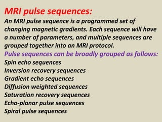
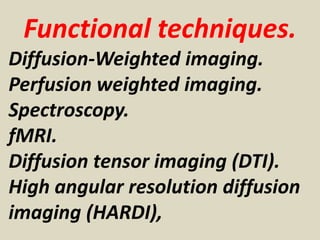
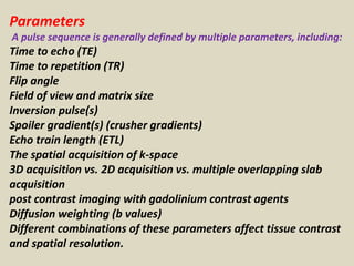


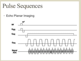


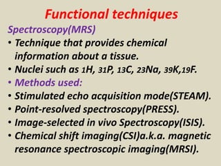

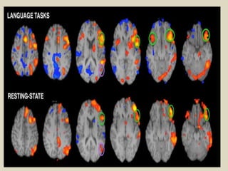
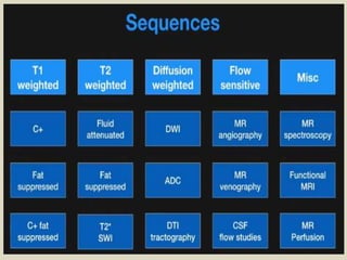


MRI pulse sequences are programmed sets of changing magnetic gradients used to generate images. There are several types of sequences, including spin echo, gradient echo, and inversion recovery sequences. Sequences are defined by parameters like time to echo, time to repetition, and flip angle. Functional techniques like diffusion-weighted imaging, perfusion imaging, and fMRI are used to evaluate brain physiology rather than just anatomy. Echo planar imaging allows for very fast image acquisition, while other sequences like spin echo provide different types of tissue contrast.


