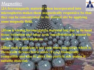
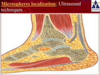
2. DEFINITION:
Magnetic micro carriers are
supramolecular particles that are small enough to circulate
through capillaries without producing embolic occlusion
(<4µm) but are sufficiently susceptible (ferromagnetic) to
become captured in micro vessel and dragged into the
adjacent tissues by magnetic fields of 0.5 to 0.8 tesla (T).
2
3. Advantages:
1. Therapeutic responses in target organs occurs
at only one tenth of free drug dose.
2. Controlled drug release within target tissues
for intervals of 30 min to 30 hr .
3. Avoidance of acute drug toxicity directed
against endothelium and normal parenchyma
cells.
4. Adaptable to any part of the body.
3
4. Disadvantages:
1. Magnetic targeting is an expensive, technical approach and
requires specialized manufacture and quality control system.
2. It needs specialized magnet for targeting, advanced techniques
for monitoring and trained personnel to perform procedures.
3. Magnets must have relatively constant gradients, in order to
avoid focal over-dosing with toxic drugs.
4. A large fraction of the magnetite which is entrapped in carriers
is deposited permanently in target tissues.
5. Due to these limitations magnetic drug targeting is likely to be
approved only for severe diseases.
4
5. Concept of targeting magnetic microspheres:
Microspheres containing magnetic material (magnetite)
are injected into an artery that supplies to a given site.
As the microspheres would be selectively and
magnetically localized at the capillary level they have free
flow access through large arteries.
Thus the microspheres would serve as the time release
capsules systems sitting in the desired location.
A magnet of sufficient field strength is thus placed
externally over the target area to localize the microspheres
at the capillary bed in this region.
www.themegallery.com LOGO
5
6. To localize microspheres in a fast moving arterial
system, greater field strength is required.
When the microspheres are first pushed against the
endothelial cells by the magnetic field, an endocytic
response was triggered with continuous magnetic influence
over certain period of time.
Microspheres migrated from endothelial cells into the
interstitial compartment and formed a depot for sustained
release over an extended period of time.
6
7. Important characteristics:
In targeting using magnetic microspheres, the
magnetite content of carrier and also magnitude of
applied magnetic field is important.
Particle size of drug carrier can affect the degree of
drug entrapment.
If a high magnetic content is incorporated, thus
amount of magnetic fields needed is reduced but the
space available for drug entrapment decreases.
7
8. Drug incorporation and magnetite has to
be delicately balanced.
Optimum magnetite content would be
between 20%-50% of drug weight in the
drug carrier complex.
8
9. Magnetite:
A ferromagnetic material when incorporated into
microspheres makes them magnetically responsive So that
they can be concentrated to the desired site by applying
some magnetic field.
Iron is strong ferromagnetic material but due to its local
tissue irritation and other toxic manifestation it cannot be
included into microspheres.
But such a problem is not seen when magnetite which is
chemically ferrous ferric oxide (Fe3o4) biologically
compatible and also its ultra fine particle size makes it
suitable material.
9
10. Super paramagnetic particles
under the influence of an external
magnetic field
Super paramagnetic particles in
absence of an external magnetic
field, monodisperse particle 10
distribution
11. Magnetic guidance:
Initially drugs were grafted on to the surface of
the magnetic particles, but it suffers from the
drawbacks like very low loading capacity and
irreversible particle aggregation under the
exposure of magnetic field.
Coating of the ferromagnetic particles with
albumin and other charged polymers decreases the
aggregation problem by making it reversible.
11
12. PREPARATION OF MAGNETIC
MICROSPHERES
•Magnetically responsive microspheres can be
prepared by using albumin as a carrier of drug and
magnetite.
•Size of microspheres is kept between 1-2 µm, so
that they can be injected into blood vessels without
problem of thrombo -embolism.
Two methods are employed for the preparation
they are
1. Phase separation emulsion polymerization
2. Continuous solvent evaporation 12
13. CONTINUOUS SOLVENT EVAPORATION
Solution in volatile organic solvent
(polymer + drug + magnet)
Auxillary Solution
Stirring
Homogenization
Stirring temp (22o-30o C)
Magnetic Microsphere
Separated by centrifugation
Freeze drying and storage at 4o C 13
15. Assembly used for separation of magnetic
microsphere from non magnetic materials
15
16. Evaluation of drug release rate in vitro
1. Dialysis method
2. Continuous column elution method
16
17. a. Dialysis methods:
Albumin microspheres were taken in a funnel, 3ml
of phosphate buffer of 7.3 pH was added.
The mouth of the funnel is covered with
cellophane paper and fastened with rubber band.
Then funnel is inverted into a beaker containing
50 ml phosphate buffer. 2.5 ml of aliquots are
withdrawn every half an hour and replaced with
fresh buffer and estimated for drug release. 17
18. b.Continuous column elution methods:
Microspheres are immobilized on a column
containing a fixed weight of glass wool (3.5 gm) as a
support material and kept at 37oC. they are
subjected to a constant flow of 50 ml phosphate
buffer, fractions are collected at equal intervals and
amount of drug release is estimated by using UV
spectroscopy.
18