Prepared by :-
Miss. Sanap Deepali S.
M. Pharm 1st Year
(Sem II).
Roll No.13
Guided By :-
Prof. A. W. Ambekar
M. Pharm
(Pharmaceutics)
Dr. Vithalrao Vikhe Patil Foundation’s
College of Pharmacy
Ahmednagar
(2018-2019)
1
CONTENTS
 Introduction
 Bodies of water
 Properties
 Preparation
 Evaluation
 Advantages
 Applications
 References 2
INTRODUCTION
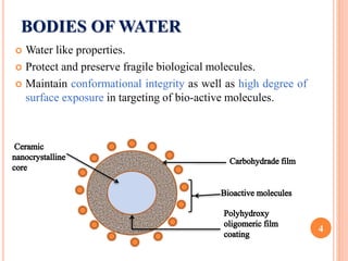
 Aquasomes can be defined as “nanoparticulate carrier
system with three layered self assembled structures
comprising of central solid nanocrystalline core coated
with polyhydroxy oligomers onto which biochemically
active molecules are adsorbed”.
 First discovered by Nir Kossovsky in 1995.
3
BODIES OF WATER
 Water like properties.
 Protect and preserve fragile biological molecules.
 Maintain conformational integrity as well as high degree of
surface exposure in targeting of bio-active molecules.
4
PROPERTIES
 Preserves the conformational integrity of bioactive
substances.
 Deliver their contents through a combination of specific
targeting, molecular shielding and slow sustained release.
 Protects the drug from harsh pH conditions & enzymatic
degradation.
 Exhibit the physical properties of colloids.
 Mechanism controlled by their surface chemistry.
 Degradation can be achieved by monocytes and
osteoclasts. 5
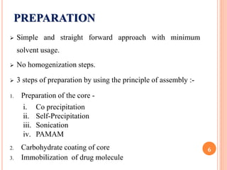
PREPARATION
 Simple and straight forward approach with minimum
solvent usage.
 No homogenization steps.
 3 steps of preparation by using the principle of assembly :-
1. Preparation of the core -
2. Carbohydrate coating of core
3. Immobilization of drug molecule
6
i. Co precipitation
ii. Self-Precipitation
iii. Sonication
iv. PAMAM
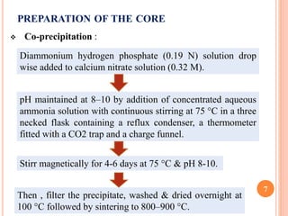
PREPARATION OF THE CORE
 Co-precipitation :
7
Diammonium hydrogen phosphate (0.19 N) solution drop
wise added to calcium nitrate solution (0.32 M).
pH maintained at 8–10 by addition of concentrated aqueous
ammonia solution with continuous stirring at 75 °C in a three
necked flask containing a reflux condenser, a thermometer
fitted with a CO2 trap and a charge funnel.
Stirr magnetically for 4-6 days at 75 °C & pH 8-10.
Then , filter the precipitate, washed & dried overnight at
100 °C followed by sintering to 800–900 °C.
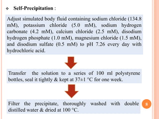
 Self-Precipitation :
8
Adjust simulated body fluid containing sodium chloride (134.8
mM), potassium chloride (5.0 mM), sodium hydrogen
carbonate (4.2 mM), calcium chloride (2.5 mM), disodium
hydrogen phosphate (1.0 mM), magnesium chloride (1.5 mM),
and disodium sulfate (0.5 mM) to pH 7.26 every day with
hydrochloric acid.
Transfer the solution to a series of 100 ml polystyrene
bottles, seal it tightly & kept at 37±1 °C for one week.
Filter the precipitate, thoroughly washed with double
distilled water & dried at 100 °C.
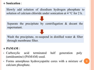
 Sonication :
 PAMAM :
• Carboxylic acid terminated half generation poly
(amidoamine) (PAMAM) used.
• Forms amorphous hydroxyapatite cores with a mixture of
calcium phosphate.
9
Slowly add solution of disodium hydrogen phosphate to
solution of calcium chloride under sonication at 4 °C for 2 h.
Separate the precipitate by centrifugation & decant the
supernatant.
Wash the precipitate, re-suspend in distilled water & filter
through membrane filter.
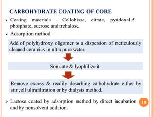
CARBOHYDRATE COATING OF CORE
 Coating materials - Cellobiose, citrate, pyridoxal-5-
phosphate, sucrose and trehalose.
 Adsorption method –
 Lactose coated by adsorption method by direct incubation
and by nonsolvent addition.
10
Add of polyhydroxy oligomer to a dispersion of meticulously
cleaned ceramics in ultra pure water.
Sonicate & lyophilize it.
Remove excess & readily desorbing carbohydrate either by
stir cell ultrafiltration or by dialysis method.
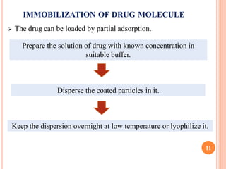
IMMOBILIZATION OF DRUG MOLECULE
 The drug can be loaded by partial adsorption.
11
Prepare the solution of drug with known concentration in
suitable buffer.
Disperse the coated particles in it.
Keep the dispersion overnight at low temperature or lyophilize it.
12
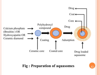
Calcium phosphate
(Brushite ) OR
Hydroxyapatite OR
Ceramic diamond
Drug
AdsorptionCoating
Polyhydroxyl
compound
Ceramic core Coated core Drug loaded
aquasome
Fig : Preparation of aquasomes
Core
Coat
Drug
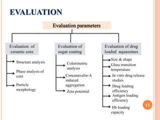
EVALUATION
13
Evaluation of
ceramic core
Evaluation of
sugar coating
Evaluation of drug
loaded aquasomes
Structure analysis
Phase analysis of
core
Particle
morphology
Colorimetric
analysis
Concanavalin-A
induced
aggregation
Zeta potential
Size & shape
Glass transition
temperature
In vitro drug release
studies
Drug loading
efficiency
Antigen loading
efficiency
Hb loading
capacity
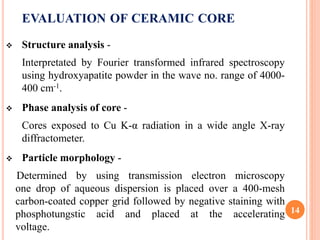
EVALUATION OF CERAMIC CORE
 Structure analysis -
Interpretated by Fourier transformed infrared spectroscopy
using hydroxyapatite powder in the wave no. range of 4000-
400 cm-1.
 Phase analysis of core -
Cores exposed to Cu K-α radiation in a wide angle X-ray
diffractometer.
 Particle morphology -
Determined by using transmission electron microscopy
one drop of aqueous dispersion is placed over a 400-mesh
carbon-coated copper grid followed by negative staining with
phosphotungstic acid and placed at the accelerating
voltage.
14
EVALUATION OF SUGAR COATING
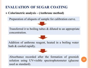
 Colorimetric analysis – (Anthrone method)
15
Preparation of aliquots of sample for calibration curve.
Transferred it to boiling tubes & diluted to an appropriate
concentration.
Addition of anthrone reagent, heated in a boiling water
bath & cooled rapidly.
Absorbance recorded after the formation of greenish
solution using UV-visible spectrophotometer (glucose
used as standard).
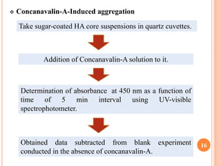
 Concanavalin-A-Induced aggregation
16
Take sugar-coated HA core suspensions in quartz cuvettes.
Addition of Concanavalin-A solution to it.
Determination of absorbance at 450 nm as a function of
time of 5 min interval using UV-visible
spectrophotometer.
Obtained data subtracted from blank experiment
conducted in the absence of concanavalin-A.
EVALUATION OF DRUG LOADED AQUASOMES
 Size & shape –
• Morphological examination - Transmission electron
microscope.
• Mean particle size & size distribution - A photon correlation
spectroscopy using a Autosizer II C apparatus and SEM.
• Chemical composition & crystalline structure - X-ray
powder diffractometry.
 Glass transition temperature –
• carbohydrates and proteins studied by using DSC analyzer.
 In-process stability studies –
• Stability and integrity of protein during the formulation of the
aquasomes determined by using sodium dodecyl
sulfatepolyacrylamide gel electrophoresis (SDS-PAGE).
17
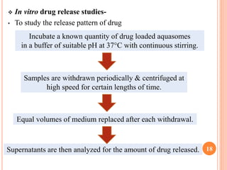
 In vitro drug release studies-
• To study the release pattern of drug
18
Incubate a known quantity of drug loaded aquasomes
in a buffer of suitable pH at 37°C with continuous stirring.
Samples are withdrawn periodically & centrifuged at
high speed for certain lengths of time.
Equal volumes of medium replaced after each withdrawal.
Supernatants are then analyzed for the amount of drug released.
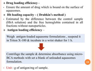
 Drug loading efficiency -
• Ensure the amount of drug which is bound on the surface of
aquasomes.
 Hb loading capacity – ( Drabkin’s method )
• Estimated by the difference between the control sample
(HbA solution) and the free hemoglobin contained in all
fractions without nanoparticles.
 Antigen loading efficiency –
• Unit : g of antigen/mg of sample.
19
Weigh antigen-loaded aquasome formulations , suspend it
in Triton X-100 & incubate in a wrist shaker for 1 h.
Centrifuge the sample & determine absorbance using micro-
BCA methods with set a blank of unloaded aquasomes
formulation.
ADVANTAGES
 Increases therapeutic efficacy of pharmaceutically active
agents.
 Avoid multiple-injection schedule.
 Offer favorable environment for proteins.
 Used for various imaging tests.
 Novel carrier for enzymes such as DNAses and
pigment/dyes.
 Act as a vaccine delivery system.
20
APPLICATION
 Oxygen carrier
 For immunotherapy
 For oral route
 For immunopotentiation
 Anti thrombic activity
 Antigen delivery
 Delivery of poorly soluble drugs
 Enzyme delivery
 Insulin delivery
 Vaccine delivery
21
REFERENCES
 Narang N. Aquasomes: Self-assembled systems for the
delivery of bioactive molecules. Asian Journal of
Pharmaceutics. April-June 2012: 95-100.
 Banerjee S, Sen KK. Aquasomes: A novel
nanoparticulate drug carrier. Journal of Drug Delivery
Science and Technology. 2018: 446–452.
22
23