Download to read offline


![• Trachea
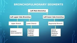
• Right and Left Principal Bronchus
• Lobar Bronchi(Secondary)[2L,3R]
• Segmental Bronchi(Tertiary)[8L,10R]
• Terminal Bronchioles(25000 in no.)
• Respiratory Bronchioles
• Alveolar ducts
• Alveolar sacs
• Alveoli
ACINUS](https://image.slidesharecdn.com/lungs-210927105730/85/Lungs-28-320.jpg)


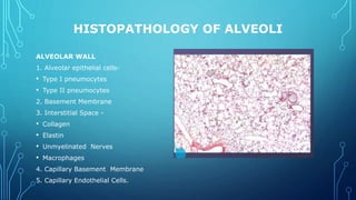
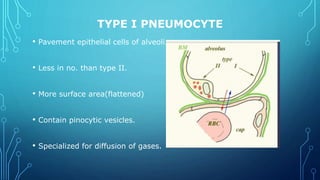


The document provides a detailed anatomical overview of the lungs, including their gross anatomy, surfaces, borders, and internal structures like the hilum, broncho-pulmonary segments, and blood supply. It discusses the histopathology of alveoli, types of pneumocytes, and the function of surfactant in reducing surface tension. Additionally, it highlights clinical significance related to segmental resection and the importance of surfactant in fetal development.


