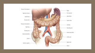
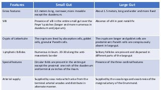
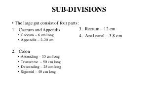
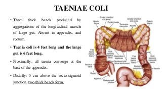
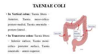
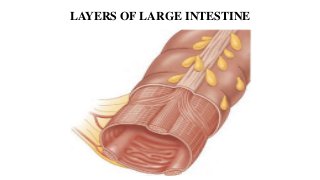
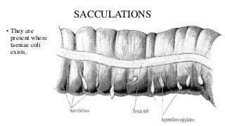
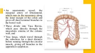
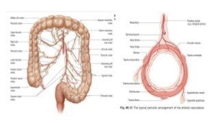
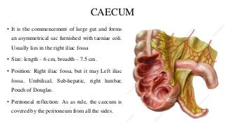
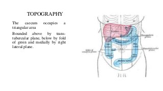
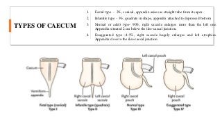
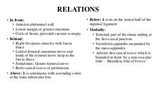
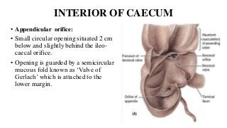
LARGE INTESTINE,
CAECUM AND APPENDIX
DR. SUNDIP CHARMODE
ASSOCIATE PROFESSOR
DEPARTMENT OF ANATOMY
AIIMS RAJKOT
INTRODUCTION
• The large intestine extends from the ileo-caecal junction to the muco-
cutaneous junction of the anal canal (pectinate line).
• In the adult, the large intestine is approximately 1–1.5 m long in vivo,
although there is considerable individual variation.
• It is formed from the distal midgut, all of the hindgut, and the
proctodeum.
DIFFERENCES FROM SMALL INTESTINE
• The large intestine differs from the small intestine in several ways:
1. It has a greater calibre;
2. For much of its course it is more fixed in position;
3. The outer longitudinal muscle layer of the colon is concentrated into
three longitudinal bands, taeniae coli; small fatty projections,
appendices epiploicae, are scattered over its free surface (although
these tend to be absent from the caecum, vermiform appendix and
rectum); and the colonic wall is puckered into sacculations
(haustrations).
Features Small Gut Large Gut
Gross features 6.5 meters long, narrower, more movable
except the duodenum.
About 1.5 meters, long and wider and more fixed
Villi Presence of villi in the entire small gut over the
Peyer’s patches (longer and more numerous in
duodenum and jejunum).
Absence of villi in post natal life
Crypts of Lieberkuhn The crypts are lined by absorptive cells, goblet
cells, granular Paneth cells.
The crypts are longer and goblet cells are
predominant. Paneth cells are conspicuously
absent in large gut.
Lymphatic follicles Numerous in ileum. 20-30 along the anti-
mesenteric border
Solitary follicles are present and dispersed in
different parts of the large gut
Special features Circular folds are present in the entire gut
except the proximal one inch of the duodenum
and terminal six inches of the ileum.
Presence of the three cardinal features
Arterial supply Supplied by vasa recta which arise from the
terminal arterial arcades and distribute in
alternate manner.
Supplied by the vasa longa and vasa brevia of the
marginal artery of the Drummond.
SUB-DIVISIONS
• The large gut consist of four parts:
1. Caecum and Appendix
• Caecum – 6 cm long
• Appendix – 2-20 cm
2. Colon
• Ascending – 15 cm long
• Transverse – 50 cm long
• Descending – 25 cm long
• Sigmoid – 40 cm long
3. Rectum – 12 cm
4. Anal canal – 3.8 cm
CHARACTERISTIC FEATURES
• It presents three cardinal features:
1. Taeniae coli
2. Sacculations
3. Appendices epiploicae
TAENIAE COLI
• Three thick bands produced by
aggregations of the longitudinal muscle
of large gut. Absent in appendix, and
rectum.
• Taenia coli is 4 feet long and the large
gut is 6 feet long.
• Proximally: all taenia converge at the
base of the appendix.
• Distally: 5 cm above the recto-sigmoid
junction, two thick bands form.
TAENIAE COLI
• In Vertical colon: Taenia libera –
Anterior, Taenia meso-colica-
postero-medial, Taenia omentalis –
postero-lateral.
• In Transverse colon: Taenia libera
– Inferior surface, Taenia meso-
colica- posterior surface, Taenia
omentalis – antero-superior.
LAYERS OF LARGE INTESTINE
SACCULATIONS
• They are
present where
taeniae coli
exists.
APPENDICES EPIPLOICAE
• They are peritoneal pouches
containing fat, and are present in
the entire gut except caecum,
appendix and rectum.
• The appendices are more
numerous in the transverse colon,
and sigmoid colon.
• An anastomotic vessel, the
marginal artery of Drummond,
which runs in the mesentery along
the inner margin of the colon and
gives off short terminal branches to
the bowel wall.
• These divide into Vasa Brevia,
which pass directly through the
muscularis externa of the colonic
wall, and
• Vasa longa, which travel through
the subserosa for a short distance
before running through the circular
muscle, giving off branches to the
appendices epiploicae.
FUNCTIONS OF LARGE GUT
1. Absorption
2. Secretion – only Mucin.
3. Reduce acidity
4. Protect from infections
CLINICAL CORRELATES OF LARGE GUT
• Congenital megacolon: Aganglionic megacolon, or Hirschsprung disease, is a
condition of unknown cause that is characterized by the absence of ganglion
cells and normal nerve fibres from the distal (or lower) 3 to 40 cm (1 to 16
inches) of the large intestine.
• Volvulus: Loop of intestine twists around itself and the mesentery that supports
it, resulting in a bowel obstruction.
• Intussusception: One segment of intestine "telescopes" inside of another,
causing an intestinal obstruction.
CAECUM
• It is the commencement of large gut and forms
an asymmetrical sac furnished with taeniae coli.
Usually lies in the right iliac fossa
• Size: length – 6 cm, breadth – 7.5 cm.
• Position: Right iliac fossa, but it may Left iliac
fossa, Umbilical, Sub-hepatic, right lumbar,
Pouch of Douglas.
• Peritoneal reflection: As as rule, the caecum is
covered by the peritoneum from all the sides.
TOPOGRAPHY
The caecum occupies a
triangular area
Bounded above by trans-
tubercular plane, below by fold
of groin and medially by right
lateral plane.
TYPES OF CAECUM
1. Foetal type - 2%, conical, appendix arises as straight tube from its apex
2. Infantile type – 3%, quadrate in shape, appendix attached to depressed bottom
3. Normal or adult type- 90%, right saccule enlarges more than the left one.
Appendix situated 2 cm below the ileo-caecal junction.
4. Exaggerated type -4-5%, right saccule hugely enlarges and left atrophies.
Appendix close to the ileo-caecal junction.
RELATIONS
• In front:
• Anterior abdominal wall
• Lower margin of greater omentum
• Coils of ileum, provided caecum is empty
• Behind:
• Right ilio-psoas muscles with fascia
iliaca
• Lateral femoral cutaneous nerve and
trunk of the femoral nerve deep to the
fascia iliaca
• Sometimes, Genito-femoral nerve
• Retro-caecal recess of peritoneum
• Above: It is continuous with ascending colon
at the trans-tubercular line.
• Below: It rests on the lateral half of the
inguinal ligament
• Medially:
• Terminal part of the ileum ending at
the ileo-caecal junction
• Vermiform appendix suspended by
the meso-appendix
• inferior ileo-caecal recess which is
bounded in front by a non-vascular
fold – ‘Bloodless fold of Treves’
BLOOD SUPPLY
• It is supplied by Anterior and Posterior Caecal branches of Inferior
division of ileo-colic artery.
• Anterior branch reaches the caecum via the superior ileo-caecal fold.
• Posterior branch supplies the base of the appendix.
• Venous drainage: Veins drain into the ileo-colic vein and hence into
the superior mesenteric vein (portal system)
LYMPHATIC DRAINAGE AND NERVE
SUPPLY
• The vessels drain into ileo-colic lymph nodes and finally into superior
mesenteric group of pre-aortic lymph nodes
• The sympathetic fibers are derived from the superior mesenteric
plexus and the pre-ganglionic fibers come from T10 to L1 segment of
spinal cord.
• The para-sympathetic fibers are derived from both vagus nerves.
INTERIOR OF CAECUM
• Ileo-caecal orifice:
• Situated in the postero-medial wall of
caecum at its junction with the ascending
colon.
• Guarded with a valve with upper and
lower lips
• Upper lip- horizontal, lower lip –
concave upwards. Frenula.
• Each lip is formed by reduplication of
mucous membrane containing
submucous tissue and thickening of the
circular muscle with an inward
projection of longitudinal muscle of
ileum.
INTERIOR OF CAECUM
• Ileo-caecal orifice:
• The lip presents ileal and caecal
surfaces.
• The ileal surface –villi; caecal surface
– no villi.
• Ileo-caecal orifice – 2.5 cm transverse
diameter.
• Regulates the flow of contents from
ileum to caecum ad prevents
regurgitation.
INTERIOR OF CAECUM
• Appendicular orifice:
• Small circular opening situated 2 cm
below and slightly behind the ileo-
caecal orifice.
• Opening is guarded by a semicircular
mucous fold known as ‘Valve of
Gerlach’ which is attached to the
lower margin.
CLINICAL CORRELATES OF CAECUM
• Acts a guide in the operation of intestinal obstruction.
• If caecum is distended, obstruction is in the large intestine.
• If caecum is empty/not-distended, obstruction is in the small intestine.
• Intussusception: Telescopically invagination of terminal part of ileum
into caecum or ascending colon produces intestinal obstruction.
APPENDIX
• It is a narrow worm like tubular diverticulum arising from the postero-
medial wall of the caecum about 2 cm below the ileo-caecal junction.
• It is suspended by a peritoneal fold – Meso-appendix.
• Appendix is devoid of taeniae coli, sacculations and appendices
epiploicae.
MEASUREMENTS
• Length – 2 cm to 20 cm. average – 9 cm.
• Length increases in young adults and diminishes after mid adulty life.
PRESENTING PARTS
1. Base – attached to postero-medial wall of caecum about 2 cm below
the ileo-caecal junction.
2. Body – narrow, tubular ad contains a canal which opens into the
caecum. The caecal opening is guarded by an incomplete mucous
fold – ‘The valve of Gerlach’
3. Tip
4. Meso-appendix
PRESENTING PARTS
1. Base – attached to postero-medial wall of caecum about 2 cm below
the ileo-caecal junction.
2. Body – narrow, tubular ad contains a canal which opens into the
caecum. The caecal opening is guarded by an incomplete mucous
fold – ‘The valve of Gerlach’
3. Tip – least vascular and is directed in various positions.
VARIOUS POSITIONS OF APPENDIX
1. Sub-caecal and Paracolic – 11
o'clock
2. Retro-caecal and Retro-colic –
commonest type – 12 o'clock
3. Splenic type – 1-2%; pre-ileal and
post-ileal variety – 2 o'clock
4. Promontoric type - 3 o'clock
5. Pelvic type - 4 o'clock
6. Mid-inguinal type - 6 o'clock
7. Ectopic type – due to error in
rotation.
BLOOD SUPPLY
• Appendicular artery – branch of inferior
division of ileo-colic artery
• The artery passes behind the terminal part of
the ileum, supplies the entire organ and
provides a recurrent branch towards the
base where it anastomoses with the posterior
caecal artery.
• It is usually an end artery.
• The vein corresponds to the artery and drains
into superior mesenteric vein.
LYMPHATIC DRAINAGE AND NERVE
SUPPLY
• Lymphatics drain into superior mesenteric lymph nodes via ileo-colic lymph
nodes.
• The Parasympathetic nerves are derived from both vagus.
• The Sympathetic nerves – derived from superior mesenteric plexus – pre-
ganglionic fibers come from – T10 segment of spinal cord.
• Inflammation of appendix, the pain is sometimes referred to umbilical
region along the distribution of dermatomes of T10 spinal cord.
CLINICAL CORRELATES OF APPENDIX
• Inflammation of appendix – Appendicitis
• Pain, vomiting, temperature.
• Pain first felt in umbilical region, then settles in right iliac region due
to local peritonitis. Associated with tenderness and rigidity.
• Anatomical factors producing the inflammation of appendix are:
1. Appendix is a blind tube and may be obstructed by a faecolith –
precipitate the attack of appendicitis
2. Supplied by an end artery
3. Presence of hiatus muscularis
4. Presence of numerous lymphatic follicles in submucous coat.
THANK YOU

Large intestine , Caecum and Appendix.pptx

  • 1.
    LARGE INTESTINE, CAECUM ANDAPPENDIX DR. SUNDIP CHARMODE ASSOCIATE PROFESSOR DEPARTMENT OF ANATOMY AIIMS RAJKOT
  • 3.
    INTRODUCTION • The largeintestine extends from the ileo-caecal junction to the muco- cutaneous junction of the anal canal (pectinate line). • In the adult, the large intestine is approximately 1–1.5 m long in vivo, although there is considerable individual variation. • It is formed from the distal midgut, all of the hindgut, and the proctodeum.
  • 4.
    DIFFERENCES FROM SMALLINTESTINE • The large intestine differs from the small intestine in several ways: 1. It has a greater calibre; 2. For much of its course it is more fixed in position; 3. The outer longitudinal muscle layer of the colon is concentrated into three longitudinal bands, taeniae coli; small fatty projections, appendices epiploicae, are scattered over its free surface (although these tend to be absent from the caecum, vermiform appendix and rectum); and the colonic wall is puckered into sacculations (haustrations).
  • 5.
    Features Small GutLarge Gut Gross features 6.5 meters long, narrower, more movable except the duodenum. About 1.5 meters, long and wider and more fixed Villi Presence of villi in the entire small gut over the Peyer’s patches (longer and more numerous in duodenum and jejunum). Absence of villi in post natal life Crypts of Lieberkuhn The crypts are lined by absorptive cells, goblet cells, granular Paneth cells. The crypts are longer and goblet cells are predominant. Paneth cells are conspicuously absent in large gut. Lymphatic follicles Numerous in ileum. 20-30 along the anti- mesenteric border Solitary follicles are present and dispersed in different parts of the large gut Special features Circular folds are present in the entire gut except the proximal one inch of the duodenum and terminal six inches of the ileum. Presence of the three cardinal features Arterial supply Supplied by vasa recta which arise from the terminal arterial arcades and distribute in alternate manner. Supplied by the vasa longa and vasa brevia of the marginal artery of the Drummond.
  • 6.
    SUB-DIVISIONS • The largegut consist of four parts: 1. Caecum and Appendix • Caecum – 6 cm long • Appendix – 2-20 cm 2. Colon • Ascending – 15 cm long • Transverse – 50 cm long • Descending – 25 cm long • Sigmoid – 40 cm long 3. Rectum – 12 cm 4. Anal canal – 3.8 cm
  • 7.
    CHARACTERISTIC FEATURES • Itpresents three cardinal features: 1. Taeniae coli 2. Sacculations 3. Appendices epiploicae
  • 8.
    TAENIAE COLI • Threethick bands produced by aggregations of the longitudinal muscle of large gut. Absent in appendix, and rectum. • Taenia coli is 4 feet long and the large gut is 6 feet long. • Proximally: all taenia converge at the base of the appendix. • Distally: 5 cm above the recto-sigmoid junction, two thick bands form.
  • 9.
    TAENIAE COLI • InVertical colon: Taenia libera – Anterior, Taenia meso-colica- postero-medial, Taenia omentalis – postero-lateral. • In Transverse colon: Taenia libera – Inferior surface, Taenia meso- colica- posterior surface, Taenia omentalis – antero-superior.
  • 10.
    LAYERS OF LARGEINTESTINE
  • 11.
    SACCULATIONS • They are presentwhere taeniae coli exists.
  • 12.
    APPENDICES EPIPLOICAE • Theyare peritoneal pouches containing fat, and are present in the entire gut except caecum, appendix and rectum. • The appendices are more numerous in the transverse colon, and sigmoid colon.
  • 13.
    • An anastomoticvessel, the marginal artery of Drummond, which runs in the mesentery along the inner margin of the colon and gives off short terminal branches to the bowel wall. • These divide into Vasa Brevia, which pass directly through the muscularis externa of the colonic wall, and • Vasa longa, which travel through the subserosa for a short distance before running through the circular muscle, giving off branches to the appendices epiploicae.
  • 15.
    FUNCTIONS OF LARGEGUT 1. Absorption 2. Secretion – only Mucin. 3. Reduce acidity 4. Protect from infections
  • 16.
    CLINICAL CORRELATES OFLARGE GUT • Congenital megacolon: Aganglionic megacolon, or Hirschsprung disease, is a condition of unknown cause that is characterized by the absence of ganglion cells and normal nerve fibres from the distal (or lower) 3 to 40 cm (1 to 16 inches) of the large intestine. • Volvulus: Loop of intestine twists around itself and the mesentery that supports it, resulting in a bowel obstruction. • Intussusception: One segment of intestine "telescopes" inside of another, causing an intestinal obstruction.
  • 18.
    CAECUM • It isthe commencement of large gut and forms an asymmetrical sac furnished with taeniae coli. Usually lies in the right iliac fossa • Size: length – 6 cm, breadth – 7.5 cm. • Position: Right iliac fossa, but it may Left iliac fossa, Umbilical, Sub-hepatic, right lumbar, Pouch of Douglas. • Peritoneal reflection: As as rule, the caecum is covered by the peritoneum from all the sides.
  • 19.
    TOPOGRAPHY The caecum occupiesa triangular area Bounded above by trans- tubercular plane, below by fold of groin and medially by right lateral plane.
  • 20.
    TYPES OF CAECUM 1.Foetal type - 2%, conical, appendix arises as straight tube from its apex 2. Infantile type – 3%, quadrate in shape, appendix attached to depressed bottom 3. Normal or adult type- 90%, right saccule enlarges more than the left one. Appendix situated 2 cm below the ileo-caecal junction. 4. Exaggerated type -4-5%, right saccule hugely enlarges and left atrophies. Appendix close to the ileo-caecal junction.
  • 21.
    RELATIONS • In front: •Anterior abdominal wall • Lower margin of greater omentum • Coils of ileum, provided caecum is empty • Behind: • Right ilio-psoas muscles with fascia iliaca • Lateral femoral cutaneous nerve and trunk of the femoral nerve deep to the fascia iliaca • Sometimes, Genito-femoral nerve • Retro-caecal recess of peritoneum • Above: It is continuous with ascending colon at the trans-tubercular line. • Below: It rests on the lateral half of the inguinal ligament • Medially: • Terminal part of the ileum ending at the ileo-caecal junction • Vermiform appendix suspended by the meso-appendix • inferior ileo-caecal recess which is bounded in front by a non-vascular fold – ‘Bloodless fold of Treves’
  • 22.
    BLOOD SUPPLY • Itis supplied by Anterior and Posterior Caecal branches of Inferior division of ileo-colic artery. • Anterior branch reaches the caecum via the superior ileo-caecal fold. • Posterior branch supplies the base of the appendix. • Venous drainage: Veins drain into the ileo-colic vein and hence into the superior mesenteric vein (portal system)
  • 23.
    LYMPHATIC DRAINAGE ANDNERVE SUPPLY • The vessels drain into ileo-colic lymph nodes and finally into superior mesenteric group of pre-aortic lymph nodes • The sympathetic fibers are derived from the superior mesenteric plexus and the pre-ganglionic fibers come from T10 to L1 segment of spinal cord. • The para-sympathetic fibers are derived from both vagus nerves.
  • 24.
    INTERIOR OF CAECUM •Ileo-caecal orifice: • Situated in the postero-medial wall of caecum at its junction with the ascending colon. • Guarded with a valve with upper and lower lips • Upper lip- horizontal, lower lip – concave upwards. Frenula. • Each lip is formed by reduplication of mucous membrane containing submucous tissue and thickening of the circular muscle with an inward projection of longitudinal muscle of ileum.
  • 25.
    INTERIOR OF CAECUM •Ileo-caecal orifice: • The lip presents ileal and caecal surfaces. • The ileal surface –villi; caecal surface – no villi. • Ileo-caecal orifice – 2.5 cm transverse diameter. • Regulates the flow of contents from ileum to caecum ad prevents regurgitation.
  • 26.
    INTERIOR OF CAECUM •Appendicular orifice: • Small circular opening situated 2 cm below and slightly behind the ileo- caecal orifice. • Opening is guarded by a semicircular mucous fold known as ‘Valve of Gerlach’ which is attached to the lower margin.
  • 27.
    CLINICAL CORRELATES OFCAECUM • Acts a guide in the operation of intestinal obstruction. • If caecum is distended, obstruction is in the large intestine. • If caecum is empty/not-distended, obstruction is in the small intestine. • Intussusception: Telescopically invagination of terminal part of ileum into caecum or ascending colon produces intestinal obstruction.
  • 28.
    APPENDIX • It isa narrow worm like tubular diverticulum arising from the postero- medial wall of the caecum about 2 cm below the ileo-caecal junction. • It is suspended by a peritoneal fold – Meso-appendix. • Appendix is devoid of taeniae coli, sacculations and appendices epiploicae.
  • 29.
    MEASUREMENTS • Length –2 cm to 20 cm. average – 9 cm. • Length increases in young adults and diminishes after mid adulty life.
  • 30.
    PRESENTING PARTS 1. Base– attached to postero-medial wall of caecum about 2 cm below the ileo-caecal junction. 2. Body – narrow, tubular ad contains a canal which opens into the caecum. The caecal opening is guarded by an incomplete mucous fold – ‘The valve of Gerlach’ 3. Tip 4. Meso-appendix
  • 31.
    PRESENTING PARTS 1. Base– attached to postero-medial wall of caecum about 2 cm below the ileo-caecal junction. 2. Body – narrow, tubular ad contains a canal which opens into the caecum. The caecal opening is guarded by an incomplete mucous fold – ‘The valve of Gerlach’ 3. Tip – least vascular and is directed in various positions.
  • 32.
    VARIOUS POSITIONS OFAPPENDIX 1. Sub-caecal and Paracolic – 11 o'clock 2. Retro-caecal and Retro-colic – commonest type – 12 o'clock 3. Splenic type – 1-2%; pre-ileal and post-ileal variety – 2 o'clock 4. Promontoric type - 3 o'clock 5. Pelvic type - 4 o'clock 6. Mid-inguinal type - 6 o'clock 7. Ectopic type – due to error in rotation.
  • 33.
    BLOOD SUPPLY • Appendicularartery – branch of inferior division of ileo-colic artery • The artery passes behind the terminal part of the ileum, supplies the entire organ and provides a recurrent branch towards the base where it anastomoses with the posterior caecal artery. • It is usually an end artery. • The vein corresponds to the artery and drains into superior mesenteric vein.
  • 34.
    LYMPHATIC DRAINAGE ANDNERVE SUPPLY • Lymphatics drain into superior mesenteric lymph nodes via ileo-colic lymph nodes. • The Parasympathetic nerves are derived from both vagus. • The Sympathetic nerves – derived from superior mesenteric plexus – pre- ganglionic fibers come from – T10 segment of spinal cord. • Inflammation of appendix, the pain is sometimes referred to umbilical region along the distribution of dermatomes of T10 spinal cord.
  • 35.
    CLINICAL CORRELATES OFAPPENDIX • Inflammation of appendix – Appendicitis • Pain, vomiting, temperature. • Pain first felt in umbilical region, then settles in right iliac region due to local peritonitis. Associated with tenderness and rigidity. • Anatomical factors producing the inflammation of appendix are: 1. Appendix is a blind tube and may be obstructed by a faecolith – precipitate the attack of appendicitis 2. Supplied by an end artery 3. Presence of hiatus muscularis 4. Presence of numerous lymphatic follicles in submucous coat.
  • 36.