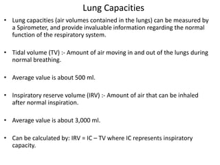
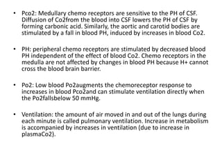
Download to read offline

![Lungs
• Cone – shaped organs located in the thoracic cavity.
• Thoracic cavity is lined with a body membrane called parietal pleura, while
the surface of lungs is covered with visceral pleura.
• The thin space between the two pleural membranes is called pleural cavity
which is filled with a clear fluid called plural fluid to minimize friction
between the tissues and to provide surface tension in the pleural cavity.
[water molecules in the pleural fluid allow the two pleural membranes to
adhere to one another, to prevent collapsing of the lungs].
• A chemical substance called surfactant secreted by the lungs also facilitate
the surface tension.](https://image.slidesharecdn.com/therespiratorysystem-210927080014/85/The-respiratory-system-4-320.jpg)
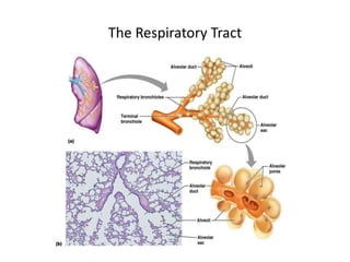
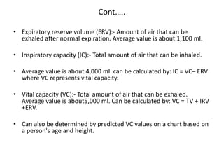
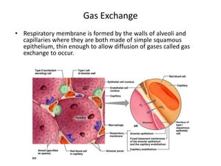
![The Bronchial tree
• tree – like branching tubes extended from the trachea. Only the primary
bronchi are external to the lungs, while the rest of the bronchial tree is
embedded in lung tissues.
• diameters of the tubes from primary bronchi to tertiary bronchi are large,
so that support with cartilage rings is necessary.
• diameter at the bronchioles is down to 1 mm where the tubes do not
need cartilage rings for support. This structure is composed of cuboidal
cells where diffusion is also not possible.
• from the alveolar duct to the alveoli, the lining tissue becomes simple
squamous epithelium where gas exchange is possible. Since there is a
much larger surface area at the alveoli, almost all gas exchange occurs at
the alveoli [300 million alveoli provide a total surface area similar to a
tennis court!].](https://image.slidesharecdn.com/therespiratorysystem-210927080014/85/The-respiratory-system-8-320.jpg)
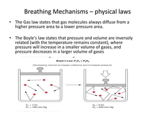
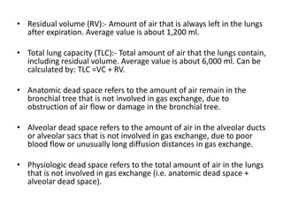
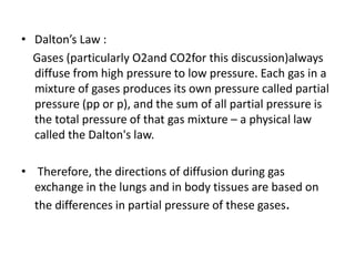
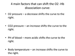
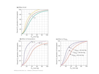
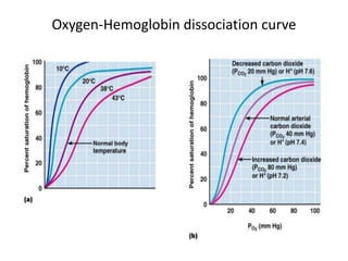
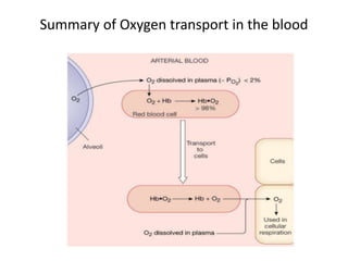
![Gas Transport - Oxygen
• 98% of O2
• is transported by binding to hemoglobin in
• erythrocytes [when O2 binds with hemoglobin (Hb)
• oxyhemoglobin (oxy-Hb) is formed which shows are a red
• pigment].
• 2% of O2
• is dissolved in the blood plasma.
• The resulting oxyhemoglobin is relatively unstable and releases
• © 2009 Ebneshahidi
• its O2
• in regions where Po2
• is low.
• More O2
• is released as the blood conc. of Co2
• increases, as the
• blood becomes more acidic, and as the blood Temp. increases.
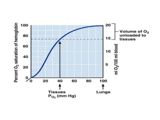
• The efficiency of oxy-Hb releasing O2
• to tissue cells during
• internal respiration is shown on the O2
• -Hb dissociation curve
• which shows a distinctive sigmoid shape.
• On this curve, as O2 partial pressure increases, the level of Hb
• saturation increases (each Hb molecule can bind up to four O2
• molecules).
• At about 40 mmHg of O2
• , roughly 75% of Hb is saturated.
• At about 80 mmHg of O2
• , close to 98% of Hb is saturated, and the
• curve becomes flattened beyound this point where only about 98 -
• 99% of Hb can be saturated no matter how high the O2 pressure is.](https://image.slidesharecdn.com/therespiratorysystem-210927080014/85/The-respiratory-system-34-320.jpg)
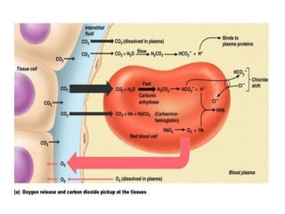
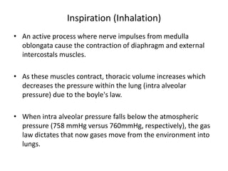
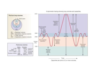
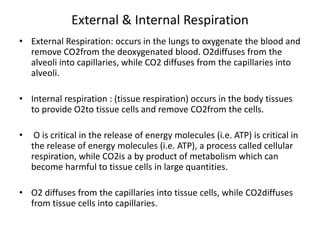
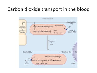
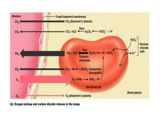
![Gas Transport – carbon dioxide
• 7% of CO2is dissolved in the blood plasma.
• 23% of CO2 binds with hemoglobin in erythrocytes. [when
CO2binds to Hb, carb-amino hemoglobin is formed which shows a
bluish pigment].
• 70% of CO2reacts with water and forms carbonic acid in
erythrocytes CO2+ H2O → H2CO3 erythrocytes CO2+ H2O →
H2CO3
• Carbonic acid is immediately broken down by the enzyme carbonic
anhydrase (CA), to become hydrogen ion and bicarbonate ion.
• H2CO3→H+ + HCO3– Where H+ quickly binds with Hb to prevent it
from affecting blood pH too drastically, and HCO3- diffuses into
blood plasma and maintains an ionic balance with chloride anion
(Cl-).](https://image.slidesharecdn.com/therespiratorysystem-210927080014/85/The-respiratory-system-42-320.jpg)


The document provides a comprehensive overview of the respiratory system, detailing its functions, anatomy, and mechanisms of breathing. It discusses the process of gas exchange, the role of different lung capacities, and factors influencing breathing control, as well as the chemical processes involved in oxygen and carbon dioxide transport. Additionally, it highlights various clinical terms related to respiratory function and conditions.


