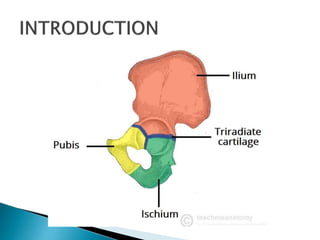
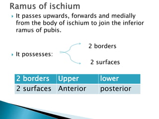
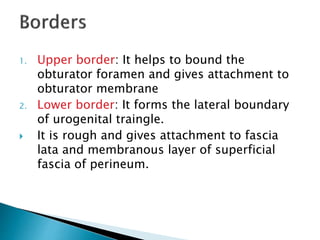
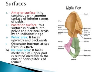
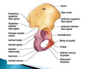
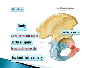
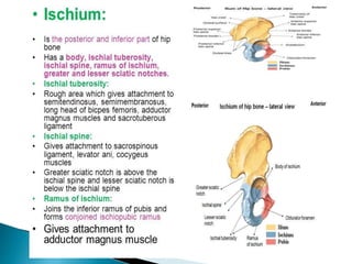
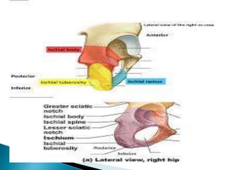
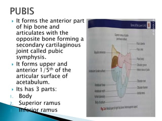
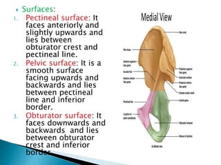
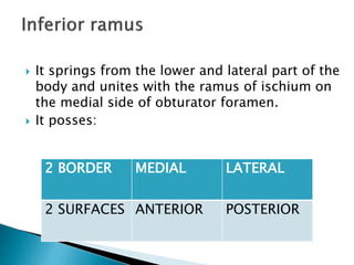
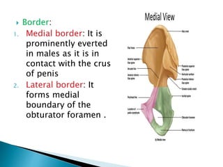
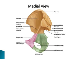
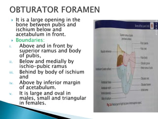
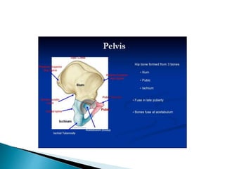
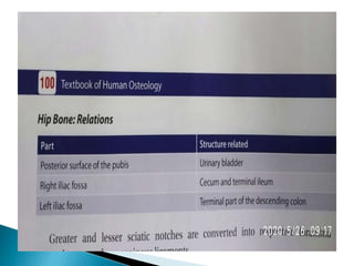
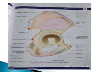
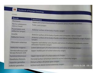
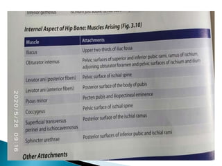
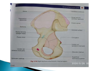
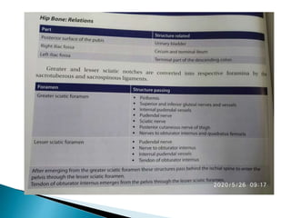
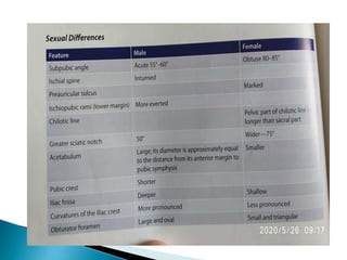
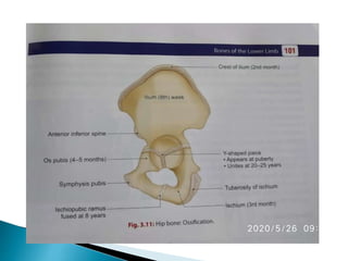
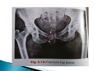
The document provides details on the anatomy of the hip bone, including its key parts and features. It describes the three parts that make up the hip bone - the ilium, ischium, and pubis. It outlines the structures and surfaces of each part, including the acetabulum, ischial tuberosity, obturator foramen, and pubic crest. Sex differences in anatomy are also briefly mentioned.
Anatomy of urinary bladder. surfaces, border of urinary bladder its relation , ligament support, peritoneal relation in male and females, pouches, blood supply of bladder, nerve supply of bladder, true and false ligament of urinary bladder,
Carpal Bone Anatomy Details PPT
Part-4 (UL Bone)
Carpal Bone names, attachments, clinical anatomy, General and specific points.
Carpal bones: 8
Like, share and comment.
Thank you
The femur or thigh bone, is the proximal bone of the hindlimb in tetrapod vertebrates. The head of the femur articulates with the acetabulum in the pelvic bone forming the hip joint, while the distal part of the femur articulates with the tibia and kneecap forming the knee joint. By most measures the femur is the strongest bone in the body. The femur is also the longest bone in the human body.
Lung Cancer: Artificial Intelligence, Synergetics, Complex System Analysis, S...Oleg Kshivets
RESULTS: Overall life span (LS) was 2252.1±1742.5 days and cumulative 5-year survival (5YS) reached 73.2%, 10 years – 64.8%, 20 years – 42.5%. 513 LCP lived more than 5 years (LS=3124.6±1525.6 days), 148 LCP – more than 10 years (LS=5054.4±1504.1 days).199 LCP died because of LC (LS=562.7±374.5 days). 5YS of LCP after bi/lobectomies was significantly superior in comparison with LCP after pneumonectomies (78.1% vs.63.7%, P=0.00001 by log-rank test). AT significantly improved 5YS (66.3% vs. 34.8%) (P=0.00000 by log-rank test) only for LCP with N1-2. Cox modeling displayed that 5YS of LCP significantly depended on: phase transition (PT) early-invasive LC in terms of synergetics, PT N0—N12, cell ratio factors (ratio between cancer cells- CC and blood cells subpopulations), G1-3, histology, glucose, AT, blood cell circuit, prothrombin index, heparin tolerance, recalcification time (P=0.000-0.038). Neural networks, genetic algorithm selection and bootstrap simulation revealed relationships between 5YS and PT early-invasive LC (rank=1), PT N0—N12 (rank=2), thrombocytes/CC (3), erythrocytes/CC (4), eosinophils/CC (5), healthy cells/CC (6), lymphocytes/CC (7), segmented neutrophils/CC (8), stick neutrophils/CC (9), monocytes/CC (10); leucocytes/CC (11). Correct prediction of 5YS was 100% by neural networks computing (area under ROC curve=1.0; error=0.0).
CONCLUSIONS: 5YS of LCP after radical procedures significantly depended on: 1) PT early-invasive cancer; 2) PT N0--N12; 3) cell ratio factors; 4) blood cell circuit; 5) biochemical factors; 6) hemostasis system; 7) AT; 8) LC characteristics; 9) LC cell dynamics; 10) surgery type: lobectomy/pneumonectomy; 11) anthropometric data. Optimal diagnosis and treatment strategies for LC are: 1) screening and early detection of LC; 2) availability of experienced thoracic surgeons because of complexity of radical procedures; 3) aggressive en block surgery and adequate lymph node dissection for completeness; 4) precise prediction; 5) adjuvant chemoimmunoradiotherapy for LCP with unfavorable prognosis.
ARTIFICIAL INTELLIGENCE IN HEALTHCARE.pdfAnujkumaranit
Artificial intelligence (AI) refers to the simulation of human intelligence processes by machines, especially computer systems. It encompasses tasks such as learning, reasoning, problem-solving, perception, and language understanding. AI technologies are revolutionizing various fields, from healthcare to finance, by enabling machines to perform tasks that typically require human intelligence.
MANAGEMENT OF ATRIOVENTRICULAR CONDUCTION BLOCK.pdfJim Jacob Roy
Cardiac conduction defects can occur due to various causes.
Atrioventricular conduction blocks ( AV blocks ) are classified into 3 types.
This document describes the acute management of AV block.
- Video recording of this lecture in English language: https://youtu.be/lK81BzxMqdo
- Video recording of this lecture in Arabic language: https://youtu.be/Ve4P0COk9OI
- Link to download the book free: https://nephrotube.blogspot.com/p/nephrotube-nephrology-books.html
- Link to NephroTube website: www.NephroTube.com
- Link to NephroTube social media accounts: https://nephrotube.blogspot.com/p/join-nephrotube-on-social-media.html
Explore natural remedies for syphilis treatment in Singapore. Discover alternative therapies, herbal remedies, and lifestyle changes that may complement conventional treatments. Learn about holistic approaches to managing syphilis symptoms and supporting overall health.
Tom Selleck Health: A Comprehensive Look at the Iconic Actor’s Wellness Journeygreendigital
Tom Selleck, an enduring figure in Hollywood. has captivated audiences for decades with his rugged charm, iconic moustache. and memorable roles in television and film. From his breakout role as Thomas Magnum in Magnum P.I. to his current portrayal of Frank Reagan in Blue Bloods. Selleck's career has spanned over 50 years. But beyond his professional achievements. fans have often been curious about Tom Selleck Health. especially as he has aged in the public eye.
Follow us on: Pinterest
Introduction
Many have been interested in Tom Selleck health. not only because of his enduring presence on screen but also because of the challenges. and lifestyle choices he has faced and made over the years. This article delves into the various aspects of Tom Selleck health. exploring his fitness regimen, diet, mental health. and the challenges he has encountered as he ages. We'll look at how he maintains his well-being. the health issues he has faced, and his approach to ageing .
Early Life and Career
Childhood and Athletic Beginnings
Tom Selleck was born on January 29, 1945, in Detroit, Michigan, and grew up in Sherman Oaks, California. From an early age, he was involved in sports, particularly basketball. which played a significant role in his physical development. His athletic pursuits continued into college. where he attended the University of Southern California (USC) on a basketball scholarship. This early involvement in sports laid a strong foundation for his physical health and disciplined lifestyle.
Transition to Acting
Selleck's transition from an athlete to an actor came with its physical demands. His first significant role in "Magnum P.I." required him to perform various stunts and maintain a fit appearance. This role, which he played from 1980 to 1988. necessitated a rigorous fitness routine to meet the show's demands. setting the stage for his long-term commitment to health and wellness.
Fitness Regimen
Workout Routine
Tom Selleck health and fitness regimen has evolved. adapting to his changing roles and age. During his "Magnum, P.I." days. Selleck's workouts were intense and focused on building and maintaining muscle mass. His routine included weightlifting, cardiovascular exercises. and specific training for the stunts he performed on the show.
Selleck adjusted his fitness routine as he aged to suit his body's needs. Today, his workouts focus on maintaining flexibility, strength, and cardiovascular health. He incorporates low-impact exercises such as swimming, walking, and light weightlifting. This balanced approach helps him stay fit without putting undue strain on his joints and muscles.
Importance of Flexibility and Mobility
In recent years, Selleck has emphasized the importance of flexibility and mobility in his fitness regimen. Understanding the natural decline in muscle mass and joint flexibility with age. he includes stretching and yoga in his routine. These practices help prevent injuries, improve posture, and maintain mobilit
The prostate is an exocrine gland of the male mammalian reproductive system
It is a walnut-sized gland that forms part of the male reproductive system and is located in front of the rectum and just below the urinary bladder
Function is to store and secrete a clear, slightly alkaline fluid that constitutes 10-30% of the volume of the seminal fluid that along with the spermatozoa, constitutes semen
A healthy human prostate measures (4cm-vertical, by 3cm-horizontal, 2cm ant-post ).
It surrounds the urethra just below the urinary bladder. It has anterior, median, posterior and two lateral lobes
It’s work is regulated by androgens which are responsible for male sex characteristics
Generalised disease of the prostate due to hormonal derangement which leads to non malignant enlargement of the gland (increase in the number of epithelial cells and stromal tissue)to cause compression of the urethra leading to symptoms (LUTS
Pulmonary Thromboembolism - etilogy, types, medical- Surgical and nursing man...VarunMahajani
Disruption of blood supply to lung alveoli due to blockage of one or more pulmonary blood vessels is called as Pulmonary thromboembolism. In this presentation we will discuss its causes, types and its management in depth.
TEST BANK for Operations Management, 14th Edition by William J. Stevenson, Ve...kevinkariuki227
TEST BANK for Operations Management, 14th Edition by William J. Stevenson, Verified Chapters 1 - 19, Complete Newest Version.pdf
Couples presenting to the infertility clinic- Do they really have infertility...Sujoy Dasgupta
Dr Sujoy Dasgupta presented the study on "Couples presenting to the infertility clinic- Do they really have infertility? – The unexplored stories of non-consummation" in the 13th Congress of the Asia Pacific Initiative on Reproduction (ASPIRE 2024) at Manila on 24 May, 2024.
4. It forms the
lower and
posterior part of
hip bone and
contributes to
form a little more
than lower and
posterior 2/5th of
the articular
surface of
acetabulum.
5. BODY
It has 2 parts :
RAMUS
BODY POSSESSES
2 ENDS UPPER LOWER *
3 BORDERS ANTERIOR LATERAL POSTERIOR
3 SURFACES FEMORAL DORSAL PELVIC
6. ENDS:
i. UPPER END: Its forms a part of acetabular
cavity and is fused with ilium and pubis.
ii. LOWER END: It gives off the ramus; forms part
of the ischial tuberosity.
Surfaces:
1) Femoral surface: It faces downwards, forwards
and laterally towards thigh and lies between
the lateral and anterior border.
2) Dorsal surface: It faces upwards, backwards
and laterally and lies between the lateral and
posterior borders. It is continuation above
with the gluteal surface of ilium.
7. It is wide near its upper part and tapers
downwards.
It is divided into an upper quadrilateral and a
lower triangular area by a transverse ridge.
a. Upper area: It is divided by an oblique ridge into
i. Upper lateral part:
ii. Upper medial part:
b. Lower area: It is divided by an irregular vertical
line into
i. Smaller medial part:
ii. Larger lateral part:
8. 3) Pelvic surface: It is a
smooth surface which
forms the lateral wall of
ischio-rectal fossa.
Borders :
i. Anterior border: forms
posterior margin of
obturator foramen.
ii. Lateral border: separates
the femoral surface from
ischial tuberosity.
iii. Posterior border:
continuous above with the
posterior border of ilium
and helps to form the
greater sciatic notch.
9. ISCHIAL SPINE: It is
a prominent
projection at the
posterior end of this
border.
LESSER SCIATIC
NOTCH: It is a
shallow notch below
the ischial spine on
the posterior border.
The notch is
converted into a
foramen by
sacrospinous
ligament.
10. It passes upwards, forwards and medially
from the body of ischium to join the inferior
ramus of pubis.
2 borders
It possesses:
2 surfaces
2 borders Upper lower
2 surfaces Anterior posterior
11. 1. Upper border: It helps to bound the
obturator foramen and gives attachment to
obturator membrane
2. Lower border: It forms the lateral boundary
of urogenital traingle.
It is rough and gives attachment to fascia
lata and membranous layer of superficial
fascia of perineum.
12. 1. Anterior surface: It is
continous with anterior
surface of inferior ramus
of pubis.
2. Posterior surface: this
surface is divided into
pelvic and perineal areas
by an indistinct ridge
a) Pelvic area: It faces
upwards and backwards.
Obturator internus arises
from this part.
b) Perineal area: It faces
medially ; its upper part
is related medially to the
crus of penis(clitoris of
female).
13.
14.
15.
16.
17. It forms the anterior part
of hip bone and
articulates with the
opposite bone forming a
secondary cartilaginous
joint called pubic
symphysis.
It forms upper and
anterior 1/5th of the
articular surface of
acetabulum.
Its has 3 parts:
1. Body
2. Superior ramus
3. Inferior ramus
18. It is flattened from before backwards and
connects the superior ramus with inferior
ramus.
It possesses:
a) 3 surfaces
Anterior
Pelvic (posterior)
symphysial
b) 1 border- pubic crest
19. Anterior surface: It faces downwards and
forwards .
Posterior surface: It forms the anterior
wall of true pelvis.
Symphysial surface: It is rough , oval and
elongated surface which articulates with
symphysial surface of the opposite pubis.
It is covered by a layer of hyaline cartilage
.
Pubic crest: It is the rounded free, upper
border of pubis.
Pubic tubercle: It is a rounded tubercle at
the lateral end of the pubic crest. It is
crossed by spermatic cord in males and
round ligament of uterus in females
20. It passes laterally and backwards from the
upper and lateral part of the body and lies
above the obturator foramen.
It possesses:
3 BORDERS OBTURATOR
CREST
PECTINEAL
LINE
INFERIOR
3 SURFACES PECTINEAL PELVIC OBTURATOR
21. Border:
1. Obturator crest: it is rounded margin which
begins from the anterior part of pubic tuberce
and passes laterally to the anterior part of
acetabular cavity.
It separetes the pectineal from the obturator
surface.
2. Pectineal line: It is sharp border which begins
from the posterior part of pubic tubercle and
passes laterally and backwards to become
continuous with the arcuate line.
It separates the pectineal line from the pelvic
surface.
3. Inferior border: it is sharp border and forms
upper boundary of obturator foramen.
22. Surfaces:
1. Pectineal surface: It
faces anteriorly and
slightly upwards and
lies between
obturator crest and
pectineal line.
2. Pelvic surface: It is a
smooth surface
facing upwards and
backwards and lies
between pectineal
line and inferior
border.
3. Obturator surface: It
faces downwards and
backwards and lies
between obturator
crest and inferior
border.
23. It springs from the lower and lateral part of the
body and unites with the ramus of ischium on
the medial side of obturator foramen.
It posses:
2 BORDER MEDIAL LATERAL
2 SURFACES ANTERIOR POSTERIOR
24. Border:
1. Medial border: It is
prominently everted
in males as it is in
contact with the crus
of penis
2. Lateral border: It
forms medial
boundary of the
obturator foramen .
25.
26. Surfaces:
1. Anterior surface: It is
continuous with anterior surface
of body.
It faces forwards and laterally
and lies between the two
borders.
2. Posterior surface: It is
continuous above with posterior
suface of the body and below
with posterior surface of the
ramus of ischium.
27. This surfaces is divided arbitrarily into 3
area by 2 indistinct ridges-
a. Medial area: It is in contact with crus of
penis.
b. Intermediate area: It is related to dorsal
nerve of penis or clitoris and internal
pudendal vessels. It may be origin to
sphincter urethrae.
c. Lateral area: gives origin to obturator
internus
28. It is also called acetabular
cavity.
It is deep cup- shaped hollow
in the hip bone which faces
laterally, downwards and
forwards.
It articulates with the head of
femur.
All 3 parts of the bone
contribute in its formation.
pubis forms upper and
anterior 1/5th of acetabulum.
Ischium forms posterior and
lower 2/5th of acetabulum
Ilium contributes to the rest
of its extent
It is covered everywhere with
articular cartilage except in
the acetabular fossa.
29. 1. Acetabular notch: It is a gap in the margin of
the cavity inferiorly.
Two ends of the notch give attachment to
transverse acetabular ligament which bridges
the gap.
2. Acetabular fossa: It is rough, non-articular
floor of the cavity.
It is covered by a pad of fat.
3. Lunate surface: It is a horse shoe-shaped
articular surface at the sides of the cavity.
It articulates with the head of femur.
30. It is a large opening in the
bone between pubis and
ischium below and
acetabulum in front.
Boundaries:
i. Above and in front by
superior ramus and body
of pubis,
ii. Below and medially by
ischio-pubic ramus
iii. Behind by body of ischium
and
iv. Above by inferior margin
of acetabulum.
v. It is large and oval in
males, small and triangular
in females.