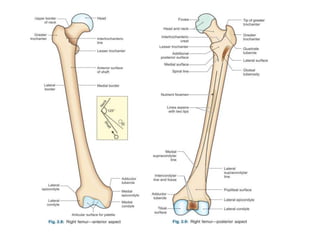
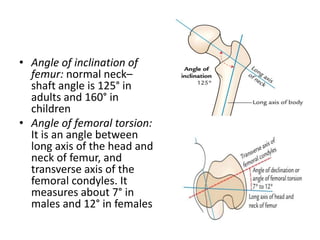
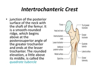
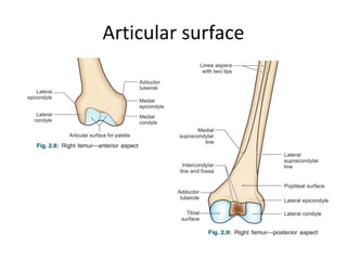
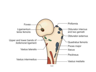
💕SONAM KUMAR💕Premium Call Girls Jaipur ↘️9257276172 ↙️One Night Stand With Lo...
Femur.pptx
1. Femur
• FEMUR: (Latin femur = thigh)
• longest and strongest bone,
• about one-fourth of the height of the individual
• upper end it articulates with the hip bone to form
the hip joint, and at the lower end it articulates
with the patella and tibia.
• transmits body weight from the hip bone to the
tibia in standing position
2. PARTS
• Upper end, shaft, and lower end
• Upper end: head, neck, and greater and
lesser trochanter.
• Shaft: gently convex anteriorly
• Lower end: medial and lateral condyles,
intercondylar fossa
• most prominent points on the condyles are
called epicondyles.
3.
4. ANATOMICAL POSITION AND SIDE
DETERMINATION:
• Its head faces upward, medially, and slightly
forward.
• Long axis of the shaft is directed downward and
medially with convexity of the shaft facing
anteriorly.
• Lower surfaces of both the condyles are in the
same horizontal plane.
• Trick-device: The anatomical position can be
achieved by balancing the neck of femur on your
index finger
5. • Angle of inclination of
femur: normal neck–
shaft angle is 125° in
adults and 160° in
children
• Angle of femoral torsion:
It is an angle between
long axis of the head and
neck of femur, and
transverse axis of the
femoral condyles. It
measures about 7° in
males and 12° in females
6. Upper End
• Head
1. forms about two-third of a sphere and
articulates with the acetabulum to form the
hip joint.
2. It presents a small pit, the fovea, just below
and behind the centre, which provides
attachment to the ligament of the head of
femur (ligamentum teres femoris).
7. Neck
• connects the head with the shaft, 3.7 cm long
• two borders and two surfaces
• upper border, concave and horizontal, meets
the shaft at the greater trochanter.
• The lower border, straight and oblique, meets
the shaft near the lesser trochanter.
• anterior surface is flat. It meets the shaft at
the intertrochanteric line, entirely
intracapsular.
8. • posterior surface is convex from above
downwards and concave from side to side, meets
the shaft at the intertrochanteric crest
• posterior surface is crossed by a horizontal
groove for the tendon of the obturator externus
to be inserted into the trochanteric fossa.
• neckshaft angle is about 125° in adults, less in
females due to their wider pelvis.
• The angle facilitates movements of the hip joint.
It is strengthened by a thickening of bone called
the calcar femorale present along its concavity.
9. • Trochanter-shaft angle is about 8° in adults.
• Angle of femoral torsion or angle of anteversion
is formed between the transverse axes of the
upper and lower ends of the femur. It is about 15°
• Blood supply: The intracapsular part of the neck
is supplied by the retinacular arteries. The
extracapsular part of the neck is supplied by the
ascending branch of the medial circumflex
femoral artery
10. Greater Trochanter
• large quadrangular prominence
located at the upper part of the
junction of neck with the shaft.
• The upper border of the
trochanter lies at the level of
the centre of the head.
• The greater trochanter has an
upper border with an apex, and
three surfaces, anterior, medial
and lateral.
• The apex is the inturned
posterior part of the posterior
border.
11. • The anterior surface is rough in its lateral part.
• The medial surface presents a rough impression
above, and a deep trochanteric fossa below.
• The lateral surface is crossed by an oblique ridge
directed downwards and forwards
• Lesser Trochanter: conical eminence directed
medially and backwards from the junction of the
posteroinferior part of the neck with the shaft.
12. Intertrochanteric Line:
• junction of the anterior surface
of the neck with the shaft of
the femur.
• a prominent roughened ridge
which begins above, at the
anterosuperior angle of the
greater trochanter as a
tubercle, and is continuous
below with the spiral line in
front of the lesser trochanter.
• The spiral line winds round the
shaft below the lesser
trochanter to reach the
posterior surface of the shaft.
13. Intertrochanteric Crest
• junction of the posterior
surface of the neck with
the shaft of the femur. It
is a smooth-rounded
ridge, which begins
above at the
posterosuperior angle of
the greater trochanter
and ends at the lesser
trochanter. The rounded
elevation, a little above
its middle, is called the
quadrate tubercle
14. Shaft
• more or less cylindrical
• narrowest in the middle, and is more
expanded inferiorly than superiorly
• convex forwards and is directed obliquely
downwards and medially
• In the middle one-third, the shaft has three
borders— medial, lateral and posterior and
three surfaces— anterior, medial and lateral.
15. • The medial and lateral
borders are rounded
and ill-defined, but
the posterior border is
in the form of a broad
roughened ridge,
called the linea aspera
(Latin rough line). The
linea aspera has
distinct medial and
lateral lips
16. • In the upper one-third of the shaft, the two
lips of the linea aspera diverge to enclose an
additional posterior surface. Thus this part
has: Four borders—medial, lateral, spiral line
and the lateral lip of the gluteal tuberosity
(Fig. 2.9) Four surfaces—anterior, medial,
lateral and posterior. The gluteal tuberosity is
a broad roughened ridge on the lateral part of
the posterior surface.
17. • In the lower one-third of the shaft also, the two
lips of the linea aspera diverge as supracondylar
lines to enclose an additional, popliteal surface.
• Thus, this part of the shaft has:
• Four borders—medial, lateral, medial
supracondylar line and lateral supracondylar line.
• Four surfaces—anterior, medial, lateral and
popliteal.
18. Lower End
• widely expanded to form two
large condyles, one medial and
one lateral.
• These form articular surface
for the knee joint.
• Anteriorly, the two condyles
are united and are in line with
the front of the shaft.
• Posteriorly, they are separated
by a deep gap, termed the
intercondylar fossa or
intercondylar notch
19. Articular Surface
• The two condyles are partially covered by a large
articular surface which is divisible into patellar and
tibial parts.
• The articular surface for patella covers the anterior
surfaces of both condyles, and extends more on the
lateral condyle than on the medial.
• Between the two condyles, the surface is grooved
vertically. It is separated from the tibial surfaces by two
faint grooves.
• The tibial surfaces cover the inferior and posterior
surfaces of the two condyles, and merge anteriorly
with the patellar surface
21. Lateral Condyle
• flat laterally, and is more in line with the shaft
• takes greater part in the transmission of body
weight to the tibia.
• stouter and stronger. The lateral aspect presents
the following.
1 A prominence called the lateral epicondyle.
2 The popliteal groove which lies just below the
epicondyle. It has a deeper anterior part and a
shallower posterior part.
3 A muscular impression posterosuperior to the
epicondyle.
22. Medial Condyle
• convex medially
• most prominent point on it is called the
medial epicondyle.
• Posterosuperior to the epicondyle there is a
projection, the adductor tubercle.
• epiphyseal line for the lower end of the femur
passes through it.
23. Intercondylar Fossa or Intercondylar
Notch
• separates the lower
and posterior parts of
the two condyles.
• limited anteriorly by
the patellar articular
surface, and posteriorly
by the intercondylar
line which separates
the notch from the
popliteal surface.
24. Attachments on the Femur:
• fovea: ligament of the head of femur or round
ligament, or ligamentum teres/ femoris
• greater trochanter.
a. piriformis inserted into the apex.
b. gluteus minimus inserted into the rough lateral part
of the anterior surface.
c. obturator internus and the two gemelli are inserted
into the upper rough impression on medial surface.
d. obturator externus is inserted into the trochanteric
fossa.
e. gluteus medius is inserted into the ridge on the
lateral surface
25.
26.
27. lesser trochanter:
• a. psoas (Greek loin) major inserted on the
apex and medial part of the rough anterior
surface
b. The iliacus is inserted on the anterior
surface of the base of the trochanter and on
the area below it
c. The smooth posterior surface of the lesser
trochanter is covered by a bursa that lies deep
to the upper horizontal fibres of the adductor
magnus.
28. intertrochanteric line
• a. Attachment to the capsular ligament of the hip joint.
b. Attachment to the upper band of the iliofemoral
ligament in its upper part
c. Attachment to the lower band of iliofemoral
ligament in its lower part.
d. Origin to the highest fibres of the vastus lateralis
from the upper end.
e. Origin to the highest fibres of the vastus medialis
from the lower end of the line.
• The quadrate tubercle receives the insertion of the
quadratus femoris
29. shaft:
a. origin of the medial head of the
gastrocnemius from the medial part of popliteal
surface.
b. vastus intermedius arises from the upper
three-fourths of the anterior and lateral
surfaces.
c. articularis genu arises just below the vastus
intermedius.
d. The lower 5 cm of the anterior surface are
related to suprapatellar bursa.
30. e. The vastus lateralis arises from the upper part of the
intertrochanteric line, anterior and inferior aspects of the
greater trochanter, the lateral margin of the gluteal
tuberosity, and the upper half of the lateral lip of the
linea aspera
f. The vastus medialis arises from the lower part of
intertrochanteric line, the spiral line, medial lip of
the linea aspera, and the medial supracondylar line
g. The deeper fibres of the lower half of the gluteus
maximus are inserted into the gluteal tuberosity.
h. The adductor longus is inserted along the medial lip of
the linea aspera between the vastus medialis
and the adductors brevis and magnus.
31. i. The adductor brevis is inserted into a line
extending from the lesser trochanter to the upper
part of the linea aspera, behind the pectineus and
the upper part of the adductor longus.
j. The adductor magnus is inserted into the medial
margin of the gluteal tuberosity, the linea aspera,
the medial supracondylar line, and the adductor
tubercle, leaving a gap/opening for the popliteal
vessels.
k. pectineus is inserted on a line extending from the
lesser trochanter to the linea aspera.
32. l. The short head of the biceps femoris arises from
the lateral lip of the linea aspera between the
vastus lateralis and the adductor magnus, and from
the upper two-thirds of the lateral supracondylar
line
m.The medial and lateral intermuscular septa are
attached to the lips of the linea aspera and to the
supracondylar lines.
They separate the extensor muscles from the
adductors medially, and from the flexors laterally
33. n. The lower end of the lateral supracondylar
line gives origin to the plantaris above and the
upper part of the lateral head of the
gastrocnemius below
o. The popliteal surface is covered with fat and
forms the floor of the popliteal fossa. The origin
of the medial head of the gastrocnemius
extends to the popliteal surface just above the
medial condyle
34.
35.
36. lateral condyle
• a. The fibular collateral ligament of the knee
joint is attached to the lateral epicondyle.
• b. The popliteus arises from the deep anterior
part of the popliteal groove.
• c. The muscular impression near the lateral
epicondyle gives origin to the lateral head of
the gastrocnemius.
37. medial condyle
a. The tibial collateral ligament of the knee joint
is attached to the medial epicondyle
b. b. The adductor tubercle receives the
insertion of the hamstring part or the ischial
head of the adductor magnus.
38. intercondylar notchare
• a. The anterior cruciate ligament is attached to
the posterior part of the medial surface of the
lateral condyle, on a smooth impression.
• b. The posterior cruciate ligament is attached to
the anterior part of the lateral surface of medial
condyle, on a smooth impression.
• c. The intercondylar line provides attachment to
the capsular ligament and laterally to the
oblique popliteal ligament.
• d. The infrapatellar synovial fold is attached to
the anterior border of the intercondylar fossa.
39. Nutrient Artery to the Femur
• derived from the second perforating artery,
branch of profunda femoris artery.
• nutrient foramen is located on the medial side
of the linea aspera, and is directed upwards
40. OSSIFICATION
• There are three
epiphyses at the
upper end and one
epiphysis at the
lower end.
• The upper
epiphyses; lesser
trochanter, greater
trochanter and
head, in that order,
fuse with the shaft
at about eighteen
years. The lower
epiphysis fuses by
the twentieth year
41. CLINICAL ANATOMY
• forced medial rotation of the thigh and leg during the
fall results in:
• a. The fracture of the shaft of femur in persons below
the age of 16 years.
• b. Bucket-handle tear of the medial meniscus between
the ages of 14 and 40 years.
• c. Pott’s (British surgeon, 1713–88) fracture of the leg
bones between the ages of 40 and 60 years.
• d. Fracture of neck of the femur over the age of 60
years. This is common in females due to osteoporosis
and degeneration of calcar femorale.
42. • arteries get injured in intracapsular fracture of
neck of femur, leading to avascular necrosis of
the head
• centre of ossification in lower end of femur
and even in upper end of tibia seen by X-ray is
used as a medicolegal evidence to prove that
the newborn (found dead) was nearly full
term and was viable