Downloaded 216 times


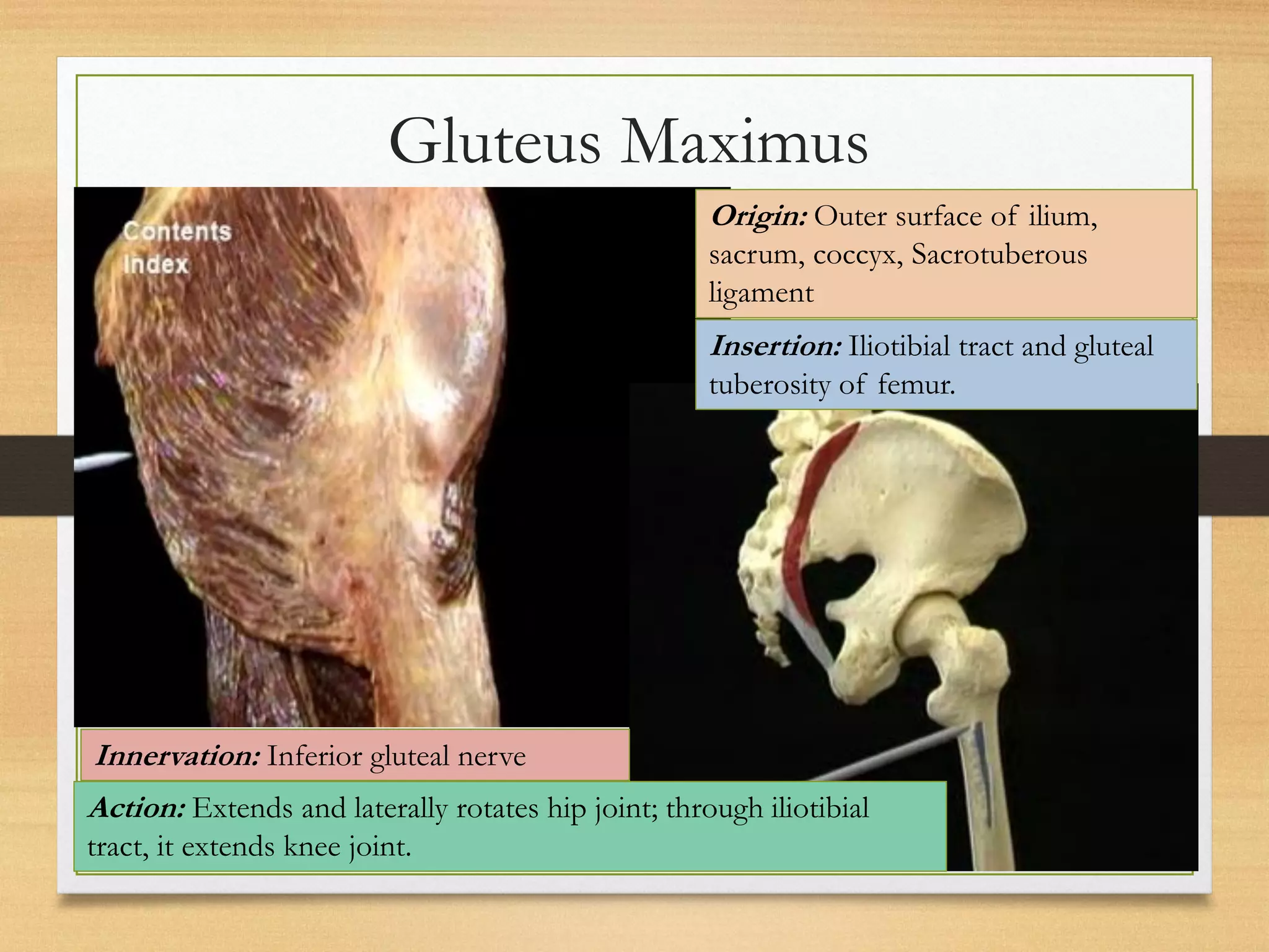


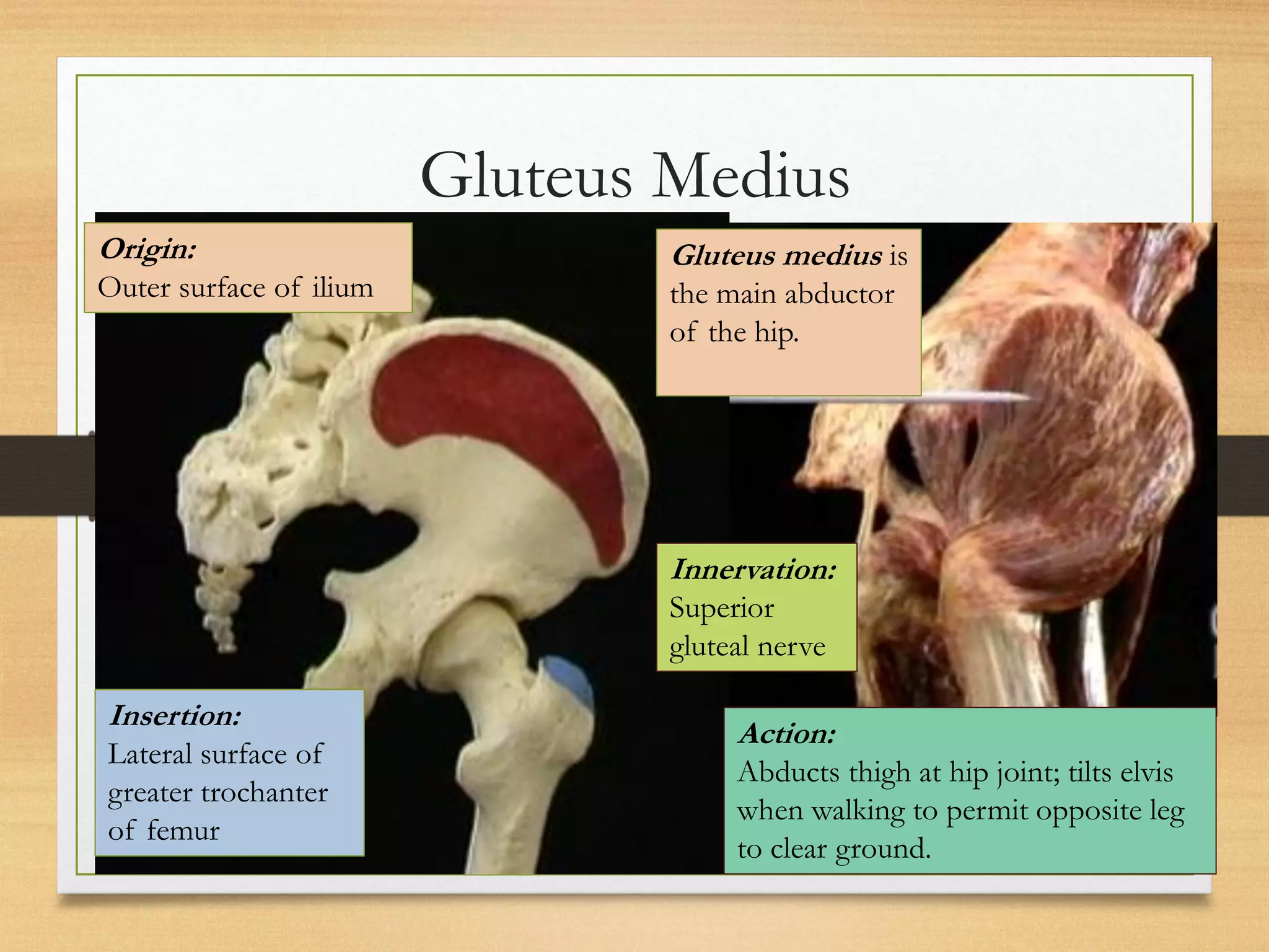
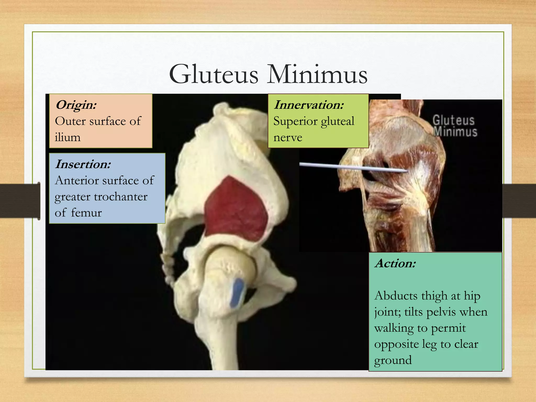
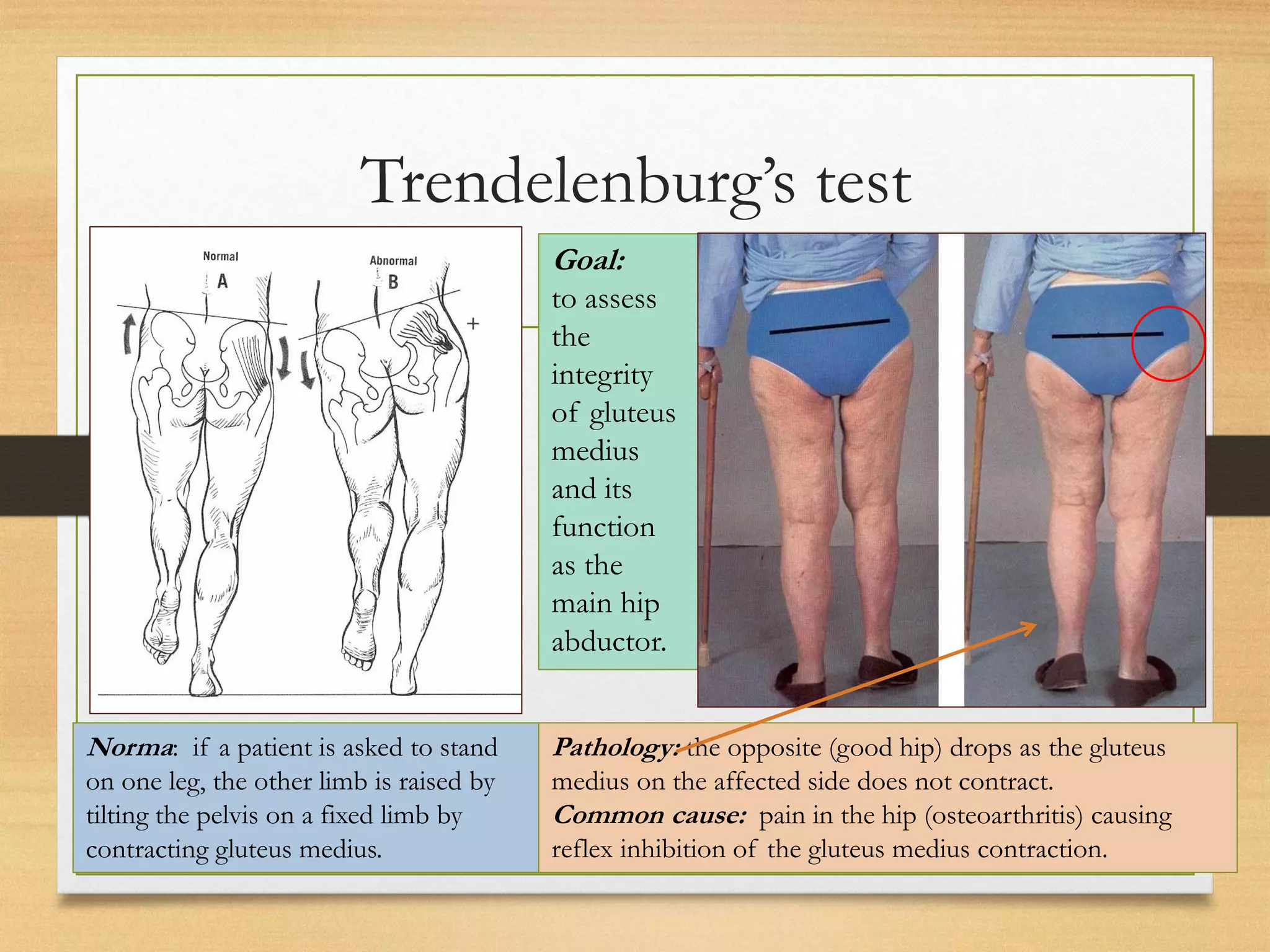


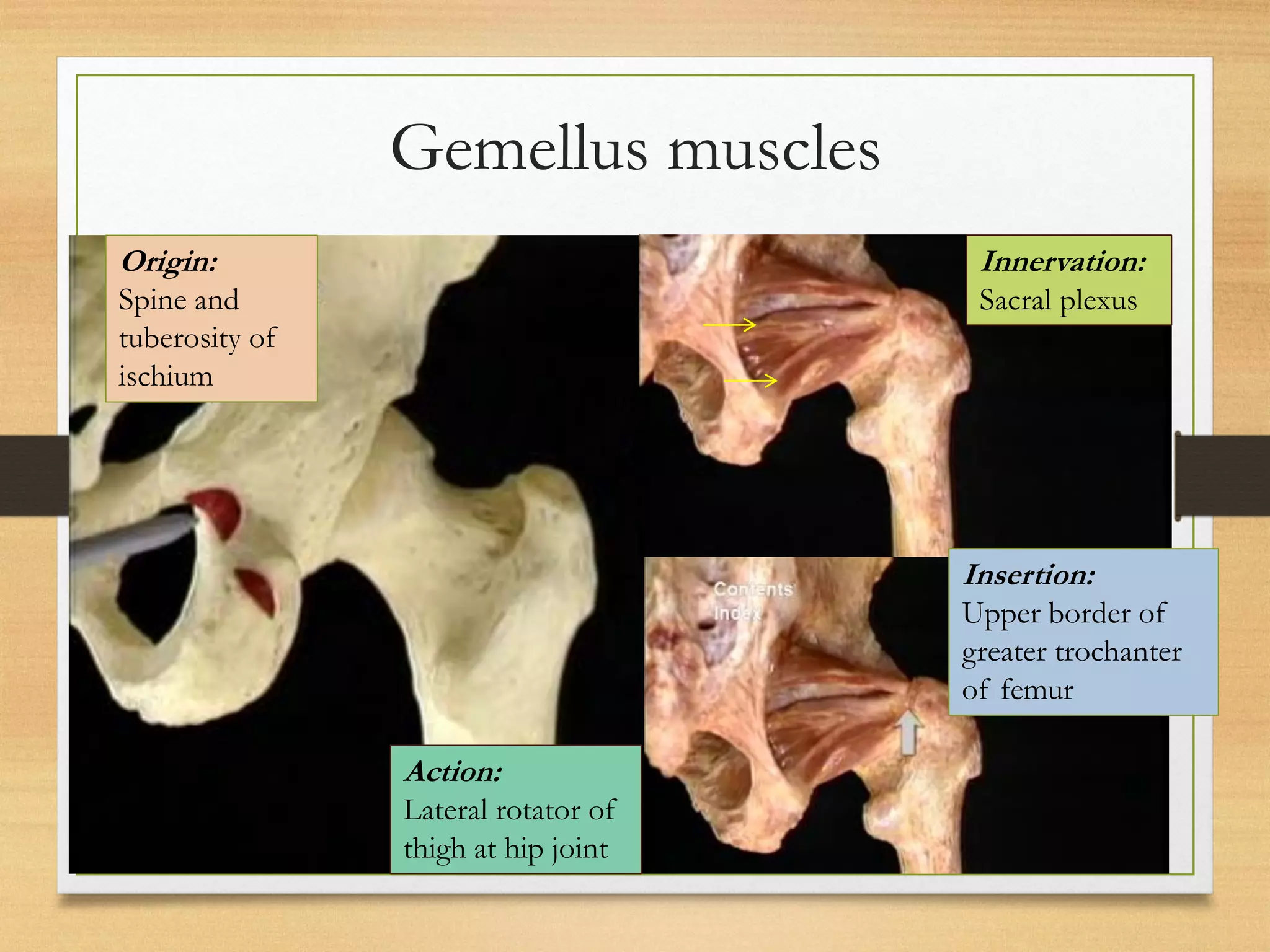
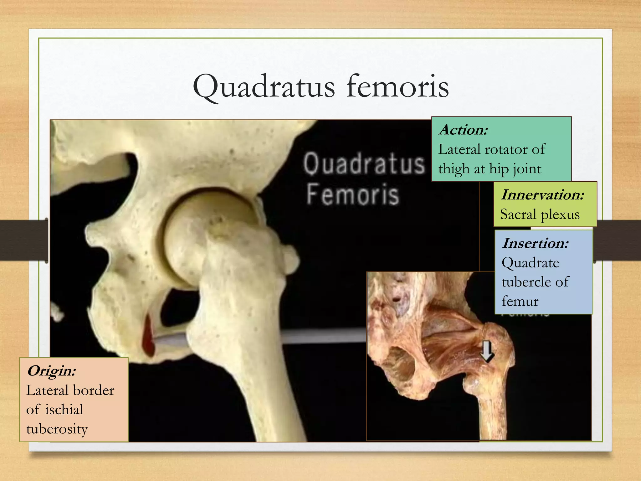


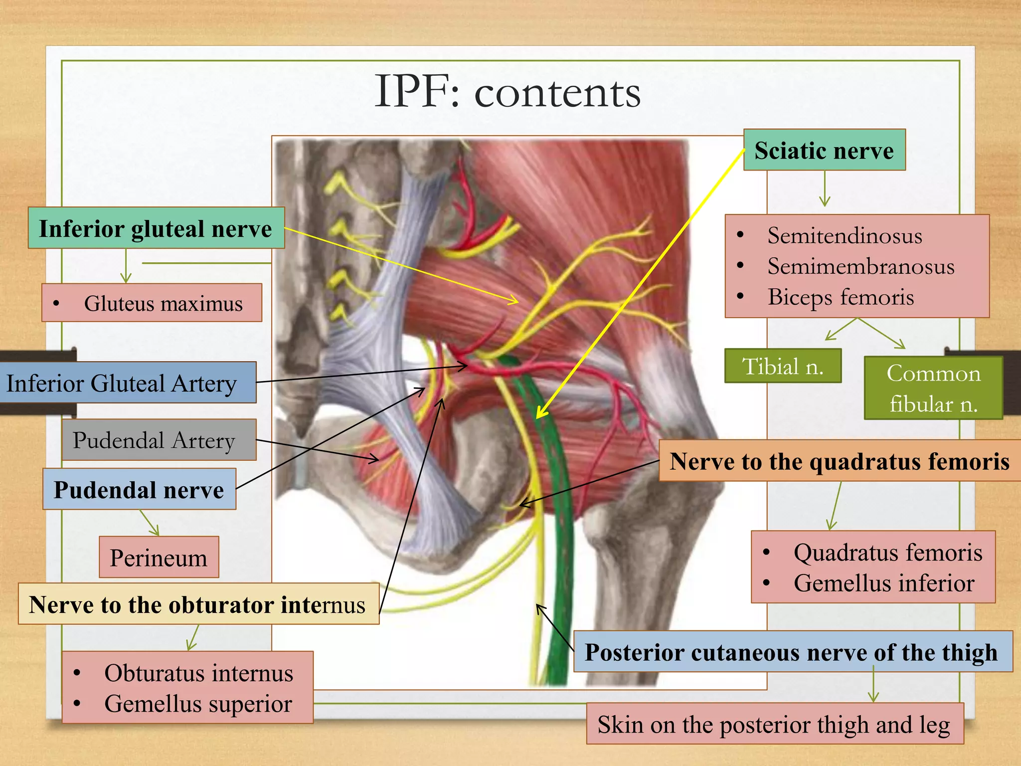


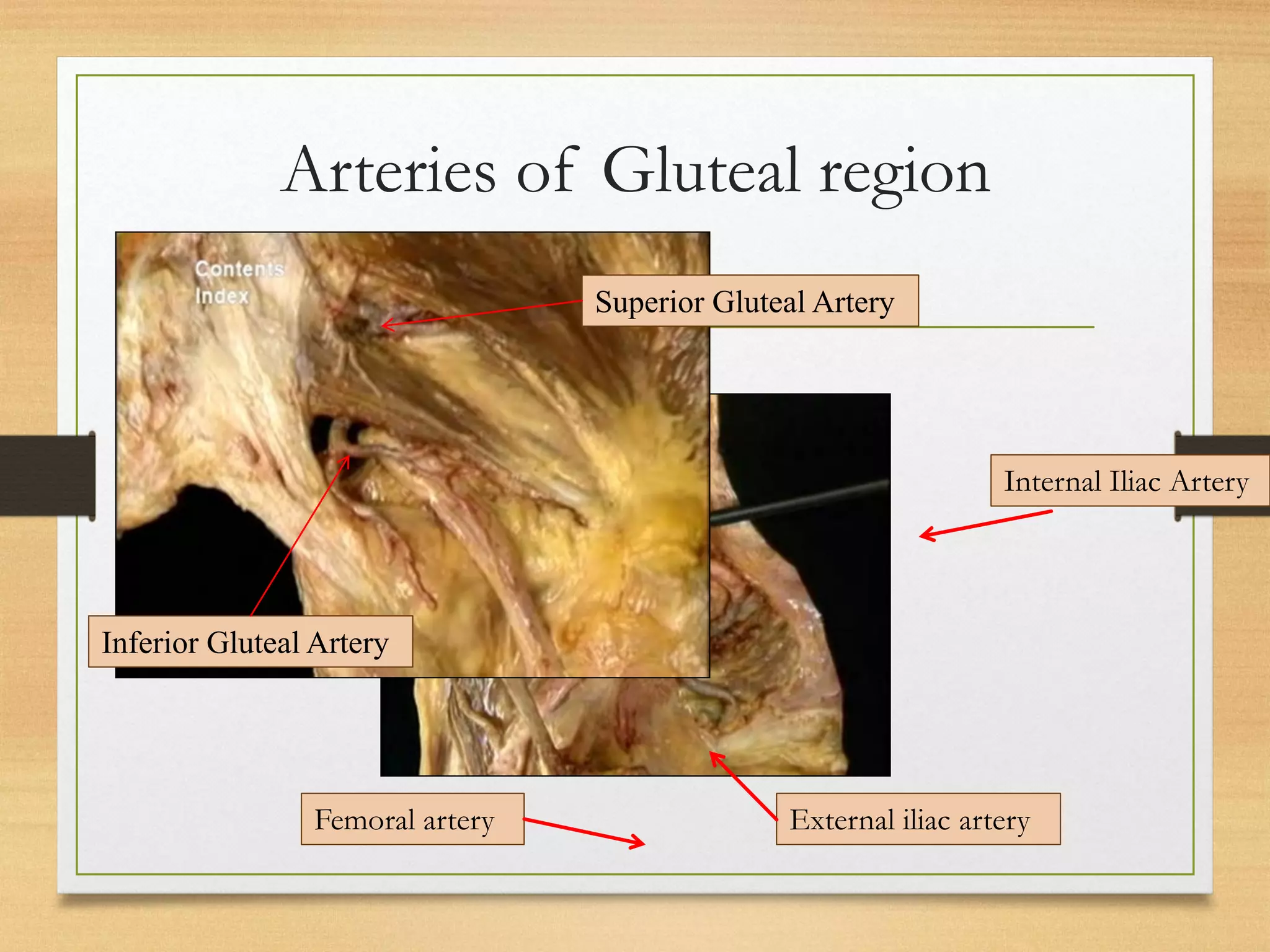
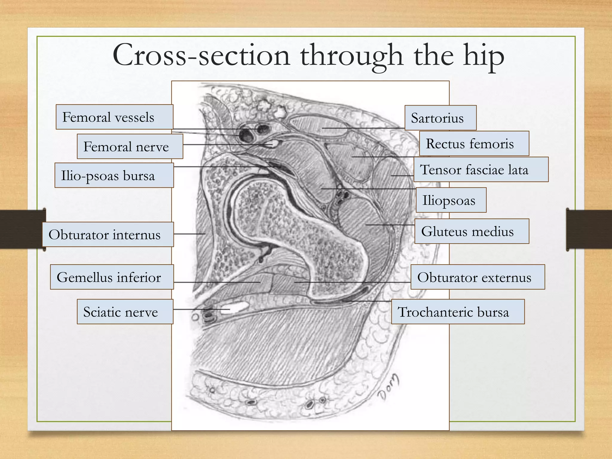


The gluteal region contains several important muscles and other structures. It is bounded superiorly by the iliac crest, laterally by the greater trochanter, and inferiorly by the gluteal folds. The gluteus maximus is the largest muscle and extends the hip. The gluteus medius and minimus abduct the thigh. Important nerves include the superior and inferior gluteal nerves. The piriformis muscle divides the gluteal region into superior and inferior compartments. The sacral plexus provides innervation to muscles in the region.