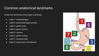
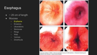
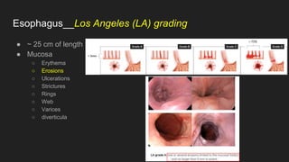
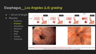
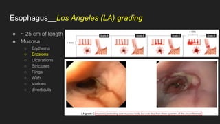
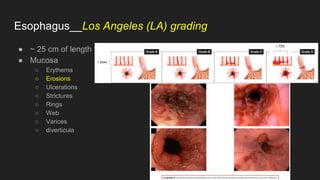
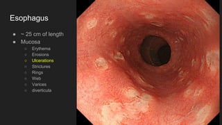
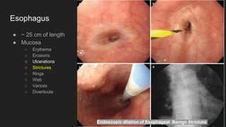
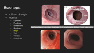
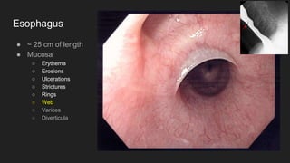
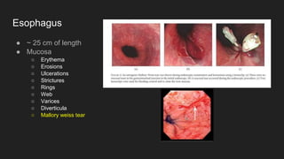
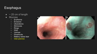
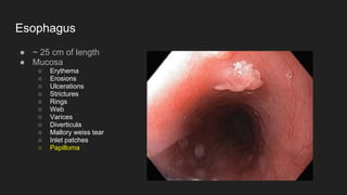
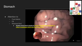
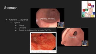
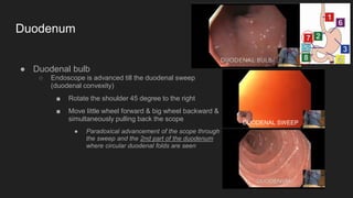
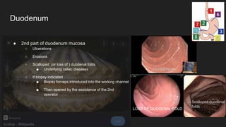
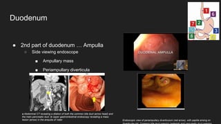
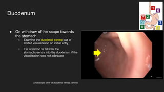
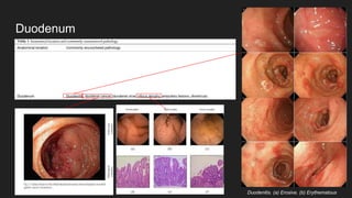
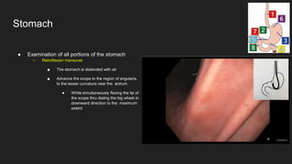
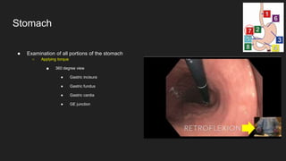
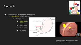
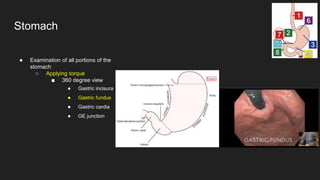
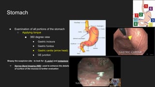
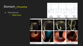
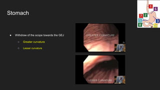
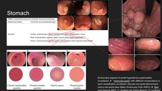
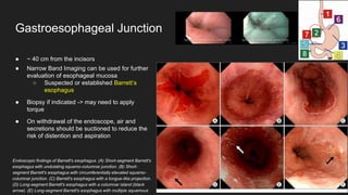
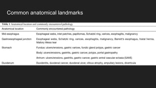
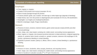
2. Common anatomical landmarks
Anatomical landmarks during upper endoscopy.
● Label 1: mid-esophagus;
● Label 2: gastroesophageal junction;
● Label 3: gastric body;
● Label 4: gastric antrum;
● Label 5: incisura;
● Label 6: gastric fundus;
● Label 7: duodenal bulb;
● Label 8: second part of duodenum.
3. Common anatomical landmarks
➔ Epiglottis and Vocal Cords
➔ Scope maneuvered post to the epiglottis &
forward to upper esophageal sphincter (which is @
thyroid cartilage ~ 15-18 cm from the incisors)
4. Common anatomical landmarks
➔ Epiglottis and Vocal Cords
➔ Scope maneuvered post to the epiglottis &
forward to upper esophageal sphincter (which is @
thyroid cartilage ~ 15-18 cm from the incisors)
◆ Gentle pressure and insufflation
5. Esophagus: May be examined in details during the scope withdraw...
● ~ 25 cm of length
● Mucosa
○ Erythema
○ Erosions
○ Ulcerations
○ Strictures
○ Rings
○ Web
○ Varices
○ Diverticula
6. Esophagus
● ~ 25 cm of length
● Mucosa
○ Erythema
○ Erosions
○ Ulcerations
○ Strictures
○ Rings
○ Web
○ Varices
○ Diverticula
7. Esophagus__Los Angeles (LA) grading
● ~ 25 cm of length
● Mucosa
○ Erythema
○ Erosions
○ Ulcerations
○ Strictures
○ Rings
○ Web
○ Varices
○ diverticula
8. Esophagus__Los Angeles (LA) grading
● ~ 25 cm of length
● Mucosa
○ Erythema
○ Erosions
○ Ulcerations
○ Strictures
○ Rings
○ Web
○ Varices
○ diverticula
9. Esophagus__Los Angeles (LA) grading
● ~ 25 cm of length
● Mucosa
○ Erythema
○ Erosions
○ Ulcerations
○ Strictures
○ Rings
○ Web
○ Varices
○ diverticula
10. Esophagus__Los Angeles (LA) grading
● ~ 25 cm of length
● Mucosa
○ Erythema
○ Erosions
○ Ulcerations
○ Strictures
○ Rings
○ Web
○ Varices
○ diverticula
11. Esophagus
● ~ 25 cm of length
● Mucosa
○ Erythema
○ Erosions
○ Ulcerations
○ Strictures
○ Rings
○ Web
○ Varices
○ diverticula
12. Esophagus
● ~ 25 cm of length
● Mucosa
○ Erythema
○ Erosions
○ Ulcerations
○ Strictures
○ Rings
○ Web
○ Varices
○ Diverticula
Endoscopic dilation of Esophageal Benign Stricture
21. Stomach
● Attention to
○ Food debris
○ FB
○ Retained fluid
■ Suction of any food in fundus to improve visualization & reduction of
reflux to esophagus which may lead to aspiration
23. ● Duodenal bulb
○ Endoscope is advanced till the duodenal sweep
(duodenal convexity)
■ Rotate the shoulder 45 degree to the right
■ Move little wheel forward & big wheel backward &
simultaneously pulling back the scope
● Paradoxical advancement of the scope through
the sweep and the 2nd part of the duodenum
where circular duodenal folds are seen
Duodenum
DUODENAL SWEEP
24. ● 2nd part of duodenum mucosa
○ Ulcerations
○ Erosions
○ Scalloped (or loss of ) duodenal folds
■ Underlying celiac diseases
○ If biopsy indicated
■ Biopsy forceps introduced into the working channel
■ Then opened by the assistance of the 2nd
operator
Duodenum
LOSS OF DUODENAL FOLD
Scalloped duodenal
folds
25. ● 2nd part of duodenum … Ampulla
○ Side viewing endoscope
■ Ampullary mass
■ Periampullary diverticula
Duodenum
Endoscopic view of periampullary diverticulum (red arrow), with papilla arising on
a Abdominal CT revealing a dilation of both the common bile duct (arrow head) and
the main pancreatic duct. b Upper gastrointestinal endoscopy revealing a mass
lesion (arrow) in the ampulla of Vater
26. ● On withdraw of the scope towards
the stomach
○ Examine the duodenal sweep cuz of
limited visualization on initial entry
○ It is common to fall into the
stomach,reentry into the duodenum if the
visualisation was not adequate
Duodenum
Endoscopic view of duodenal sweep (arrow)
28. ● Examination of all portions of the stomach
○ Retroflexion maneuver
■ The stomach is distended with air
■ Advance the scope to the region of angularis,
to the lesser curvature near the antrum
● While simultaneously flexing the tip of
the scope thru dialing the big wheel in
downward direction to the maximum
extent
Stomach
29. ● Examination of all portions of the stomach
○ Applying torque
■ 360 degree view
● Gastric incisura
● Gastric fundus
● Gastric cardia
● GE junction
Stomach
30. ● Examination of all portions of the stomach
○ Applying torque with your right hand
■ 360 degree view
● Gastric incisura
(arrow)
● Gastric fundus
● Gastric cardia
● GE junction
Stomach
Endoscopic view of pyloric ring (arrow head)
Normal: Round, reactive and patent
31. ● Examination of all portions of the
stomach
○ Applying torque
■ 360 degree view
● Gastric incisura
● Gastric fundus
● Gastric cardia
● GE junction
Stomach
32. ● Examination of all portions of the stomach
○ Applying torque
■ 360 degree view
● Gastric incisura
● Gastric fundus
● Gastric cardia (arrow head)
● GE junction
Biopsy the suspicion site: to look for H. pylori and metaplasia
➢ Narrow Band Imaging (NBI): used to enhance the details
of surface of the mucosa & further evaluation
Stomach
33. ● Examination of all portions of the stomach
○ Applying torque
■ 360 degree view
● Gastric incisura
● Gastric fundus
● Gastric cardia (arrow head)
● GE junction
Biopsy the suspicion site: to look for H. pylori and metaplasia
➢ Narrow Band Imaging (NBI): used to enhance the details
of surface of the mucosa & further evaluation
Stomach
35. ● Withdraw of the scope towards the GEJ
○ Greater curvature
○ Lesser curvature
Stomach
36. Stomach
Endoscopic aspects of portal hypertensive gastropathy
considered. A: "cherryred spots" with different nomenclature in
each classification as follows: discrete red spots-McCormack;
cherry-red spots-New Italian Endoscopy Club (NIEC); B: Red-
point lesions-NIEC; C: Isolated red marks-Baveno; D: confluent
38. ● ~ 40 cm from the incisors
● Narrow Band Imaging can be used for further
evaluation of esophageal mucosa
○ Suspected or established Barrett’s
esophagus
● Biopsy if indicated -> may need to apply
torque
● On withdrawal of the endoscope, air and
secretions should be suctioned to reduce the
risk of distention and aspiration
Gastroesophageal Junction
Endoscopic findings of Barrett's esophagus. (A) Short-segment Barrett's
esophagus with undulating squamo-columnar junction. (B) Short-
segment Barrett's esophagus with circumferentially elevated squamo-
columnar junction. (C) Barrett's esophagus with a tongue-like projection.
(D) Long-segment Barrett's esophagus with a columnar island (black
arrow). (E) Long-segment Barrett's esophagus with multiple squamous
39. ● Prague classification
Gastroesophageal Junction
In the following illustration, the circumferential segment (C) is 3 cm and the tongue an additional 2 cm,
so that M is 5 cm (3 cm circumferential + 2 cm tongue = 5 cm maximum Barrett’s extent, M). The
length of the Barrett’s is thus C3M5. A short Barrett’s segment only forming a 1-cm tongue is reported
as C0M1. A circular Barrett’s that is 2 cm long without tongues — i.e., with a relatively straight proximal
boundary —is reported as C2M2.