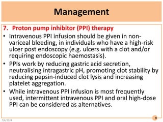
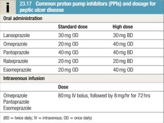
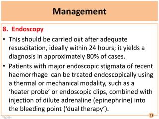
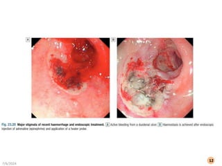
1
BY:HUSSEIN JASSIM
GASTROENTEROLOGY
GI BLEEDING
Gastrointestinal bleeding
• Acute upper gastrointestinal bleeding
• This is the most common gastrointestinal emergency, with an estimated incidence
of 134 per 100000 of the population in the UK; the mortality of patients admitted
to hospital is around 10%.
• Clinical assessment:
• Haematemesis is red with clots when bleeding is rapid and profuse,
or black (‘coffee grounds’) when less severe.
• Syncope may occur and is caused by hypotension from intravascular volume
depletion.
• Symptoms of anaemia suggest chronic bleeding.
• Melaena is the passage of black, tarry stools containing altered blood; it is usually
caused by bleeding from the upper gastrointestinal tract, although
haemorrhage from the right side of the colon is occasionally responsible.
• The characteristic colour and smell are the result of the action of digestive
enzymes and of bacteria on haemoglobin.
• Severe acute upper gastrointestinal bleeding can sometimes cause maroon or
bright red stool.
7/6/2024
2
Hematochezia: The passage of bright red (fresh) blood through the anus PLUS hemodynamic instability
7/6/2024
3
7/6/2024
4
Management
1. Intravenous access: The first step is to gain intravenous access, ideally using two
large-borecannulae.
2. Initial clinical assessment:
• Define circulatory status. Severe bleeding causes tachycardia, hypotension and
oliguria. The patient is cold and sweating, and may be agitated.
• Seek evidence of liver disease. Jaundice, cutaneous stigmata,
hepatosplenomegaly and ascites may be present in decompensated cirrhosis.
• Identify comorbidity. The presence of cardiorespiratory, cerebrovascular or renal
disease is important, both because these may be worsened by acute bleeding and
because they increase the hazards of endoscopy and surgical operations.
• These comorbidities are, therefore, a common cause of death following acute
gastrointestinal haemorrhage, even after successful haemostasis.
7/6/2024
5
Management
3. Basic investigations:
• Full blood count: anaemia, but the haemoglobin concentration
may be normal.
• Thrombocytopenia may be a clue to the presence of
hypersplenism in chronic liver disease.
• Urea and electrolytes: This test may show evidence of renal
failure.
• Liver function tests: These may show evidence of chronic liver
disease.
• Prothrombin time: Check when there is a clinical suggestion of
liver disease or patients are anticoagulated.
• Cross-matching: At least 2 units of blood should be cross-
matched if a significant bleed is suspected.
7/6/2024
6
Management
4. Resuscitation
• Intravenous crystalloid fluids should be given to raise the
blood pressure, with a 500ml bolus recommended over less
than 15minutes in haemodynamically unstable patients.
• In most patients, blood should be transfused when
haemoglobin is less than 70g/L, although transfusion should
be considered at higher levels in those with haemodynamic
instability or ischaemic heart disease.
5. Oxygen
• Oxygen saturations should be monitored with pulse
oximetry, with a target saturation of 94%–98% and oxygen
prescribed as required.
7/6/2024
7
Management
6. Antithrombotic drugs
• An increasing number of individuals present with an upper
gastrointestinal bleed while using antithrombotic
medication.
• Aspirin can be continued during an upper gastrointestinal
bleed. P2Y12-receptor antagonists (e.g. clopidogrel) should
be temporarily stopped (unless prescribed following
coronary artery stenting), as well as warfarin and direct oral
anticoagulant therapy.
• However, early reintroduction of these medications should
occur after haemostasis has been achieved to reduce
thrombotic events and death.
7/6/2024
8
Management
7. Proton pump inhibitor (PPI) therapy
• Intravenous PPI infusion should be given in non-
variceal bleeding, in individuals who have a high-risk
ulcer post endoscopy (e.g. ulcers with a clot and/or
requiring endoscopic haemostasis).
• PPIs work by reducing gastric acid secretion,
neutralising intragastric pH, promoting clot stability by
reducing pepsin-induced clot lysis and increasing
platelet aggregation.
• While intravenous PPI infusion is most frequently
used, intermittent intravenous PPI and oral high-dose
PPI can be considered as alternatives.
7/6/2024
9
7/6/2024
10
Management
8. Endoscopy
• This should be carried out after adequate
resuscitation, ideally within 24 hours; it yields a
diagnosis in approximately 80% of cases.
• Patients with major endoscopic stigmata of recent
haemorrhage can be treated endoscopically using
a thermal or mechanical modality, such as a
‘heater probe’ or endoscopic clips, combined with
injection of dilute adrenaline (epinephrine) into
the bleeding point (‘dual therapy’).
7/6/2024
11
7/6/2024
12
Management
9. Monitoring
• Patients should be closely observed, with hourly measurements of pulse, blood pressure,
oxygen saturations and urine output.
10. Radiology and surgery
• Patients who have recurrent bleeding, where endoscopic attempts at haemostasis have
failed, should be considered for radiological or surgical intervention.
• If available, angiographic control of bleeding is generally preferred to surgery in older, frail
patients. If surgery is required, the choice of operation depends on the site and diagnosis
of the bleeding lesion.
• Duodenal ulcers are treated by under-running, with or without pyloroplasty. Under-
running for gastric ulcers can also be carried out (a biopsy must be taken to exclude
carcinoma).
• Local excision may be performed, but when neither is possible, partial gastrectomy is
required.
11. Eradication
• Following treatment for ulcer bleeding, all patients should avoid non-steroidal anti-
inflammatory drugs (NSAIDs) and those who test positive for H. pylori infection should
receive eradication therapy.
• Successful eradication should be confirmed by urea breath or faecal antigen testing.
7/6/2024
13
Lower gastrointestinal bleeding
• The incidence of lower gastrointestinal bleeding is around 30–90
per 100000 of the population in the UK, with an in-hospital
mortality of around 4%.
• This may be caused by haemorrhage from the colon, anal canal
or small bowel.
7/6/2024
14
Severe acute lower gastrointestinal bleeding
• This presents with profuse red or maroon diarrhoea
and with hypovolaemic shock.
• If available, CT angiography should be performed
initially to localise the site of blood loss.
• If the bleeding source is identified, then catheter
angiography with embolisation should be performed.
• If no source of bleeding is found then a colonoscopy
should be performed.
• Some patients presenting with an apparent severe
lower GI bleed are ultimately found to have a
significant upper GI bleed.
7/6/2024
15
Severe acute lower gastrointestinal bleeding
• The commonest cause of lower GI bleeding is
diverticular disease, with up to two-thirds of cases
being classified as severe.
• Bleeding from diverticular disease is often due to
erosion of an artery within the mouth of a
diverticulum.
• Multiple endoscopic options are available, with
endoscopic clipping either alone or after the
injection of dilute adrenaline (epinephrine)
considered as first-line treatment in the UK.
7/6/2024
16
Angiodysplasia
• Angiodysplasia is a disease of older adults, in which
vascular malformations develop within the GI tract,
commonly in the caecum.
• Bleeding can be acute and profuse; it usually stops
spontaneously, but commonly recurs.
• Diagnosis is often difficult.
• Colonoscopy may reveal characteristic vascular spots and, in
the acute phase, angiography can show bleeding into the
intestinal lumen and an abnormal large, draining vein.
• The treatment of choice is endoscopic thermal ablation, but
resection of the affected bowel may be required if bleeding
continues.
7/6/2024
17
Bowel ischaemia
• Bowel ischaemia due to occlusion of the inferior
mesenteric artery can present with abdominal colic
and rectal bleeding.
• It should be considered in patients (particularly
older patients) who have evidence of generalised
atherosclerosis.
• The diagnosis is made at colonoscopy.
• Resection is required only in the presence of
peritonitis.
7/6/2024
18
Meckel’s diverticulum
• Meckel’s diverticulum with ectopic gastric
epithelium may ulcerate and erode into a major
artery.
• The diagnosis should be considered in children or
adolescents who present with profuse or recurrent
lower gastrointestinal bleeding.
• A Meckel’s 99mTc-pertechnetate scan is
sometimes positive, but the diagnosis is commonly
made only by laparotomy, at which time the
diverticulum is excised.
7/6/2024
19
Subacute or chronic lower gastrointestinal bleeding
• This can occur at all ages and is usually due to
haemorrhoids or anal fissure.
• Haemorrhoidal bleeding is bright red and occurs
during or after defecation.
• Proctoscopy can be used to make the diagnosis, but
individuals who have altered bowel habit and those
who present over the age of 40 years should undergo
colonoscopy to exclude coexisting colorectal cancer.
• Anal fissure should be suspected when fresh rectal
bleeding and anal pain occur during defecation.
7/6/2024
20
Major gastrointestinal bleeding of unknown cause
• In some patients who present with major gastrointestinal
bleeding,upper endoscopy, colonoscopy and CT angiography
may fail to reveal a diagnosis.
• Wireless capsule endoscopy is increasingly used in such
patients. The diagnostic yield is highest when performed as
close as possible to the bleeding episode, particularly within
the first 48 hours of presenting with bleeding.
• Wireless capsule endoscopy is often used to define a source
of bleeding prior to enteroscopy with push or double
balloon enteroscopy being used to visualise the small
intestine and treat the bleeding source.
• When all else fails, laparotomy with on-table endoscopy is
indicated.
7/6/2024
21
7/6/2024
22
Chronic occult gastrointestinal bleeding
• In this context, occult means that blood or its breakdown
products are present in the stool but cannot be seen by the
naked eye.
• Occult bleeding may reach 200mL per day and cause iron
deficiency anaemia.
• Any cause of gastrointestinal bleeding may be responsible,
but the most important is colorectal cancer, particularly
carcinoma of the caecum, which may produce no
gastrointestinal symptoms.
• In clinical practice, investigation of the upper and lower
gastrointestinal tract should be considered whenever a
patient presents with unexplained iron deficiency anaemia.
7/6/2024
23

3-Gastrointestinal bleeding Approach.pdf

  • 1.
  • 2.
    Gastrointestinal bleeding • Acuteupper gastrointestinal bleeding • This is the most common gastrointestinal emergency, with an estimated incidence of 134 per 100000 of the population in the UK; the mortality of patients admitted to hospital is around 10%. • Clinical assessment: • Haematemesis is red with clots when bleeding is rapid and profuse, or black (‘coffee grounds’) when less severe. • Syncope may occur and is caused by hypotension from intravascular volume depletion. • Symptoms of anaemia suggest chronic bleeding. • Melaena is the passage of black, tarry stools containing altered blood; it is usually caused by bleeding from the upper gastrointestinal tract, although haemorrhage from the right side of the colon is occasionally responsible. • The characteristic colour and smell are the result of the action of digestive enzymes and of bacteria on haemoglobin. • Severe acute upper gastrointestinal bleeding can sometimes cause maroon or bright red stool. 7/6/2024 2 Hematochezia: The passage of bright red (fresh) blood through the anus PLUS hemodynamic instability
  • 3.
  • 4.
  • 5.
    Management 1. Intravenous access:The first step is to gain intravenous access, ideally using two large-borecannulae. 2. Initial clinical assessment: • Define circulatory status. Severe bleeding causes tachycardia, hypotension and oliguria. The patient is cold and sweating, and may be agitated. • Seek evidence of liver disease. Jaundice, cutaneous stigmata, hepatosplenomegaly and ascites may be present in decompensated cirrhosis. • Identify comorbidity. The presence of cardiorespiratory, cerebrovascular or renal disease is important, both because these may be worsened by acute bleeding and because they increase the hazards of endoscopy and surgical operations. • These comorbidities are, therefore, a common cause of death following acute gastrointestinal haemorrhage, even after successful haemostasis. 7/6/2024 5
  • 7.
    Management 3. Basic investigations: •Full blood count: anaemia, but the haemoglobin concentration may be normal. • Thrombocytopenia may be a clue to the presence of hypersplenism in chronic liver disease. • Urea and electrolytes: This test may show evidence of renal failure. • Liver function tests: These may show evidence of chronic liver disease. • Prothrombin time: Check when there is a clinical suggestion of liver disease or patients are anticoagulated. • Cross-matching: At least 2 units of blood should be cross- matched if a significant bleed is suspected. 7/6/2024 6
  • 8.
    Management 4. Resuscitation • Intravenouscrystalloid fluids should be given to raise the blood pressure, with a 500ml bolus recommended over less than 15minutes in haemodynamically unstable patients. • In most patients, blood should be transfused when haemoglobin is less than 70g/L, although transfusion should be considered at higher levels in those with haemodynamic instability or ischaemic heart disease. 5. Oxygen • Oxygen saturations should be monitored with pulse oximetry, with a target saturation of 94%–98% and oxygen prescribed as required. 7/6/2024 7
  • 9.
    Management 6. Antithrombotic drugs •An increasing number of individuals present with an upper gastrointestinal bleed while using antithrombotic medication. • Aspirin can be continued during an upper gastrointestinal bleed. P2Y12-receptor antagonists (e.g. clopidogrel) should be temporarily stopped (unless prescribed following coronary artery stenting), as well as warfarin and direct oral anticoagulant therapy. • However, early reintroduction of these medications should occur after haemostasis has been achieved to reduce thrombotic events and death. 7/6/2024 8
  • 10.
    Management 7. Proton pumpinhibitor (PPI) therapy • Intravenous PPI infusion should be given in non- variceal bleeding, in individuals who have a high-risk ulcer post endoscopy (e.g. ulcers with a clot and/or requiring endoscopic haemostasis). • PPIs work by reducing gastric acid secretion, neutralising intragastric pH, promoting clot stability by reducing pepsin-induced clot lysis and increasing platelet aggregation. • While intravenous PPI infusion is most frequently used, intermittent intravenous PPI and oral high-dose PPI can be considered as alternatives. 7/6/2024 9
  • 11.
  • 12.
    Management 8. Endoscopy • Thisshould be carried out after adequate resuscitation, ideally within 24 hours; it yields a diagnosis in approximately 80% of cases. • Patients with major endoscopic stigmata of recent haemorrhage can be treated endoscopically using a thermal or mechanical modality, such as a ‘heater probe’ or endoscopic clips, combined with injection of dilute adrenaline (epinephrine) into the bleeding point (‘dual therapy’). 7/6/2024 11
  • 13.
  • 14.
    Management 9. Monitoring • Patientsshould be closely observed, with hourly measurements of pulse, blood pressure, oxygen saturations and urine output. 10. Radiology and surgery • Patients who have recurrent bleeding, where endoscopic attempts at haemostasis have failed, should be considered for radiological or surgical intervention. • If available, angiographic control of bleeding is generally preferred to surgery in older, frail patients. If surgery is required, the choice of operation depends on the site and diagnosis of the bleeding lesion. • Duodenal ulcers are treated by under-running, with or without pyloroplasty. Under- running for gastric ulcers can also be carried out (a biopsy must be taken to exclude carcinoma). • Local excision may be performed, but when neither is possible, partial gastrectomy is required. 11. Eradication • Following treatment for ulcer bleeding, all patients should avoid non-steroidal anti- inflammatory drugs (NSAIDs) and those who test positive for H. pylori infection should receive eradication therapy. • Successful eradication should be confirmed by urea breath or faecal antigen testing. 7/6/2024 13
  • 15.
    Lower gastrointestinal bleeding •The incidence of lower gastrointestinal bleeding is around 30–90 per 100000 of the population in the UK, with an in-hospital mortality of around 4%. • This may be caused by haemorrhage from the colon, anal canal or small bowel. 7/6/2024 14
  • 16.
    Severe acute lowergastrointestinal bleeding • This presents with profuse red or maroon diarrhoea and with hypovolaemic shock. • If available, CT angiography should be performed initially to localise the site of blood loss. • If the bleeding source is identified, then catheter angiography with embolisation should be performed. • If no source of bleeding is found then a colonoscopy should be performed. • Some patients presenting with an apparent severe lower GI bleed are ultimately found to have a significant upper GI bleed. 7/6/2024 15
  • 17.
    Severe acute lowergastrointestinal bleeding • The commonest cause of lower GI bleeding is diverticular disease, with up to two-thirds of cases being classified as severe. • Bleeding from diverticular disease is often due to erosion of an artery within the mouth of a diverticulum. • Multiple endoscopic options are available, with endoscopic clipping either alone or after the injection of dilute adrenaline (epinephrine) considered as first-line treatment in the UK. 7/6/2024 16
  • 18.
    Angiodysplasia • Angiodysplasia isa disease of older adults, in which vascular malformations develop within the GI tract, commonly in the caecum. • Bleeding can be acute and profuse; it usually stops spontaneously, but commonly recurs. • Diagnosis is often difficult. • Colonoscopy may reveal characteristic vascular spots and, in the acute phase, angiography can show bleeding into the intestinal lumen and an abnormal large, draining vein. • The treatment of choice is endoscopic thermal ablation, but resection of the affected bowel may be required if bleeding continues. 7/6/2024 17
  • 19.
    Bowel ischaemia • Bowelischaemia due to occlusion of the inferior mesenteric artery can present with abdominal colic and rectal bleeding. • It should be considered in patients (particularly older patients) who have evidence of generalised atherosclerosis. • The diagnosis is made at colonoscopy. • Resection is required only in the presence of peritonitis. 7/6/2024 18
  • 20.
    Meckel’s diverticulum • Meckel’sdiverticulum with ectopic gastric epithelium may ulcerate and erode into a major artery. • The diagnosis should be considered in children or adolescents who present with profuse or recurrent lower gastrointestinal bleeding. • A Meckel’s 99mTc-pertechnetate scan is sometimes positive, but the diagnosis is commonly made only by laparotomy, at which time the diverticulum is excised. 7/6/2024 19
  • 21.
    Subacute or chroniclower gastrointestinal bleeding • This can occur at all ages and is usually due to haemorrhoids or anal fissure. • Haemorrhoidal bleeding is bright red and occurs during or after defecation. • Proctoscopy can be used to make the diagnosis, but individuals who have altered bowel habit and those who present over the age of 40 years should undergo colonoscopy to exclude coexisting colorectal cancer. • Anal fissure should be suspected when fresh rectal bleeding and anal pain occur during defecation. 7/6/2024 20
  • 22.
    Major gastrointestinal bleedingof unknown cause • In some patients who present with major gastrointestinal bleeding,upper endoscopy, colonoscopy and CT angiography may fail to reveal a diagnosis. • Wireless capsule endoscopy is increasingly used in such patients. The diagnostic yield is highest when performed as close as possible to the bleeding episode, particularly within the first 48 hours of presenting with bleeding. • Wireless capsule endoscopy is often used to define a source of bleeding prior to enteroscopy with push or double balloon enteroscopy being used to visualise the small intestine and treat the bleeding source. • When all else fails, laparotomy with on-table endoscopy is indicated. 7/6/2024 21
  • 23.
  • 24.
    Chronic occult gastrointestinalbleeding • In this context, occult means that blood or its breakdown products are present in the stool but cannot be seen by the naked eye. • Occult bleeding may reach 200mL per day and cause iron deficiency anaemia. • Any cause of gastrointestinal bleeding may be responsible, but the most important is colorectal cancer, particularly carcinoma of the caecum, which may produce no gastrointestinal symptoms. • In clinical practice, investigation of the upper and lower gastrointestinal tract should be considered whenever a patient presents with unexplained iron deficiency anaemia. 7/6/2024 23