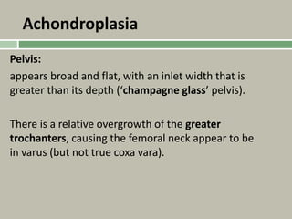
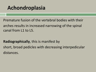
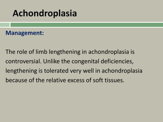
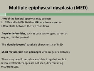
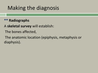
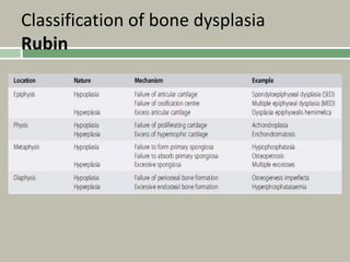
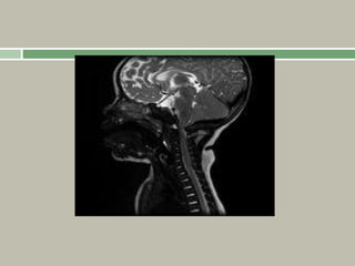
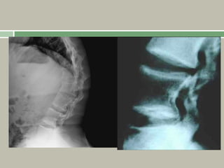
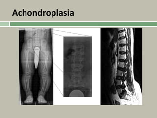
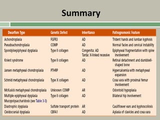
1. A skeletal dysplasia is a congenital abnormality of bone growth or development that results in structural abnormalities of the bones.
2. Making a diagnosis involves taking a thorough history and physical examination, including measurements of height, limb lengths, and facial features. Radiographs can identify which bones are affected.
3. Achondroplasia is the most common skeletal dysplasia, caused by a mutation in the FGFR3 gene, and is characterized by disproportionate short stature, frontal bossing, trident hands, genu varum, and foramen magnum stenosis.
This presentation gives a detailed study of achondroplasia-caused conditions, with CT-imaging and other detailed figures. It also elaborates management techniques and recommended consultation for the disease.
TRAUMATOLOGY OF LOWER LIMB WITH MECHANISM OF INJURY, CLASSIFICATION, RADIOLOGY, NON OPERATIVE/ OPERATIVE TREATMENT, POTENTIAL PROBLEMS AND PREVENTIVE MEASURES
Skeletal dysplasia musculoskeletal radiology is very concise and it cover the all-important topic of skeletal dysplasia with their characteristic feature and radiological findings with a proper radiographic image. Starting from classification and approach. It includes nosology classification. Thanks.
Degenerative Marrow Changes (Signal intensity changes) adjacent to the endplates of degenerated discs are a common observation on MR images.
This is a teaching lecture given twice by Prof. Dr. Mohamed Mohi Eldin, professor of neurosurgery, in the weekly conference of kasr El Aini Neurosurgery Department, Cairo University, November 2010 and January 2013.
Presentation contain etiology, blood supply of femoral head & neck,pathogenesis ,classification system ,clinical features,diagnosis,managment, pelvic & femoral osteotomies in detail
In many fetal skeletal dysplasias ,the skin and s/c tissue continues to grow at a rate proportionately greater than the long bones resulting in relatively thickened skin folds (on occasion mistaken for hydrops fetalis ) .
Polyhydraminos –common .cause –variable combination of the following –oesophageal compression by the small chest ,GI abnormalities ,micrognathia ,or hypotonia .
1- Why isn't Jamie allowed unlimited use of aspirin for pain- 2- Why d.docxKevinjrHWatsono
1. Why isn't Jamie allowed unlimited use of aspirin for pain? 2. Why does Jamie's pain subside after a period of inactivity? 3. Why is Sever disease more likely to occur in a child who is active than in one who is sedentary? 4. Why does Sever disease occur in children rather than adults? 5. Describe the pattern of pain appearance and remission that can be expected if Jamie uses heel pads only intermittently. 6. Why are Jamie's X rays normal? If Jamie had calcaneal spurs, how would the X rays have differed? 7. In craniosynostosis, why would premature closure of the sagittal suture restrict the lateral growth of the skull? 8. If you were a pediatrician, what measurements of the skull would you take to distinguish different forms of craniosynostossis from each other? 9. About 50% of children with cranioynostossi exhibit mental retardation. Explain the probable connection between the skeletal deformity and the neurological effect. 10. Explain why Paget disease could cause abnormal pressure on a spinal nerve. Objectives In this chapter we will study - foor developmental disorders of the skull-microcephaly, macrocephaly, acrania, and craniosynostosis; - two developmental disorders of the face and jaw-cleft palate and mandible disorders: - developmental disorders of the vertebral column, incloding spina bifida and aboormal spine curvatures; - some of the causes of lower back pain later in life; and - two disorders of the pelvis-rachitic pelvis and pelvis asyametry. - three disorders of the legs - congenital short fenur, genu varum, and genu valgum: - skeletal disorders of the foet, specifically foot deformities and heel pain. Diagnosing Skeletal Disorders Craniosymosiosis This chapter focuses on disonders of some of the groups of booes Normal human babies are born with unfused e that compose the skeletoa, and examines skeletal problems at the able to shift enough to allow their heads to organ and system level as opposed to the tissue level treated in canal. The cranial booes become rigidly joi the previous chapter. You may find it helpfal to refer to a buman 2 years after birth. Craniosynostonis oecurs asatomy textbook to review the skeleton. The methods used to di- of the cranial sutures fuses prematurely durin agnose the disorders diseussed in this chapter are largely the same months of life. It occurs in about 5 out of 100 as those deseribed in the previous chapter. twice as often in males as in females. Developmental Disorders of the Skull Premature closure of a suture results in I Developmental defects that occur daring the formation of the tures that remain open. For example, if the o bones of the skull can be so minor that they have little or no effect prematurely, the head cannot grow normally or so major that they cause death. This discussion focuses ce two direction but shows excessive promth in a la defects of the skull: acrania and crasiosynostosis. pendicular to the sagittal suture. As a result, Acrania mally wide (left to right) and sh.
New Directions in Targeted Therapeutic Approaches for Older Adults With Mantl...i3 Health
i3 Health is pleased to make the speaker slides from this activity available for use as a non-accredited self-study or teaching resource.
This slide deck presented by Dr. Kami Maddocks, Professor-Clinical in the Division of Hematology and
Associate Division Director for Ambulatory Operations
The Ohio State University Comprehensive Cancer Center, will provide insight into new directions in targeted therapeutic approaches for older adults with mantle cell lymphoma.
STATEMENT OF NEED
Mantle cell lymphoma (MCL) is a rare, aggressive B-cell non-Hodgkin lymphoma (NHL) accounting for 5% to 7% of all lymphomas. Its prognosis ranges from indolent disease that does not require treatment for years to very aggressive disease, which is associated with poor survival (Silkenstedt et al, 2021). Typically, MCL is diagnosed at advanced stage and in older patients who cannot tolerate intensive therapy (NCCN, 2022). Although recent advances have slightly increased remission rates, recurrence and relapse remain very common, leading to a median overall survival between 3 and 6 years (LLS, 2021). Though there are several effective options, progress is still needed towards establishing an accepted frontline approach for MCL (Castellino et al, 2022). Treatment selection and management of MCL are complicated by the heterogeneity of prognosis, advanced age and comorbidities of patients, and lack of an established standard approach for treatment, making it vital that clinicians be familiar with the latest research and advances in this area. In this activity chaired by Michael Wang, MD, Professor in the Department of Lymphoma & Myeloma at MD Anderson Cancer Center, expert faculty will discuss prognostic factors informing treatment, the promising results of recent trials in new therapeutic approaches, and the implications of treatment resistance in therapeutic selection for MCL.
Target Audience
Hematology/oncology fellows, attending faculty, and other health care professionals involved in the treatment of patients with mantle cell lymphoma (MCL).
Learning Objectives
1.) Identify clinical and biological prognostic factors that can guide treatment decision making for older adults with MCL
2.) Evaluate emerging data on targeted therapeutic approaches for treatment-naive and relapsed/refractory MCL and their applicability to older adults
3.) Assess mechanisms of resistance to targeted therapies for MCL and their implications for treatment selection
Title: Sense of Taste
Presenter: Dr. Faiza, Assistant Professor of Physiology
Qualifications:
MBBS (Best Graduate, AIMC Lahore)
FCPS Physiology
ICMT, CHPE, DHPE (STMU)
MPH (GC University, Faisalabad)
MBA (Virtual University of Pakistan)
Learning Objectives:
Describe the structure and function of taste buds.
Describe the relationship between the taste threshold and taste index of common substances.
Explain the chemical basis and signal transduction of taste perception for each type of primary taste sensation.
Recognize different abnormalities of taste perception and their causes.
Key Topics:
Significance of Taste Sensation:
Differentiation between pleasant and harmful food
Influence on behavior
Selection of food based on metabolic needs
Receptors of Taste:
Taste buds on the tongue
Influence of sense of smell, texture of food, and pain stimulation (e.g., by pepper)
Primary and Secondary Taste Sensations:
Primary taste sensations: Sweet, Sour, Salty, Bitter, Umami
Chemical basis and signal transduction mechanisms for each taste
Taste Threshold and Index:
Taste threshold values for Sweet (sucrose), Salty (NaCl), Sour (HCl), and Bitter (Quinine)
Taste index relationship: Inversely proportional to taste threshold
Taste Blindness:
Inability to taste certain substances, particularly thiourea compounds
Example: Phenylthiocarbamide
Structure and Function of Taste Buds:
Composition: Epithelial cells, Sustentacular/Supporting cells, Taste cells, Basal cells
Features: Taste pores, Taste hairs/microvilli, and Taste nerve fibers
Location of Taste Buds:
Found in papillae of the tongue (Fungiform, Circumvallate, Foliate)
Also present on the palate, tonsillar pillars, epiglottis, and proximal esophagus
Mechanism of Taste Stimulation:
Interaction of taste substances with receptors on microvilli
Signal transduction pathways for Umami, Sweet, Bitter, Sour, and Salty tastes
Taste Sensitivity and Adaptation:
Decrease in sensitivity with age
Rapid adaptation of taste sensation
Role of Saliva in Taste:
Dissolution of tastants to reach receptors
Washing away the stimulus
Taste Preferences and Aversions:
Mechanisms behind taste preference and aversion
Influence of receptors and neural pathways
Impact of Sensory Nerve Damage:
Degeneration of taste buds if the sensory nerve fiber is cut
Abnormalities of Taste Detection:
Conditions: Ageusia, Hypogeusia, Dysgeusia (parageusia)
Causes: Nerve damage, neurological disorders, infections, poor oral hygiene, adverse drug effects, deficiencies, aging, tobacco use, altered neurotransmitter levels
Neurotransmitters and Taste Threshold:
Effects of serotonin (5-HT) and norepinephrine (NE) on taste sensitivity
Supertasters:
25% of the population with heightened sensitivity to taste, especially bitterness
Increased number of fungiform papillae
HOT NEW PRODUCT! BIG SALES FAST SHIPPING NOW FROM CHINA!! EU KU DB BK substit...GL Anaacs
Contact us if you are interested:
Email / Skype : kefaya1771@gmail.com
Threema: PXHY5PDH
New BATCH Ku !!! MUCH IN DEMAND FAST SALE EVERY BATCH HAPPY GOOD EFFECT BIG BATCH !
Contact me on Threema or skype to start big business!!
Hot-sale products:
NEW HOT EUTYLONE WHITE CRYSTAL!!
5cl-adba precursor (semi finished )
5cl-adba raw materials
ADBB precursor (semi finished )
ADBB raw materials
APVP powder
5fadb/4f-adb
Jwh018 / Jwh210
Eutylone crystal
Protonitazene (hydrochloride) CAS: 119276-01-6
Flubrotizolam CAS: 57801-95-3
Metonitazene CAS: 14680-51-4
Payment terms: Western Union,MoneyGram,Bitcoin or USDT.
Deliver Time: Usually 7-15days
Shipping method: FedEx, TNT, DHL,UPS etc.Our deliveries are 100% safe, fast, reliable and discreet.
Samples will be sent for your evaluation!If you are interested in, please contact me, let's talk details.
We specializes in exporting high quality Research chemical, medical intermediate, Pharmaceutical chemicals and so on. Products are exported to USA, Canada, France, Korea, Japan,Russia, Southeast Asia and other countries.
MANAGEMENT OF ATRIOVENTRICULAR CONDUCTION BLOCK.pdfJim Jacob Roy
Cardiac conduction defects can occur due to various causes.
Atrioventricular conduction blocks ( AV blocks ) are classified into 3 types.
This document describes the acute management of AV block.
Ozempic: Preoperative Management of Patients on GLP-1 Receptor Agonists Saeid Safari
Preoperative Management of Patients on GLP-1 Receptor Agonists like Ozempic and Semiglutide
ASA GUIDELINE
NYSORA Guideline
2 Case Reports of Gastric Ultrasound
Ethanol (CH3CH2OH), or beverage alcohol, is a two-carbon alcohol
that is rapidly distributed in the body and brain. Ethanol alters many
neurochemical systems and has rewarding and addictive properties. It
is the oldest recreational drug and likely contributes to more morbidity,
mortality, and public health costs than all illicit drugs combined. The
5th edition of the Diagnostic and Statistical Manual of Mental Disorders
(DSM-5) integrates alcohol abuse and alcohol dependence into a single
disorder called alcohol use disorder (AUD), with mild, moderate,
and severe subclassifications (American Psychiatric Association, 2013).
In the DSM-5, all types of substance abuse and dependence have been
combined into a single substance use disorder (SUD) on a continuum
from mild to severe. A diagnosis of AUD requires that at least two of
the 11 DSM-5 behaviors be present within a 12-month period (mild
AUD: 2–3 criteria; moderate AUD: 4–5 criteria; severe AUD: 6–11 criteria).
The four main behavioral effects of AUD are impaired control over
drinking, negative social consequences, risky use, and altered physiological
effects (tolerance, withdrawal). This chapter presents an overview
of the prevalence and harmful consequences of AUD in the U.S.,
the systemic nature of the disease, neurocircuitry and stages of AUD,
comorbidities, fetal alcohol spectrum disorders, genetic risk factors, and
pharmacotherapies for AUD.
micro teaching on communication m.sc nursing.pdfAnurag Sharma
Microteaching is a unique model of practice teaching. It is a viable instrument for the. desired change in the teaching behavior or the behavior potential which, in specified types of real. classroom situations, tends to facilitate the achievement of specified types of objectives.
Couples presenting to the infertility clinic- Do they really have infertility...Sujoy Dasgupta
Dr Sujoy Dasgupta presented the study on "Couples presenting to the infertility clinic- Do they really have infertility? – The unexplored stories of non-consummation" in the 13th Congress of the Asia Pacific Initiative on Reproduction (ASPIRE 2024) at Manila on 24 May, 2024.
Explore natural remedies for syphilis treatment in Singapore. Discover alternative therapies, herbal remedies, and lifestyle changes that may complement conventional treatments. Learn about holistic approaches to managing syphilis symptoms and supporting overall health.
ARTIFICIAL INTELLIGENCE IN HEALTHCARE.pdfAnujkumaranit
Artificial intelligence (AI) refers to the simulation of human intelligence processes by machines, especially computer systems. It encompasses tasks such as learning, reasoning, problem-solving, perception, and language understanding. AI technologies are revolutionizing various fields, from healthcare to finance, by enabling machines to perform tasks that typically require human intelligence.
Lung Cancer: Artificial Intelligence, Synergetics, Complex System Analysis, S...Oleg Kshivets
RESULTS: Overall life span (LS) was 2252.1±1742.5 days and cumulative 5-year survival (5YS) reached 73.2%, 10 years – 64.8%, 20 years – 42.5%. 513 LCP lived more than 5 years (LS=3124.6±1525.6 days), 148 LCP – more than 10 years (LS=5054.4±1504.1 days).199 LCP died because of LC (LS=562.7±374.5 days). 5YS of LCP after bi/lobectomies was significantly superior in comparison with LCP after pneumonectomies (78.1% vs.63.7%, P=0.00001 by log-rank test). AT significantly improved 5YS (66.3% vs. 34.8%) (P=0.00000 by log-rank test) only for LCP with N1-2. Cox modeling displayed that 5YS of LCP significantly depended on: phase transition (PT) early-invasive LC in terms of synergetics, PT N0—N12, cell ratio factors (ratio between cancer cells- CC and blood cells subpopulations), G1-3, histology, glucose, AT, blood cell circuit, prothrombin index, heparin tolerance, recalcification time (P=0.000-0.038). Neural networks, genetic algorithm selection and bootstrap simulation revealed relationships between 5YS and PT early-invasive LC (rank=1), PT N0—N12 (rank=2), thrombocytes/CC (3), erythrocytes/CC (4), eosinophils/CC (5), healthy cells/CC (6), lymphocytes/CC (7), segmented neutrophils/CC (8), stick neutrophils/CC (9), monocytes/CC (10); leucocytes/CC (11). Correct prediction of 5YS was 100% by neural networks computing (area under ROC curve=1.0; error=0.0).
CONCLUSIONS: 5YS of LCP after radical procedures significantly depended on: 1) PT early-invasive cancer; 2) PT N0--N12; 3) cell ratio factors; 4) blood cell circuit; 5) biochemical factors; 6) hemostasis system; 7) AT; 8) LC characteristics; 9) LC cell dynamics; 10) surgery type: lobectomy/pneumonectomy; 11) anthropometric data. Optimal diagnosis and treatment strategies for LC are: 1) screening and early detection of LC; 2) availability of experienced thoracic surgeons because of complexity of radical procedures; 3) aggressive en block surgery and adequate lymph node dissection for completeness; 4) precise prediction; 5) adjuvant chemoimmunoradiotherapy for LCP with unfavorable prognosis.
These lecture slides, by Dr Sidra Arshad, offer a quick overview of physiological basis of a normal electrocardiogram.
Learning objectives:
1. Define an electrocardiogram (ECG) and electrocardiography
2. Describe how dipoles generated by the heart produce the waveforms of the ECG
3. Describe the components of a normal electrocardiogram of a typical bipolar leads (limb II)
4. Differentiate between intervals and segments
5. Enlist some common indications for obtaining an ECG
Study Resources:
1. Chapter 11, Guyton and Hall Textbook of Medical Physiology, 14th edition
2. Chapter 9, Human Physiology - From Cells to Systems, Lauralee Sherwood, 9th edition
3. Chapter 29, Ganong’s Review of Medical Physiology, 26th edition
4. Electrocardiogram, StatPearls - https://www.ncbi.nlm.nih.gov/books/NBK549803/
5. ECG in Medical Practice by ABM Abdullah, 4th edition
6. ECG Basics, http://www.nataliescasebook.com/tag/e-c-g-basics
Tom Selleck Health: A Comprehensive Look at the Iconic Actor’s Wellness Journeygreendigital
Tom Selleck, an enduring figure in Hollywood. has captivated audiences for decades with his rugged charm, iconic moustache. and memorable roles in television and film. From his breakout role as Thomas Magnum in Magnum P.I. to his current portrayal of Frank Reagan in Blue Bloods. Selleck's career has spanned over 50 years. But beyond his professional achievements. fans have often been curious about Tom Selleck Health. especially as he has aged in the public eye.
Follow us on: Pinterest
Introduction
Many have been interested in Tom Selleck health. not only because of his enduring presence on screen but also because of the challenges. and lifestyle choices he has faced and made over the years. This article delves into the various aspects of Tom Selleck health. exploring his fitness regimen, diet, mental health. and the challenges he has encountered as he ages. We'll look at how he maintains his well-being. the health issues he has faced, and his approach to ageing .
Early Life and Career
Childhood and Athletic Beginnings
Tom Selleck was born on January 29, 1945, in Detroit, Michigan, and grew up in Sherman Oaks, California. From an early age, he was involved in sports, particularly basketball. which played a significant role in his physical development. His athletic pursuits continued into college. where he attended the University of Southern California (USC) on a basketball scholarship. This early involvement in sports laid a strong foundation for his physical health and disciplined lifestyle.
Transition to Acting
Selleck's transition from an athlete to an actor came with its physical demands. His first significant role in "Magnum P.I." required him to perform various stunts and maintain a fit appearance. This role, which he played from 1980 to 1988. necessitated a rigorous fitness routine to meet the show's demands. setting the stage for his long-term commitment to health and wellness.
Fitness Regimen
Workout Routine
Tom Selleck health and fitness regimen has evolved. adapting to his changing roles and age. During his "Magnum, P.I." days. Selleck's workouts were intense and focused on building and maintaining muscle mass. His routine included weightlifting, cardiovascular exercises. and specific training for the stunts he performed on the show.
Selleck adjusted his fitness routine as he aged to suit his body's needs. Today, his workouts focus on maintaining flexibility, strength, and cardiovascular health. He incorporates low-impact exercises such as swimming, walking, and light weightlifting. This balanced approach helps him stay fit without putting undue strain on his joints and muscles.
Importance of Flexibility and Mobility
In recent years, Selleck has emphasized the importance of flexibility and mobility in his fitness regimen. Understanding the natural decline in muscle mass and joint flexibility with age. he includes stretching and yoga in his routine. These practices help prevent injuries, improve posture, and maintain mobilit
2. Introduction
** A skeletal dysplasia can be defined as a congenital
structural abnormality of bone, resulting in
disturbances of normal growth or development of the
trunk or extremities.
3. Making the diagnosis
History
Antenatal or postnatal complications including
abortions, Developmental history,
Past medical history and Problems with:
eyes,
Hearing and
Respiratory problems.
Family history.
4. Making the diagnosis
Examination
Standing height – short stature
(defined as a height that is less than the Third percentile
for the chronologic age).
Sitting height – compare with standing height on growth
charts to determine whether shortening is proportionate
(short trunk and limbs) or disproportionate (short limbs or
trunk only).
5. Making the diagnosis
** Determine pattern of limb shortening based upon
the segment that is most affected.
Spinal examination – sagittal or coronal plane
deformity.
*Lower-limb alignment.
Examination for flexion contractures of hip, knee
and elbow.
6. Making the diagnosis
** Facies:
Forehead (e.g. frontal bossing of achondroplasia),
Trefoil (triangular) facies of osteogenesis imperfecta,
Dentition (abnormalities may indicate a
collagenopathy).
8. Making the diagnosis
** Radiographs
A skeletal survey will establish:
The bones affected,
The anatomic location (epiphysis, metaphysis or
diaphysis).
11. Achondroplasia
Basic science
Autosomal dominant inheritance with complete
penetrance.
90% of cases are due to spontaneous mutations,
which have been linked with paternal age greater
than 36 years
12. Achondroplasia
The mutation is a glycine-arginine substitution in the
gene encoding the fibroblast growth factor
receptor-3 (FGFR-3)
on chromosome 4p
14. Achondroplasia
The soft tissues of the limb, including the muscles,
are relatively spared, giving the appearance that
they are excessive for the length of the limbs.
There is often also ligamentous laxity.
15. Achondroplasia
The typical facies include frontal bossing, maxillary
hypoplasia and a depression of the nasal bridge.
There may be flexion contractures of the elbow,
which can be the result of dislocation of the
radial head.
16. Achondroplasia
Relative shortening of the middle finger gives the
appearance that all fingers are the same length
(‘starfish
hand’). An abnormally increased separation of the
middle and ring fingers gives the hand a ‘trident’
appearance.
There is relative overgrowth of the fibula in relation
to the tibia, believed to be the cause of tibia vara,
genu varum and ankle varus
17.
18. Achondroplasia
Foramen magnum hypoplasia can cause
craniocervical stenosis, which may cause
hypotonia, sleep apnoea or even sudden death.
Thoracolumbar kyphosis is commonly seen in
infants, but usually resolves with growth and
maturity as muscle tone improves.
19. Achondroplasia
While stenosis of the spinal canal occurs in all
patients
with achondroplasia (secondary to thickening of the
pedicles, hypertrophy of the facets and enlarged
laminae), not all will develop symptoms due to this.
Most of those who become symptomatic will do so
by
the Third decade of life.
23. Achondroplasia
Radiographic features
The tubular bones (including those of the hand and
feet) appear short, but with a relative increase in
their density and diameter.
The metaphyses are widened, with physes that are
U- or V-shaped.
The epiphyses are unaffected.
24. Achondroplasia
Pelvis:
appears broad and flat, with an inlet width that is
greater than its depth (‘champagne glass’ pelvis).
There is a relative overgrowth of the greater
trochanters, causing the femoral neck appear to be
in varus (but not true coxa vara).
25. Achondroplasia
Premature fusion of the vertebral bodies with their
arches results in increased narrowing of the spinal
canal from L1 to L5.
Radiographically, this is manifest by
short, broad pedicles with decreasing interpedicular
distances.
27. Achondroplasia
Management:
The role of limb lengthening in achondroplasia is
controversial. Unlike the congenital deficiencies,
lengthening is tolerated very well in achondroplasia
because of the relative excess of soft tissues.
28. A single lengthening can achieve a 35% increase in
the original bone length (7–15 cm). This does not
result in a height within the normal range.
Even after multiple staged lengthening of both
femora and tibiae
Still had a more negative body image than normal
control subjects,
leading many to question the benefits of such
complex reconstruction.
29. The lower-limb angular deformities are rarely
associated with degenerative changes in the knees,
so surgery is
recommended only for those with symptomatic or
cosmetically unacceptable deformities.
Spinal decompression may be required for those
with symptomatic stenosis.
30. Growth hormone has been shown to increase
height, but as it has a greater effect on the spine
than the lower limbs, it accentuates the
disproportionate stature.
31. Hypochondroplasia
A rare form of short stature
resembling achondroplasia, but it is generally less
severe.
It is 30 times less common than achondroplasia.
Hypochondroplasia and achondroplasia are allelic
disorders, as both are the result of defects at the
same gene locus (the FGFR-3 gene); the difference
is the specific genetic mutation.
32. Hypochondroplasia
It is inherited in an autosomal dominant fashion.
Unlike achondroplasia, there is variability in the
mutation that causes hypochondroplasia, and thus in
the clinical expression (phenotype).
33. Pseudoachondroplasia
** Unlike achondroplasia, pseudoachondroplasia
(PSACH) affects the epiphyses and the metaphyses.
Those with the condition have short limbs, ligament
laxity and often develop premature osteoarthritis.
Prevalence is 4 per 1 million.
34. Pseudoachondroplasia
Basic sciences
Although autosomal recessive forms are believed to
exist, PSACH is usually transmitted as a dominant trait;
both forms have mild and severe phenotypes.
The defect affects the gene encoding cartilage oligomeric
matrix protein (COMP).
35. Pseudoachondroplasia
The mutation in the COMP gene results in
intracellular retention of COMP within chondrocytes,
which leads to Premature cell death, resulting in
decreased physeal growth.
A deficiency of the extracellular COMP, which
predisposes articular cartilage to degenerative changes.
Similar mutations of the COMP gene are found in some
forms of (MED).
36. Pseudoachondroplasia
Clinical features
Not present at birth
Become apparent in the first 3 years of life with rhizomelic
shortening.
Unlike achondroplasia, the skull and faces are unaffected.
Angular deformity of the lower limbs is a common feature,
with both genu varum and valgum possible.
38. Pseudoachondroplasia
The ligamentous laxity associated with this condition can
accentuate the deformities; this needs to be accounted
for in considering corrective osteotomy.
There is often incongruity between the femoral head,
which is often flattened, and the acetabulum, which is
often dysplastic.
The epiphyseal deformities and joint incongruency often
result in premature osteoarthritis.
41. Radiographic findings
Odontoid hypoplasia together with the ligamentous
laxity can result in cervical atlantoaxial instability
(AAI), which can cause symptoms ranging from
increased fatigability to myelopathy.
Radiographic investigations for C1–2 instability are
therefore necessary in the pre-operative assessment of
any child.
42. Radiographic findings
Spine
Platyspondyly, with anterior beaking and irregular
endplates,
Normal interpedicular distance
Odontoid hypoplasia
Atlantoaxial instability may be seen on flexion or
extension views.
43. Radiographic findings
Synchronous and symmetric involvement of the femoral
epiphyses, without development of lucencies,
differentiates this from Perthes disease.
The femoral heads become flattened and enlarged, which
may result in hip subluxation.
Knees can demonstrate varus or valgus malalignment.
44. Pseudoachondroplasia
Management
Atlantoaxial fusion for cervical instability when present.
Corrective osteotomies are usually required for lower-limb
malalignment.
Valgus osteotomy of the proximal femur may improve
both congruity of the joint, and abductor function.
Salvage acetabular augmentation (e.g. shelf) may improve
femoral head coverage.
Arthroplasty is technically demanding due to the joint
dysplasia and small bone sizes. Customized implants are
usually required.
45. Pseudoachondroplasia Vs Achondroplasia
Differs from achondroplasia by
Normal facies on physical exam
Multiple joint deformities
Severe early dysplasia of the hips
Associated with cervical instability due to odontoid
hypoplasia
Absence of spinal stenosis
46. Spondyloepiphyseal dysplasia (SED)
Disproportionate short stature demonstrating
progressive involvement of the spine and epiphyses of
the long bones.
The congenita (SEDC) type is present at birth
whereas the milder tarda (SEDT) type presents later in
childhood.
47. Spondyloepiphyseal dysplasia (SED)
Congenita (SEDC)
Basic science
Caused by a mutation in the COL2A1 gene, which
encodes the α1 chain of Type II collagen.
Autosomal dominant fashion, but most
cases are spontaneous mutations.
48. Spondyloepiphyseal dysplasia (SED)
Clinical features:
Short trunk, rhizomelic and mesomelic dwarfism,
Cervical spine instability (may cause myelopathy or
respiratory problems),
Barrel chest
Hip flexion contractures with associated lumbar lordosis,
Coxa vara
Genu valgum
Congenital talipes equinovarus (CTEV),
The epiphyseal deformity predisposes to premature
osteoarthritis,
50. Radiographic findings
Delayed appearance of the epiphyses (femoral heads
appear age 5 years),
Flattened irregular epiphyses,
Platyspondyly, possibly with a kyphoscoliosis,
Odontoid hypoplasia,
Atlantoaxial instability may be seen on flexion or
extension views,
Coxa vara, if severe, may result in femoral neck
discontinuity.
51. Spondyloepiphyseal dysplasia (SED)
Management:
Posterior cervical fusion for atlantoaxial instability.
Scoliosis may require bracing, but the response is
variable.
Proximal femoral valgus osteotomy if:
Progressive varus deformity,
Neck-shaft angle less than 100°,
Hilgenreiner epiphyseal angle is greater than 60°,
Fairbank’s triangle present,
** Note: proximal femoral valgus osteotomy will
increase the genu valgum deformity.
52. Spondyloepiphyseal dysplasia (SED)
Extension osteotomy of the proximal femur may be
required for flexion deformities of the hips.
Distal femoral varus osteotomies may be required for
genu valgum.
53. Spondyloepiphyseal dysplasia (SED)
It is unclear whether corrective osteotomies prevent
the development of degenerative joint disease.
In one study,
almost all patients with SED reported activity-related
pain,
even though half had undergone previous orthopaedic
surgical procedures.
54. Spondyloepiphyseal dysplasia (SED)
1, 2: Odontoid
hypoplasia and subtle anatomic axis
instability.
4:Pelvis, delayed ossification and irregular
epiphyses of the hips with overgrown
trochanters.
Fairbank’s triangle is present
3,5: Kyphoscoliosis with irregular vertebral
epiphyses.
56. Spondyloepiphyseal dysplasia tarda
(SEDT)
Later childhood or adolescence with
mild short stature or hip pain.
Inheritance is X-linked recessive (the SEDL gene) or
autosomal recessive.
The dysplasia of the femoral heads may be
confused with Perthes disease. Symmetric, synchronous
involvement of the hips, platyspondyly and
abnormalities of the other epiphyses helps make the
differentiation.
Odontoid hypoplasia and consequent atlantoaxial
instability may require posterior cervical fusion
57. Spondyloepiphyseal dysplasia tarda
(SEDT)
Valgus proximal femoral osteotomy, with acetabular
augmentation, aims to prevent premature arthritis, but
the outcomes remain unknown
Hip arthroplasty with custom implants may be required,
even in early adult life.
58. Multiple epiphyseal dysplasia (MED)
This was first described by Fairbank as dysplasia
epiphysealis multiplex.
It is characterized by a delay in the appearance of
the epiphyses, which, once formed, are irregular.
59. Multiple epiphyseal dysplasia (MED)
Basic science
Commonly, autosomal dominant inheritance (rarely,
autosomal recessive).
Genetic mutation affects the COMP gene
on chromosome 19 (similar to PSACH).
The primary pathological abnormality in MED is irregular
enchondral ossification of the epiphyses, with areas of
degeneration. The articular cartilage becomes misshapen
because of a lack of underlying osseous support.
60. Multiple epiphyseal dysplasia (MED)
The femoral and humeral epiphyses are the most
commonly affected, but the short tubular bones of the
hands and feet may also be involved.
61. Multiple epiphyseal dysplasia (MED)
Clinical features
Not recognizable at birth,
and is often not diagnosed until adolescence.
Presenting features can include:
Delayed walking,
Pain,
Limp and waddling gait,
Joint stiffness or flexion contractures (Hips, Knees)
Short stature
63. Multiple epiphyseal dysplasia (MED)
Radiographic findings:
Delayed appearance ossification centres: small and
irregular.
Reductions in epiphyseal and carpal height can help
make the diagnosis.
Changes are most commonly seen in the proximal
femur, and need to be differentiated from bilateral
Legg–Calvé–Perthes disease (LCPD).
64. MED Vs LCPD
The following are suggestive of MED:
Symmetric, synchronous changes bilaterally,
Acetabular changes,
Absence of metaphyseal cysts (seen in LCPD),
Epiphyseal irregularities in the knees or shoulders
65. Multiple epiphyseal dysplasia (MED)
AVN of the femoral epiphysis may be seen
in LCPD and in MED. Neither MRI nor bone scan can
differentiate between the two conditions.
Angular deformities, such as coxa vara or genu varum or
valgum, may be present.
The ‘double-layered’ patella is characteristic of MED.
Short metacarpals and phalanges with irregular epiphyses.
There may be mild vertebral endplate irregularities, but
severe vertebral changes are not seen, differentiating
MED from SED.
66. Top left: Radiograph of hips, showing metachronous appearance of epiphysis similar
to Legg–Calvé–Perthes disease. Top
right: Skyline view of knee, showing dislocated patella, and also the characteristic
double-layered patella. Bottom: CT of patellae showing double-layered patella.
67.
68. Multiple epiphyseal dysplasia (MED)
Management:
Physiotherapy, to maintain joint range of motion.
Although the radiographic changes are similar to
Perthes disease, there is no evidence to support
containment orthoses or surgery in MED.
69. Multiple epiphyseal dysplasia (MED)
Painful hinge abduction may require proximal femoral
valgus osteotomy.
Corrective osteotomies of the femur may be required
to correct lower-limb malalignment.
Osteoarthritis may require arthroplasty
70. Diastrophic dysplasia
Diastrophic’ is derived from the Greek word for
‘twisted’.
Associated with severe short stature,
Rigid congenital talipes equinovarus (CTEV), scoliosis
and ‘hitchhiker’s thumb’.
71. Diastrophic dysplasia
Basic science:
Autosomal recessive inheritance.
The mutation affects the DD sulfate transporter (DTDST)
gene on chromosome 5.
The impaired function of the sulfate transporter ultimately
leads to:
Stunted enchondral growth,
Susceptibility of articular cartilage to early degenerative
change.
72. Diastrophic dysplasia
Clinical features:
Diagnosed at birth by:
‘Cauliflower ear’,
‘Hitchhiker’s thumb’.
The newborn child is very short statured, with short limbs.
A shortened triangular first metacarpal causes radial
subluxation of the metacarpophalangeal joint of the
thumb, causing it to lie almost perpendicular to the index
finger (‘hitchhiker’s thumb’).
The CTEV deformities are marked and rigid.
Most patients have stiff joints, often with flexion
contractures of the hips, knees and elbows, but most
patients are ambulatory.
73. Diastrophic dysplasia
There is congenital dislocation of the hips in 25% of
patients.
Patellae and radial head dislocations are commonly
seen.
With increasing mobility, the child may develop a
kyphoscoliosis, which is often rigid and progressive
Skewfoot (serpentine or Z foot)tarsometatarsal
adductus and valgus hindfoot
74. Radiographic features
Delayed appearance of the epiphyses, which, once formed,
are flattened and irregular,
Short broad long bones with flared metaphyses,
Short triangular first metacarpals and metatarsals,
A saucer-like indentation is often seen on the proximal
femur,
Coxa vara,
Genu valgum.
76. Dislocated right hip with abnormal proximal
and distal femoral epiphyses. 2, 3: Severe club
foot deformities.
4, 5: Elbow contractures and radial head
dislocation
77.
78.
79. Diastrophic dysplasia
Management:
The foot deformities are usually resistant to non-operative
correction, and require open surgical release.
The hip dislocations are teratologic; while open reduction
can be performed, they may be best left alone.
Flexion contractures of the knee and hip may require soft
tissue
release, or even extension osteotomy.
Severe and progressive spinal deformity may necessitate
surgery during early childhood.
80. Chondrodysplasia punctata
Group of dysplasias characterized by
multiple punctate calcifications within the unossified
cartilage at the ends of the long bones, tarsal bones, pelvis
and vertebrae.
These calcifications disappear within the first year of life.
The most common form of this condition is Conradi–
Hünermann syndrome, which is inherited as an X-linked
dominant trait.
81. Chondrodysplasia punctata
The clinical features include:
Short stature, possibly with rhizomelic limb shortening.
Ichthyosiform erythroderma (dry, scaly skin),
Heart defects
Cataracts.
82. Chondrodysplasia punctata
Radiograph of an older child with
Conradi–Hünermann syndrome shows
speckled ossification features of right
hip. This was associated with bilateral coxa
vara.
83. Metaphyseal chondrodysplasia
Failure of normal mineralization of the zone of
provisional calcification leads
to widened physes and enlarged, cupped metaphyses
(similar to rickets).
The physeal pathology interferes with normal
longitudinal bone growth, causing short stature and
angular deformities (particularly coxa vara and
genu varum).
The epiphyses are spared, so arthritis is not common.
There are several different types.
84. Metaphyseal chondrodysplasia
Jansen’s type
The rarest but most severe type; usually apparent
at birth.
It is due to a mutation of the parathyroid-hormone or
parathyroid-hormone-related peptide receptor gene.
It is associated with severe hypercalcaemia and
hypophosphatasia despite normal parathyroid hormone
(PTH) levels.
85. Metaphyseal chondrodysplasia
Schmid type
The most common type
Autosomal dominant inheritance,
Genetic mutation on chromosome 6 affects type X
collagen (COL10A1)
Skeletal changes develop after weight-bearing at age
3–5 years.
87. McKusick type
common associated with:
atlantoaxial instability secondary to odontoid
hypoplasia.
Ankle deformity due to fibular overgrowth
associated with:
abnormal immunocompetence
increased risk for malignancies
intenstinal malabsorption
megacolon
88.
89.
90.
91. Osteopoikilosis
An autosomal dominant condition
asymptomatic, and requires no treatment.
Characterized by clusters of 2–10 mm oval radiodensities within cancellous bone.
Approximately 10% of patients have associated yellow
subcutaneous nodules (the skin and bone changes
together are called dermatofibrosis lenticularis
disseminata).
Confused with bone metastases; bone scans cannot differentiate, as the lesions may
show increased or no uptake.
Over time, the bone lesions may increase in size or regress
and disappear.
Malignant change, though rare, has been described.
92. Osteopoikilosis
Plain X-radiographs
and MRI show numerous white densities of
similar
size spread throughout all the bones. It must
be
differentiated from osteoblastic metastasis; it
tends
to present with larger and more irregular
densities
in less of a uniform pattern. Another
differentiating
factor is age, with osteoblastic metastasis
mostly
affecting older people, and osteopoikilosis
being
found in people of up to 20 years of age.
93. Melorheostosis
Rare dysplasia is not thought to be a genetic
disorder.
Characterized by a ‘flowing’ hyperostosis of the cortex,
The radiographic: dripping wax.
Either (monostotic) or (polyostotic).
There are no reported cases of involvement of skull or
facial bones.
It is associated with osteopoikilosis, NF,
tuberous sclerosis, scleroderma and RA.
It usually presents in childhood or adolescence with bone
pain and flexion tissue contractures.
95. Infantile cortical hyperostosis (Caffey
disease)
Self-limiting condition mimicking infection
It usually presents before the age of 5 months:
Fever and irritability.
Localized deep soft-tissue mass, most often over the
mandible, but also frequently over the ulna, tibia and
clavicle.
Multifocal involvement is common.
Radiographs demonstrate periosteal new bone
formation over the diaphysis of the affected bone
96. Infantile cortical hyperostosis (Caffey
disease)
Radiographs show periosteal reactions and
widening of bones seen in multiple segments.
97. Infantile cortical hyperostosis (Caffey
disease)
(ESR) and level of alkaline phosphatase may be elevated.
The condition may occur in the prenatal period, and if
occurring before 35 weeks’ gestation, can be fatal.
Corticosteroids can be used in severe cases to treat the
acute systemic symptoms, but do not affect the bony
changes.
Complete spontaneous recovery usually occurs within
6–9 months.
98. Cleidocranial dysostosis
A dysplasia of the bones formed by
intramembranous ossification (including clavicles,
pelvis and cranium).
The pattern of inheritance is autosomal dominant,
but one-third of new cases are spontaneous
mutations.
99. Cleidocranial dysostosis
Clinical features
Present before the age of 2 years and include:
Hypoplasia or absence of one or both clavicles (the lateral
end is most commonly affected),
There may be similar underdevelopment of the
associated muscles (e.g. sternocleidomastoid and anterior
deltoid),
Pectus excavatum
Hypoplastic scapula, which may demonstrate winging,
Rarely, there may be upper-limb pain or numbness due to
irritation of the brachial plexus,
There are associations with syringomyelia and Wilms’
tumour.
100. Radiographic features
Hypoplastic or absent clavicles,
Multiple wormian bones in the skull,
Widened symphysis pubis and sacroiliac joints,
Coxa vara,
Scoliosis and spondylolysis,
Hypoplastic/absent terminal phalanges,
Increased length of the second metacarpals.
104. Treatment
Orthopaedic management:
Proximal femoral valgus osteotomy,
Excision of clavicular fragments to decompress the
irritated brachial plexus,
Scapulothoracic arthrodesis for painful scapular
winging,
Scoliosis treatment as per adolescent idiopathic
scoliosis.
105. Kniest's Dysplasia
Form of disproportionate short-trunk dwarfisma
Type II collagenopathy
Autosomal dominant
Defect in COL2A1 (missense, splice or deletion)
defect leads to abnormal Type II collagen,
chondrocyte degeneration, cytoplasmic inclusions
106. Kniest's Dysplasia
orthopaedic manifestations
short stature
disproportionate short-trunk dwarfism
joint stiffness / contractures
dumbell-shaped femora
hypoplastic pelvis and spine
scoliosis & kyphosis
early osteoarthritis
111. Mucopolysaccharidoses
A group of 13 metabolic syndromes caused by the
absence or malfunctioning of lysosomal
enzymes which break down glycosaminoglycans
Main forms include:
Morquio syndrome and Sanfilippo syndrome most
common
Hurler syndrome
Hunter syndrome
119. Morquio Syndrome
Radiographs:
Thickened skull
Wide ribs
Vertebral beaking
Coxa vara with unossified femoral head
Bullet-shaped metacarpals
Odontoid hypoplasia leading to cervical
instability (obtain flexion-extension x-rays)
Thoracic kyphosis with platyspondia
120.
121. Treatment
Decompression and cervical fusion
myelopathy with C1-C2 instability
Realignment osteotomies
restores plumb alignment to limb
malalignment commonly recurs
Guided growth
attractive alternative to osteotomies, but there is a lack of
evidence
122. Dysplasia Epiphysealis Hemimelica
(Trevor's Disease)
Development of a benign cartilage lesion in the
epiphysis
Osteochondroma that occurs in the epiphysis
Extremely rare incidence is estimated
at ~ 1:1,000,000
123. Dysplasia Epiphysealis Hemimelica
(Trevor's Disease)
Most common in knee or ankle
Usually only involves one joint
Caused by a defect in the Groove of Ranvier
Asymmetrical limb deformity due to localized
overgrowth of cartilage