1


Routine Assays
in the Coagulation Laboratory
Larry Smith, PhD
Director, Coagulation/Hemostasis Laboratories
Assistant Attending
2


Objectives
•   Review of the traditional coagulation cascade
•   Discuss routine assays frequently ordered
•   Describe how these assay work
•   Discuss interpretation and clinical utility
3


aPTT

            PT


                 Fibrinogen


       D-Dimer
4


Prothrombin Time (PT)
•   Armand Quick 1935
•   Measures the time required for fibrin clot to
    form when plasma is added to [TF + Ca2+] mixture           Ca2+
•   TF combines with FVII to form the “extrinsic”               +
    tenase complex                                        Thromboplastin
•   Measures                                                   (TF)
       a. FVII of the extrinsic pathway
       b. FX, FV, FII, FI of the common pathway           Patient Plasma
       c. Measures 3 of the vitamin K-dependent
            factors
          • II, VII, X – does not measure IX
•   PT is prolonged in
       – Deficiencies of I, II, V, VII, X
       – Liver Disease
       – OAT
         – Increased sensitivity to reduction in VKDF’s
      – Increased FDP’s, antibiotics
      – High dose heparin therapy and DTI’s
•   PT is shortened following treatment with rVIIa
•   Most common use of PT  monitoring OAT
5

  Monitoring OAT
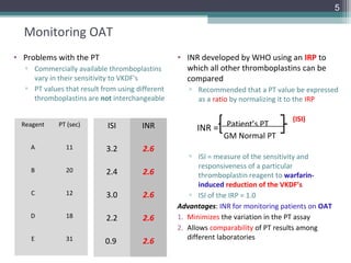
• Problems with the PT                            • INR developed by WHO using an IRP to
   ▫ Commercially available thromboplastins         which all other thromboplastins can be
     vary in their sensitivity to VKDF’s            compared
   ▫ PT values that result from using different      ▫ Recommended that a PT value be expressed
     thromboplastins are not interchangeable           as a ratio by normalizing it to the IRP

                                                                                    (ISI)
  Reagent    PT (sec)        ISI       INR             INR = Patient’s PT
                                                             GM Normal PT
    A          11           3.2         2.6
                                                     ▫ ISI = measure of the sensitivity and
                                                         responsiveness of a particular
    B          20           2.4         2.6              thromboplastin reagent to warfarin-
                                                         induced reduction of the VKDF’s
    C          12           3.0         2.6          ▫ ISI of the IRP = 1.0
                                                  Advantages: INR for monitoring patients on OAT
    D          18           2.2         2.6       1. Minimizes the variation in the PT assay
                                                  2. Allows comparability of PT results among
    E          31                                    different laboratories
                            0.9         2.6
6

   Limitations of Warfarin Therapy
   • PT/INR
     Limitations                 Consequence
     Slow onset of action        Need to overlap with a parenteral        Narrow therapeutic index
                                 anticoagulant
     Early procoagulant effect   Early prothrombotic effect

     Multiple drug/food          Frequent monitoring required
     interactions
     Genetic variation in        Variable dose requirement
     metabolism
     ADRs                        ~177,000/yr, ~30,000 ER visits/yr, ~3%
                                    major bleeds, ~0.6% fatal bleeds
     Narrow therapeutic index    Bleeding or thrombosis


         •   INR should ONLY be used for patients who stabilized on OAT
         •   INR should NEVER be used as a substitute for the PT in patients who are NOT on OAT
               1. Exponential nature of the INR may obscure interpretation of a mildly prolonged PT
WHY???            that may be suggestive of a coagulopathy
               2. Sufficient studies have NOT been done to demonstrate how well the INR correlates
                  with diagnosis or outcome
7

    aPTT
•   PTT – Langdell, Wagner and Brinkhous 1953
•   aPTT – Proctor and Rappaport – 1961
                                                             Ca2+
•   Measures
     a. Time required for fibrin clot to form when a
         plasma is incubated with an                          PL
         [activator + partial thromboplastin + Ca2+ ]          +
     a. Activation of the contact factors                  activator
        PK, HMWK, FXII, FXI
     a. Intrinsic pathway factors                          Plasma
        FXII, XI, IX, VIII
     a. Common pathway
        FX, V, II, I

•   Prolonged in
      1. Deficiencies of all factors except VII and XIII
      2. Presence of inhibitors
         Specific inhibitors – FVIII and FIX
         Nonspecific inhibitors—LA, Heparin, DTI’s
•   Shortened
      ▫ Elevated FVIII

•   Monitor Heparin Therapy
8


  Laboratory Monitoring of UFH
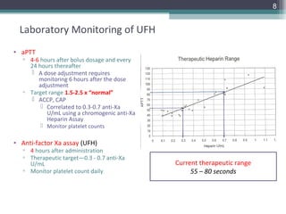
• aPTT
   ▫ 4-6 hours after bolus dosage and every
     24 hours thereafter
      A dose adjustment requires
        monitoring 6 hours after the dose
        adjustment
   ▫ Target range 1.5-2.5 x “normal”
      ACCP, CAP
          Correlated to 0.3-0.7 anti-Xa
           U/mL using a chromogenic anti-Xa
           Heparin Assay
          Monitor platelet counts

• Anti-factor Xa assay (UFH)
   ▫ 4 hours after administration
   ▫ Therapeutic target—0.3 - 0.7 anti-Xa
     U/mL                                     Current therapeutic range
   ▫ Monitor platelet count daily                  55 – 80 seconds
9

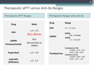
Therapeutic aPTT versus Anti-Xa Ranges

Therapeutic aPTT Ranges               Therapeutic Ranges with anti-Xa

                                      Drug           Range
     Drug                 Ratio
                                      UFH            0.3 – 0.7 U/mL
                      1.5 – 2.5
     UFH                                             2x/day
                   55.0 – 80.0 sec                        0.5 – 1.1 U/mL
                                      LMWH           1x/day
                        N/A                               1.1 – 2.0 u/mL
    LWMH
                   Not sensitive to
Pentasaccharide
                       LMWH
                                                     Prophylaxis of VTE
                                                           Trough – 0.14 – 0.19
  Argatroban          1.5 – 3.0       Fondaparinux
                                                           Peak – 0.39 – 0.50
                                                     Treatment of VTE
                                                           Trough – 0.46 – 0.62
    Lepirudin                                              Peak – 1.20 – 1.26
                      1.5 - 2.5
   (Refludan)
Reagent Factor Sensitivity
  • aPTT is sensitive to deficiencies of contact and intrinsic factors
  • MILD deficiency may result in a normal aPTT
11

    Fibrinogen
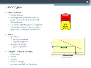
• Clauss Technique
  ▫ Functional assay
  ▫ Fibrinogen concentration is inversely      High conctr
      proportional to the thrombin time of      thrombin
      diluted plasma
  ▫ A reference (standard) curve is prepared      1:10
                                                dilution
      using known fibrinogen concentrations     plasma
      versus their respective thrombin times

•   Detects
    ▫ Quantitative
         Hypofibrinogenemia
         Hyperfibrinogenemia
    ▫ Qualitative deficiency
         Dysfibrinogenemia

•   Acute phase protein  elevated in
    ▫ Inflammation
    ▫ Trauma
    ▫ Infection
    ▫ Increases with age
    ▫ Associated with CVD and thrombosis
12

Fibrinogen
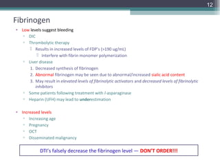
• Low levels suggest bleeding
   ▫ DIC
   ▫ Thrombolytic therapy
       Results in increased levels of FDP’s (>190 ug/mL)
           Interfere with fibrin monomer polymerization
   ▫ Liver disease
      1. Decreased synthesis of fibrinogen
      2. Abnormal fibrinogen may be seen due to abnormal/increased sialic acid content
      3. May result in elevated levels of fibrinolytic activators and decreased levels of fibrinolytic
         inhibitors
   ▫ Some patients following treatment with l-asparaginase
   ▫ Heparin (UFH) may lead to underestimation

• Increased levels
   ▫ Increasing age
   ▫ Pregnancy
   ▫ OCT
   ▫ Disseminated malignancy

             DTI’s falsely decrease the fibrinogen level — DON’T ORDER!!!
13

Thrombin Time / Reptilase Time
Thrombin Time
• Measure the conversion of fibrinogen to fibrin
• Cleaves fibrinopeptides A and B
• Screen for heparin contamination
• Prolonged
   ▫ Heparin therapy (UFH)
   ▫ Hypofibrinogenemia, Dysfibrinogenemia
   ▫ Paraproteins (Amyloidosis, Myeloma)
   ▫ Severe LD
   ▫ Elevated FDP’s
   ▫ DTI’s
   ▫ Bovine thrombin glues

Reptilase time
• Cleaves fibrinopeptide A only
   • Unaffected by Heparin
   • Unaffected by bovine thrombin glues
   • Same as above
14


D-Dimer
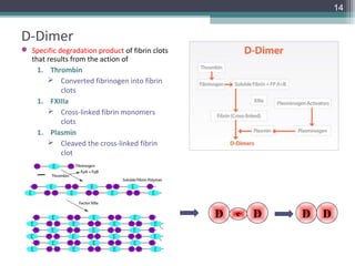
 Specific degradation product of fibrin clots
  that results from the action of
    1. Thrombin
        Converted fibrinogen into fibrin
            clots
    1. FXIIIa
        Cross-linked fibrin monomers
            clots
    1. Plasmin
        Cleaved the cross-linked fibrin
            clot


                                                 D   e   D   D   D
15


D-Dimer
• Monoclonal antibody raised
  against specific epitopes on D-
  dimer that react with cross-linked
  fibrin
   ▫ Does not react with
       Fibrinogen degradation
         products
       Non-cross-linked fibrin
         degradation products
   ▫ Ensures high specificity for D-
     dimer as a biomarker of fibrin
     formation and stabilization
16

Clinical Utility
• Diagnosis of VTE in combination with pretest clinical probability
  ▫ High negative predictive value for exclusion of DVT
  ▫ Poor positive predictive value for DVT
       Elevated in conditions unrelated to thrombosis
       Almost all patients with acute disease will have elevated D-dimer levels

• Clinical use
  ▫ Identification of individuals at increased risk thrombotic events (arterial and venous)
  ▫ Identification of individuals at increased risk of recurrent VTE
        Elevated levels following discontinuation of anticoagulant therapy
  ▫ Establishing of optimal duration of secondary prophylaxis after a first episode of VTE
  ▫ Pregnancy monitoring
  ▫ Diagnosis/monitoring of DIC
        Sensitive but NOT specific marker for DIC


        1. A positive D-dimer is NOT specific for VTE
        2. Negative D-dimer is highly unlikely for VTE
        3. The greatest utility of D-dimer is its negative predictive value for VTE!
17


    Fibrin(ogen) Degradation Products

• Patient plasma mixed with latex particles
  coated with monoclonal anti-FDP
  antibodies

• Positive FDP assay indicates
    ▫ Fibrin and/or fibrinogen is being
      degraded by plasmin

• Elevated FDPs
  ▫ Dysfibrinogenemia
  ▫ LD, DIC, DVT, PE, MI
  ▫ Thrombolytic therapy
  ▫ Primary and secondary
     fibrinogenolysis

•   What would the D-dimer and FSP levels be in a person who
    has a congenital FXIII deficiency?
•   What would the D-dimer and FSP levels be in individuals
    with FXIII deficiency due to DIC?
18


Algorithm of Mixing Tests


                  Prolongation of the Coagulation
                    Time of the Screening Test


              Mixing: Patient Plasma + Normal Plasma


            Correction                       No Correction


     Factor Deficiency      Lupus Anticoagulant        Specific Factor Inhibitor
19


Mixing Studies
• Look for the presence of:
  ▫ Factor Deficiency versus Inhibitor

            Step 1
            Mix and Run
                           TUBE 1       TUBE 2        TUBE 3
                                      300 uL PNP
                          1 mL PNP   300 uL patient    1 mL           Run immediately
                                        plasma        patient
                                                      plasma


                                                                    Incubate @ 37oC
            Step 2
            Mix and Run

                          TUBE 1       TUBE 2          TUBE 3
                                     300 uL PNP                  Run after 1 hour incubation
                           PNP         300 uL          Patient
                                       patient         plasma
                                       plasma
20


Interpreting Mixing Studies
 If the 50/50 corrects after the immediate and remains corrected after the 60 minute
  incubation
     Factor Deficiency
     Follow-up with specific factor assays

 If the 50/50 corrects after the immediate, but prolongs after the 60 minute incubation
     Time-dependent inhibitor – usually a specific factor inhibitor
     ~ 15% of LA’s may be time-and-temperature dependent
     Follow-up with a specific factor assay and specific factor inhibitor assay

 If the 50/50 prolongs after the immediate and remains prolonged after the 60 minute
  incubation
     Nonspecific inhibitor such as a lupus anticoagulant
     Follow-up with Lupus anticoagulant assay

 Mixing studies on samples minimally prolonged (<3 sec) may produce confusing results
    Addition of normal plasma sometimes dilutes a weak inhibitor  lead to a “false”
      correction
21


Mixing Study Panel
• Look for the presence of:
  ▫ Factor Deficiency versus Inhibitor   • Thrombin Time
                                           ▫ Rule-out the presence of
  aPTT Mixing Study Panel                    heparin

  1.aPTT Patient                         • PTT-FS
  2.50/50 mix            immediate
                                           ▫ Lupus-insensitive reagent for
  3.Normal Plasma
                                             the aPTT
  4.aPTT Patient                               Not prolonged in the
  5.50/50 mix            60 minutes
                                                presence of LA
  6.Normal Plasma                              Sensitive to FD

  7.Thrombin Time
  8.PTT-FS*
22


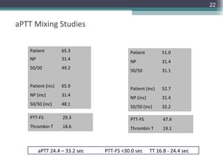
aPTT Mixing Studies

    Patient         65.3                  Patient         51.0
    NP              31.4
                                          NP              31.4
    50/50           49.2
                                          50/50           31.1


    Patient (inc)   65.9
                                          Patient (inc)   52.7
    NP (inc)        31.4
                                          NP (inc)        31.4
    50/50 (inc)     48.1
                                          50/50 (inc)     32.2

    PTT-FS          29.3                  PTT-FS           47.4
    Thrombin T      18.6                  Thrombin T       19.1


         aPTT 24.4 – 33.2 sec   PTT-FS <30.0 sec     TT 16.8 - 24.4 sec
23


Lupus Anticoagulant/APAs
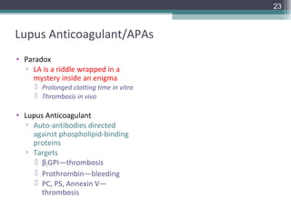
• Paradox
  ▫ LA is a riddle wrapped in a
    mystery inside an enigma
      Prolonged clotting time in vitro
      Thrombosis in vivo

• Lupus Anticoagulant
  ▫ Auto-antibodies directed
    against phospholipid-binding
    proteins
  ▫ Targets
      β2GPI—thrombosis
      Prothrombin—bleeding
      PC, PS, Annexin V—
       thrombosis
24


ISTH Criteria for Lupus Anticoagulant Testing

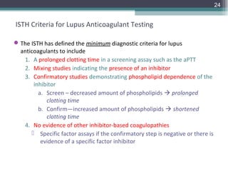
 The ISTH has defined the minimum diagnostic criteria for lupus
  anticoagulants to include
   1. A prolonged clotting time in a screening assay such as the aPTT
   2. Mixing studies indicating the presence of an inhibitor
   3. Confirmatory studies demonstrating phospholipid dependence of the
       inhibitor
         a. Screen – decreased amount of phospholipids  prolonged
            clotting time
         b. Confirm—increased amount of phospholipids  shortened
            clotting time
   4. No evidence of other inhibitor-based coagulopathies
       Specific factor assays if the confirmatory step is negative or there is
          evidence of a specific factor inhibitor
25


ISTH Criteria for Lupus Anticoagulant Testing

• Updated ISTH guidelines (2009) ISTH
  ▫ Pengo V, Tripodi A, Reber G, Rand JH, Ortel TL, Galli M, de Groot PG.
    Update of the guidelines for lupus anticoagulant detection. J Thromb
    Haemost 2009; 7: 1737–40

  ▫ Choice of tests
   1. Two tests based on different principles
   2. dRVVT should be the first test considered
   3. Seconds test should be a sensitive aPTT (low phospholipids and silica
      as activator)
   4. LA should be considered as positive if one of the two tests gives a
      positive result
26


  Detection of LA

• Assays
   dRVVT*      Clot-based assays
                                     SCT                dPT
   SCT*                                    Why do we see so few LA’s
                                            on the extrinsic side???
   HEX
   Kaolin CT
   dPT                             DRVVT
27


dRVVT Screen (Normal plasma)

                        X

          dRVVT

                       Xa

                                Prothrombin

                       Xa
   Phospholipid
                  Va   Ca2+
      (PF3)
                                Thrombin


                   Fibrinogen           Fibrin
28


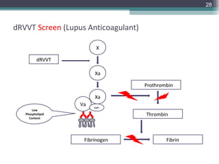
dRVVT Screen (Lupus Anticoagulant)

                        X

          dRVVT

                       Xa

                                     Prothrombin

                       Xa
                  Va   Ca2+
       Low
   Phospholipid                      Thrombin
     Content


                   Fibrinogen                Fibrin
29


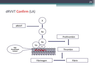
dRVVT Confirm (LA)

                          X

          dRVVT

                         Xa

                                  Prothrombin

                         Xa
                  Va     Ca2+
      High
   Phospholipid                   Thrombin
     Content


                     Fibrinogen           Fibrin
30


  Assays to Detect APA’s

• All LA’s are antiphospholipid antibodies, BUT not all APA’s are LA’s

• Antiphospholipid antibodies
                                          ELISA-based assays
       Anticardiolipin antibodies        (IgG, IgM, IgA)
      β2-Glycoprotein I antibodies


• Systematic reviews have consistently reported
   ▫ LA is a stronger risk factor than aCL aβ2GPI for both arterial and venous
     thrombosis and obstetric complications
   ▫ aCL and aβ2GPI only show some significant association with thrombosis
     and obstetric complications at high titer
   ▫ Independent of LA and aCL neither aβ2GPI nor antiprothrombin
     antibodies are associated with arterial or venous thrombosis
31


  Patients with LA on Warfarin

• LA can influence PT/INR  can lead to INRs that do not accurately reflect the true level
  of anticoagulation
     a. Use of the INR (to standardize PTs) may be invalid for some patients with LA
     b. To prevent supratherapeutic or subtherapeutic anticoagulation  these patients
        must be individually monitored with a test that is insensitive to LA

                                      Ann Intern Med. 1997;127:177-185


• Chromogenic Factor X
  • Therapeutic Range = 23-47%
       • [INR 4.0 (23%) – INR 2.0 (47%)]
32


The End