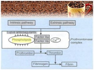
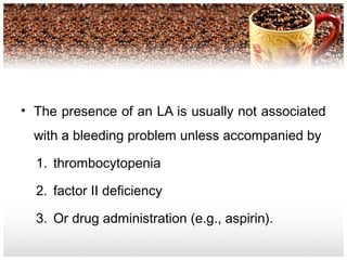
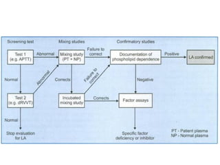
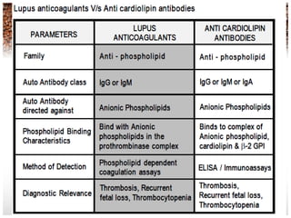
Lupus AnticoagulantsLupus Anticoagulants
ConleyConley
General Background
• Lupus anticoagulants (LA) were first described
in the year 1952 by Conley and HartmannConley and Hartmann in two
patients suffering from (SLE).
• It was given the name “Lupus” since it was
initially recognized in patients with SLE and the
name “Anticoagulant” due it`s invitro effect on
APTT and because the two patients diagnosed
as SLE developed bleeding complications.
• Lupus Anticoagulant (also known as lupus
antibody, LA, or lupus inhibitors) is an
immunoglobulin that binds to phospholipids
and proteins associated with the cell
membrane.
• Lupus anticoagulant is a misnomer as it is
actually a prothrombotic agent.
General Background
• It is now clear that Lupus anticoagulant is also
encountered in patients without SLE and is
associated with thrombosis rather than
abnormal bleeding.
• Thus LA is neither a syndrome nor a disease
but a laboratory phenomenon detected by
prolonged results with phospholipid dependent
clot based assays.
Lupus Anticoagulants
• Lupus Anticoagulants are phospholipid
“interfering” antibodies, which prolong
clotting tests that are responsive to
procoagulant phospholipid concentration
such as APTT.
• Lupus Anticoagulants are not corrected
by mixing with normal plasma but may be
corrected by the addition of
phospholipid.
Lupus Anticoagulants
• lupus anticoagulants are called nonspecific inhibitors.
• They are Auto antibodies
• Chronic lupus anticoagulants confer a 30% risk of
arterial or venous thrombosis; thus every acute care
laboratory must provide a means for their detection.
• The presence of an LA is usually not associated
with a bleeding problem unless accompanied by
1. thrombocytopenia
2. factor II deficiency
3. Or drug administration (e.g., aspirin).
Laboratory Diagnosis of Lupus
Anticoagulants
• The traditional screening methods used
for the laboratory diagnosis of APS
• Anticoagulants can be broadly
classified as follows:
A.Immunological assays
B. Clot based assays incorporating
phospholipids in the reagent
sysytem
A. Immunological Assays
• Anti-beta-2 glycoprotein I (anti-β2 GPI) and
Anticardiolipin antibodies (ACAs) are
antiphospholipid immunoglobulins that are IgG,
IgM.
• Antiphospholipid antibodies are a
heterogeneous group of autoantibodies
including ACA, LA, beta-2 glycoprotein-l
(β2GPI).
• Some patients withSome patients with elevated anti- β2GPI and/or
ACAs have been reported to also have an LA.have an LA.
Anti-Phospholipid Syndrom
• Several studies have shown that patients with
the LA and the closely related anti-
β2GPI/ACA are prone to
1. Recurrent venous and arterial thrombosis,
2. Recurrent spontaneous abortions,
This tendency has been described as the
antiphospholipid syndrome (APS).
• The criteria for diagnosing this syndrome are
divided into clinical and laboratory groups.
Anti-Phospholipid Antibody Assays
• APS is present if at least one of the
clinical criteriaclinical criteria and one of the lab.lab.
criteriacriteria that follow are met:
1.Clinical criteria: vascular thrombosis or
pregnancy morbidity
Laboratory criteria
A. Lupus anticoagulant present in plasma on two
or more occasions at least 12 weeks apart.
B. Anticardiolipin antibody, IgG or IgM positive
in medium or high titer on two or more
occasions at least 12 weeks apart
C. Anti-β2GPl antibody, IgG or IgM present on
two or more occasions at least 12 weeks
apart.
A. Immunological Assays
• Individuals with Lupus anticoagulants may also show the
presence of other anti phospholipid antibodies.
• The most frequent finding is the presence of Anti-
cardiolipin antibodies.
• The commonly employed method is the ELISA technique
• An important point to note is that Lupus anticoagulants
and anti-cardiolipin antibodies differ in their phospholipid-
binding characteristic hence detection of anti cardiolipin
antibodies is not specific for the presence of Lupus
anticoagulants though they may be present in the same
patient.
B. Clot based assays
1. APTT ( Activated Partial Thromboplastin
Time)
2. KCT (Kaolin Clotting Time)
3. PNP (Platelet Neutralization procedure)
4. dRVVT
APTT ( Activated Partial
Thromboplastin Time)
Since Lupus anticoagulants bind to the
phospholipid complex they do prolong
phospholipid based coagulation assays.
Logically the activated partial
thromboplastin time (APTT) is prolonged
and this property has been used for the
detection of LA using APTT reagents.
APTT shortcomings
• Different reagents have varying sensitivity
for the presence of Lupus anticoagulants on
the basis of their phospholipid composition.
• The APTT test is affected by the presence
of inhibitors to Factor VIII, IX and XI. In
order to rule out the presence of inhibitors
usually mixing studies are performed.
APTT
• The APTT test is also the test of choice for
monitoring heparin therapy. This reduces the
specificity of the APTT to LA.
• The presence of heparin can be ruled out by using a
thrombin time test. If thrombin time test shows
normal values, then the sample does not contain
heparin.
• But if the thrombin time test is abnormal, then
heparin neutralization test is used which includes
protamine sulphate.
KCT (Kaolin Clotting Time)
KCT is similar to APTT, the difference being
that KCT reagent is devoid of phospholipids and
incorporates Kaolin as contact activator.
The test is performed on a range of mixture of
normal and patient’s plasma.
Different patterns of response are obtained
indicating the presence of Lupus anticoagulants
or the deficiency of one or more coagulation
factors.
KCT (Kaolin Clotting Time(
The KCT test though sensitive is not specific
for LA, additionally:
•The test shows prolonged results with factor
VIII, IX, XI & XII deficiency or corresponding
inhibitors
•The test is also highly sensitive for the
presence of heparin
``1111
Patient Plasma normal Plasma
0% 100%
10% 90%
20% 80%
50% 50%
80% 20%
90% 10%
• A ratio of ≥1.2 is considered positive for a
lupus anticoagulant although reference ranges
should be established locally.
Platelet Neutralization procedure
• The PNP is based on the ability of
platelets to significantly correct in vitro
coagulation abnormalities.
• The disrupted platelet membranes
present in the freeze-thawed platelet
suspension neutralize phospholipid
antibodies present in the plasma of
patients with LA.
PNP
• After the patient plasma is mixed with
the freeze-thawed platelet suspension,
the APTT will be shortened when
compared with the original baseline
APTT. But if an inhibitor is directed
against specific coagulation factor, the
clotting time is not shortened.
PNP
InterpretationInterpretation
•A correction of the baseline APTT of a defined
amount of time (i.e., 3 to 5 seconds or more) by
the platelet suspension as compared with the
control is indicative of the presence of an LA.
The PNP test though useful did not gain wide
usage Due to limited stability the platelet
preparations loose their activities on storage
hence do not show reproducible results.
DDilute RRussell’s VViper VVenom TTime
• The test of choice for screening and
confirmation of LA
–Indicating the phospholipid dependence
of LA
–Achieving maximum sensitivty for the
precence of LA’s.
dRVVTdRVVT
• In general dRVVT based tests comprise of:In general dRVVT based tests comprise of:
– SCREENING REAGENT, containing limited
amount of phospholipids with RVV (Russell’s
Viper Venom)
– CONFIRMATION REAGENT, containing
additional phospholipids with same amount
of Russell’s Viper Venom, to confirm the
presence of phospholipid dependent Lupus
anticoagulants
Principle of dRVVT for LA
detection
• Russell’s Viper Venom directly activates
Factor V and X in presence of phospholipid
and calcium ions, bypassing Factor VII of the
extrinsic pathway and the contact and
antihaemophilic factors of the intrinsic
pathway.
• In normal plasma, in the absence of lupus
anticoagulants, Factor V and X is directly
activated by Russell’s Viper Venom, which in
presence of phospholipid and calcium ion leads
to clot formation.
Principle of dRVVT for LA
detection
• In patients with LA, autoantibodies bind
the epitopes of reagent phospholipids
thereby preventing the activation of
prothrombinase complex. This results in
a prolongation of clotting time with
SCREENING reagent.
• The CONFIRMATION reagent
incorporates additional phospholipids to
neutralize the LA.
• Once LA are neutralized clot formation
proceeds relatively uninterrupted
achieving a lower clotting time, to prove
the phospholipid dependence of the
autoantibodies
Principle of dRVVT for LA
detection
Interpretation of results with dRVVT
test
• If SCREEN TIME is prolonged, to confirm the
presence of lupus anticoagulants the plasma sample is
tested with CONFIRMATION REAGENT.
• If CONFIRM TIME results shows a lower clotting
time as compared to SCREEN TIME, it indicates the
presence of phospholipid dependant Lupus
Anticoagulants.
• Also the results can be expressed as ratio,
Interpretation of results with dRVVT
test
• The results expressed, as ratio is further
useful in classifying the patient as normal,
moderate, high and very high LA.
• N.V: 0.9-1.05
• If results of the ratio are borderline, mixing
studies may be done further with the sample
specimen.
Lupus anticoagulants

Lupus anticoagulants

  • 1.
  • 2.
  • 3.
    General Background • Lupusanticoagulants (LA) were first described in the year 1952 by Conley and HartmannConley and Hartmann in two patients suffering from (SLE). • It was given the name “Lupus” since it was initially recognized in patients with SLE and the name “Anticoagulant” due it`s invitro effect on APTT and because the two patients diagnosed as SLE developed bleeding complications.
  • 4.
    • Lupus Anticoagulant(also known as lupus antibody, LA, or lupus inhibitors) is an immunoglobulin that binds to phospholipids and proteins associated with the cell membrane. • Lupus anticoagulant is a misnomer as it is actually a prothrombotic agent.
  • 5.
    General Background • Itis now clear that Lupus anticoagulant is also encountered in patients without SLE and is associated with thrombosis rather than abnormal bleeding. • Thus LA is neither a syndrome nor a disease but a laboratory phenomenon detected by prolonged results with phospholipid dependent clot based assays.
  • 6.
    Lupus Anticoagulants • LupusAnticoagulants are phospholipid “interfering” antibodies, which prolong clotting tests that are responsive to procoagulant phospholipid concentration such as APTT. • Lupus Anticoagulants are not corrected by mixing with normal plasma but may be corrected by the addition of phospholipid.
  • 8.
    Lupus Anticoagulants • lupusanticoagulants are called nonspecific inhibitors. • They are Auto antibodies • Chronic lupus anticoagulants confer a 30% risk of arterial or venous thrombosis; thus every acute care laboratory must provide a means for their detection.
  • 9.
    • The presenceof an LA is usually not associated with a bleeding problem unless accompanied by 1. thrombocytopenia 2. factor II deficiency 3. Or drug administration (e.g., aspirin).
  • 11.
    Laboratory Diagnosis ofLupus Anticoagulants • The traditional screening methods used for the laboratory diagnosis of APS • Anticoagulants can be broadly classified as follows: A.Immunological assays B. Clot based assays incorporating phospholipids in the reagent sysytem
  • 12.
    A. Immunological Assays •Anti-beta-2 glycoprotein I (anti-β2 GPI) and Anticardiolipin antibodies (ACAs) are antiphospholipid immunoglobulins that are IgG, IgM. • Antiphospholipid antibodies are a heterogeneous group of autoantibodies including ACA, LA, beta-2 glycoprotein-l (β2GPI). • Some patients withSome patients with elevated anti- β2GPI and/or ACAs have been reported to also have an LA.have an LA.
  • 14.
    Anti-Phospholipid Syndrom • Severalstudies have shown that patients with the LA and the closely related anti- β2GPI/ACA are prone to 1. Recurrent venous and arterial thrombosis, 2. Recurrent spontaneous abortions, This tendency has been described as the antiphospholipid syndrome (APS). • The criteria for diagnosing this syndrome are divided into clinical and laboratory groups.
  • 15.
    Anti-Phospholipid Antibody Assays •APS is present if at least one of the clinical criteriaclinical criteria and one of the lab.lab. criteriacriteria that follow are met: 1.Clinical criteria: vascular thrombosis or pregnancy morbidity
  • 16.
    Laboratory criteria A. Lupusanticoagulant present in plasma on two or more occasions at least 12 weeks apart. B. Anticardiolipin antibody, IgG or IgM positive in medium or high titer on two or more occasions at least 12 weeks apart C. Anti-β2GPl antibody, IgG or IgM present on two or more occasions at least 12 weeks apart.
  • 17.
    A. Immunological Assays •Individuals with Lupus anticoagulants may also show the presence of other anti phospholipid antibodies. • The most frequent finding is the presence of Anti- cardiolipin antibodies. • The commonly employed method is the ELISA technique • An important point to note is that Lupus anticoagulants and anti-cardiolipin antibodies differ in their phospholipid- binding characteristic hence detection of anti cardiolipin antibodies is not specific for the presence of Lupus anticoagulants though they may be present in the same patient.
  • 18.
    B. Clot basedassays 1. APTT ( Activated Partial Thromboplastin Time) 2. KCT (Kaolin Clotting Time) 3. PNP (Platelet Neutralization procedure) 4. dRVVT
  • 19.
    APTT ( ActivatedPartial Thromboplastin Time) Since Lupus anticoagulants bind to the phospholipid complex they do prolong phospholipid based coagulation assays. Logically the activated partial thromboplastin time (APTT) is prolonged and this property has been used for the detection of LA using APTT reagents.
  • 20.
    APTT shortcomings • Differentreagents have varying sensitivity for the presence of Lupus anticoagulants on the basis of their phospholipid composition. • The APTT test is affected by the presence of inhibitors to Factor VIII, IX and XI. In order to rule out the presence of inhibitors usually mixing studies are performed.
  • 21.
    APTT • The APTTtest is also the test of choice for monitoring heparin therapy. This reduces the specificity of the APTT to LA. • The presence of heparin can be ruled out by using a thrombin time test. If thrombin time test shows normal values, then the sample does not contain heparin. • But if the thrombin time test is abnormal, then heparin neutralization test is used which includes protamine sulphate.
  • 22.
    KCT (Kaolin ClottingTime) KCT is similar to APTT, the difference being that KCT reagent is devoid of phospholipids and incorporates Kaolin as contact activator. The test is performed on a range of mixture of normal and patient’s plasma. Different patterns of response are obtained indicating the presence of Lupus anticoagulants or the deficiency of one or more coagulation factors.
  • 23.
    KCT (Kaolin ClottingTime( The KCT test though sensitive is not specific for LA, additionally: •The test shows prolonged results with factor VIII, IX, XI & XII deficiency or corresponding inhibitors •The test is also highly sensitive for the presence of heparin
  • 24.
    ``1111 Patient Plasma normalPlasma 0% 100% 10% 90% 20% 80% 50% 50% 80% 20% 90% 10%
  • 25.
    • A ratioof ≥1.2 is considered positive for a lupus anticoagulant although reference ranges should be established locally.
  • 26.
    Platelet Neutralization procedure •The PNP is based on the ability of platelets to significantly correct in vitro coagulation abnormalities. • The disrupted platelet membranes present in the freeze-thawed platelet suspension neutralize phospholipid antibodies present in the plasma of patients with LA.
  • 27.
    PNP • After thepatient plasma is mixed with the freeze-thawed platelet suspension, the APTT will be shortened when compared with the original baseline APTT. But if an inhibitor is directed against specific coagulation factor, the clotting time is not shortened.
  • 28.
    PNP InterpretationInterpretation •A correction ofthe baseline APTT of a defined amount of time (i.e., 3 to 5 seconds or more) by the platelet suspension as compared with the control is indicative of the presence of an LA. The PNP test though useful did not gain wide usage Due to limited stability the platelet preparations loose their activities on storage hence do not show reproducible results.
  • 29.
    DDilute RRussell’s VViperVVenom TTime • The test of choice for screening and confirmation of LA –Indicating the phospholipid dependence of LA –Achieving maximum sensitivty for the precence of LA’s.
  • 30.
    dRVVTdRVVT • In generaldRVVT based tests comprise of:In general dRVVT based tests comprise of: – SCREENING REAGENT, containing limited amount of phospholipids with RVV (Russell’s Viper Venom) – CONFIRMATION REAGENT, containing additional phospholipids with same amount of Russell’s Viper Venom, to confirm the presence of phospholipid dependent Lupus anticoagulants
  • 31.
    Principle of dRVVTfor LA detection • Russell’s Viper Venom directly activates Factor V and X in presence of phospholipid and calcium ions, bypassing Factor VII of the extrinsic pathway and the contact and antihaemophilic factors of the intrinsic pathway. • In normal plasma, in the absence of lupus anticoagulants, Factor V and X is directly activated by Russell’s Viper Venom, which in presence of phospholipid and calcium ion leads to clot formation.
  • 33.
    Principle of dRVVTfor LA detection • In patients with LA, autoantibodies bind the epitopes of reagent phospholipids thereby preventing the activation of prothrombinase complex. This results in a prolongation of clotting time with SCREENING reagent.
  • 34.
    • The CONFIRMATIONreagent incorporates additional phospholipids to neutralize the LA. • Once LA are neutralized clot formation proceeds relatively uninterrupted achieving a lower clotting time, to prove the phospholipid dependence of the autoantibodies Principle of dRVVT for LA detection
  • 35.
    Interpretation of resultswith dRVVT test • If SCREEN TIME is prolonged, to confirm the presence of lupus anticoagulants the plasma sample is tested with CONFIRMATION REAGENT. • If CONFIRM TIME results shows a lower clotting time as compared to SCREEN TIME, it indicates the presence of phospholipid dependant Lupus Anticoagulants. • Also the results can be expressed as ratio,
  • 36.
    Interpretation of resultswith dRVVT test • The results expressed, as ratio is further useful in classifying the patient as normal, moderate, high and very high LA. • N.V: 0.9-1.05 • If results of the ratio are borderline, mixing studies may be done further with the sample specimen.

Editor's Notes

  • #13 LA antibodies are directed against plasma proteins bound to anionic phospholipids aCL antibodies are directed against phospholipids bound to proteins Cardiolipin= mitochondrial phospholipid Causes a biologic false positive test for syphilis Can be IgA, M, or G (subclasses 1-4) IgG (esp G2) associated with a greater risk of APS Anti 2GPI antibodies are directed against a plasma protein that binds phospholipid with high affinity