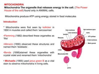
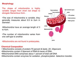
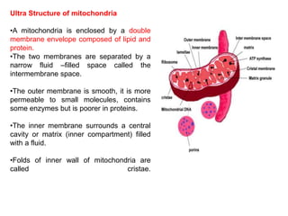
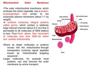
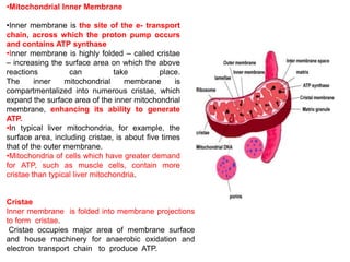
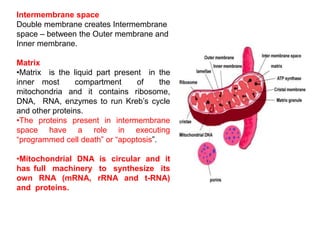
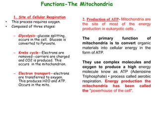
Dr. Manikandan Kathirvel M.Sc., Ph.D., (NET)
Assistant Professor,
Department of Life Sciences,
Kristu Jayanti College (Autonomous),
Reaccredited with "A++" Grade by NAAC
K. Narayanapura, Kothanur (PO)
Bengaluru
Structure and function of
Mitochondria
MITOCHONDRIA
Mitochondria The organelle that releases energy in the cell. (The Power
House of the cell) found only in ANIMAL cells.
Mitochondria produce ATP using energy stored in food molecules
Introduction
* Mitochondria were first seen by kollicker in
1850 in muscles and called them ‘sarcosomes’
•Flemming (1882) described these organelles as
‘filia’
•Altmann (1890) observed these structures and
named them ‘bioblasts’.
•Benda (1898)stained these organelles with
crystal violet and renamed them ‘mitochondria’
• Michaelis (1900) used janus green B as a vital
stain to observe mitochondria in living cells.
Morphology
The shape of mitochondria is highly
variable ranges from short rod shape to
elongate filamentous form .
•The size of mitochondria is variable ,they
generally measures about 0.5 to 2um in
diameter.
•Mitochondria have an average length of 3
to 4um.
•The number of mitochondria varies from
one cell type to another.
•Mitochondria are not found in prokaryotes.
Chemical Composition
• Mitochondria consists of protein-70 percent & lipids -25 -30percent.
•Mitochondria contain 0.5percent of RNA & traces of DNA .
•Mitochondrial DNA comprises about 1 percent of total cell DNA
•Mitochondria contain enzymes for oxidation, phosphorylation &electron transfer.
Ultra Structure of mitochondria
•A mitochondria is enclosed by a double
membrane envelope composed of lipid and
protein.
•The two membranes are separated by a
narrow fluid –filled space called the
intermembrane space.
•The outer membrane is smooth, it is more
permeable to small molecules, contains
some enzymes but is poorer in proteins.
•The inner membrane surrounds a central
cavity or matrix (inner compartment) filled
with a fluid.
•Folds of inner wall of mitochondria are
called cristae.
Mitochondrial Outer Membrane
•The outer mitochondrial membrane, which
encloses the entire organelle, has a protein-
to-phospholipid ratio similar to the
eukaryotic plasma membrane (about 1:1 by
weight).
•It contains numerous integral proteins
called porins, which contain a relatively
large internal channel (about 2-3 nm) that is
permeable to all molecules of 5000 daltons
or less. Thus Porin allows free movement
of molecules less than 5000 da within
and outside mitochondria.
Where as larger molecules or proteins
moves into the mitochondria through
transporters involving signal peptides
known as “mitochondrial targeting
sequence”.
Larger molecules, for example most
proteins, can only traverse the outer
membrane by active transport.
Cristae
Inner membrane is folded into membrane projections
to form cristae.
Cristae occupies major area of membrane surface
and house machinery for anaerobic oxidation and
electron transport chain to produce ATP.
•Mitochondrial Inner Membrane
•Inner membrane is the site of the e- transport
chain, across which the proton pump occurs
and contains ATP synthase
•Inner membrane is highly folded – called cristae
– increasing the surface area on which the above
reactions can take place.
The inner mitochondrial membrane is
compartmentalized into numerous cristae, which
expand the surface area of the inner mitochondrial
membrane, enhancing its ability to generate
ATP.
•In typical liver mitochondria, for example, the
surface area, including cristae, is about five times
that of the outer membrane.
•Mitochondria of cells which have greater demand
for ATP, such as muscle cells, contain more
cristae than typical liver mitochondria.
Intermembrane space
Double membrane creates Intermembrane
space – between the Outer membrane and
Inner membrane.
Matrix
•Matrix is the liquid part present in the
inner most compartment of the
mitochondria and it contains ribosome,
DNA, RNA, enzymes to run Kreb’s cycle
and other proteins.
•The proteins present in intermembrane
space have a role in executing
“programmed cell death” or “apoptosis”.
•Mitochondrial DNA is circular and it
has full machinery to synthesize its
own RNA (mRNA, rRNA and t-RNA)
and proteins.
2. Production of ATP- Mitochondria are
the site of most of the energy
production in eukaryotic cells .
The primary function of
mitochondria is to convert organic
materials into cellular energy in the
form of ATP.
They use complex molecules and
oxygen to produce a high energy
molecule know as ATP (Adenosine
Triphosphate) • process called aerobic
respiration. Energy production the
mitochondria has been called
the "powerhouse of the cell".
Functions-The Mitochondria
1. Site of Cellular Respiration
• This process requires oxygen.
• Composed of three stages:
– Glycolysis--glucose splitting,
occurs in the cell. Glucose is
converted to Pyruvate.
– Krebs cycle--Electrons are
removed--carriers are charged
and CO2 is produced. This
occurs in the mitochondrion.
– Electron transport--electrons
are transferred to oxygen.
This produces H2O and ATP.
Occurs in the mito.
Inheritance
(mitochondrial DNA, unlike nuclear DNA, is
inherited exclusively from the mother, while
nuclear DNA is inherited from both parents.
Mitochondria are found in eggs but not the
head of sperm cells, therefore mtDNA is only
inherited from mothers. mtDNA does not rec
ombine during meiosis. The process of
recombination in nuclear DNA mixes sections
of DNA from the mother and father, creating
a garbled genetic history.)
3. All the mitochondria in your body came from your mother. • Mitochondria are
not part of the genetic code in the nucleus of your cells.
Fathers only give genes to their children. • Mothers give genes and cytoplasm to
their children in their egg cells. • Since mitochondria are in the cytoplasm and
reproduce themselves they only are inherited from mothers. Geneticists have
used this curious feature of mitochondria to study maternal family lines and
rates of evolution. Used to track tree of a family.
Functions-The Mitochondria
4. Generation of Reactive Oxygen Species (ROS) in immune cells to kill
infectious agents.
5. Mitochondria also play an important role in many metabolic tasks, such as: •
Apoptosis-Programmed cell death • Cellular proliferation • Heme synthesis •
Steroid synthesis.
Role in programmed cell death or “apoptosis”. Apoptosis is the programmed cell
death involving a series of events involving cellular metalloprotease known as
caspases.
6. Mitochondria are very abundant in cells that require lots of energy. • Ex:-
Muscle
7. Unique • Mitochondria have their own circular DNA • have their own
Ribosomes. (The DNA in the cell nucleus does not code for the construction of
mitochondria. )
8. Heat production (enabling the organism to stay warm).
9. Some mitochondrial functions are performed only in specific types of cells. For
example, mitochondria in liver cells contain enzymes that allow them to detoxify
ammonia, a waste product of protein metabolism. A mutation in the genes
regulating any of these functions can result in a variety of mitochondrial
diseases.
Functions-The Mitochondria
Mitochondrial genome
The mitochondrial genome is circular, whereas the nuclear genome is linear.
The mitochondrial genome is built of 16,569 DNA base pairs, whereas the
nuclear genome is made of 3.3 billion DNA base pairs. The mitochondrial
genome contains 37 genes that encode 13 proteins, 22 tRNAs, and 2 rRNAs.
Mitochondrial DNA replication – D loop Model
1. The mitochondrial genome is a circle, 16.6 kb of DNA.
2. The two strands are notably different in base composition, leading to one strand being
“heavy” (the H strand) and the other light (the L strand).
3. Both strands encode genes, although more are on the H strand.
4. The D loop is also the site where most of replication and transcription is controlled.
1. Transcription in human mitochondria is driven by a DNA-dependant RNA
polymerase called POLRMT, which is structurally similar to RNA
polymerases in T3 and T7 bacteriophages.
2. This includes high sequence homology to the C-terminal catalytic core of the
enzyme.
3. At the N-terminal domain, POLRMT also contains two pentatricopeptide repeat
(PPR) domains, commonly found in RNA-associated proteins, where they are
required for site-specific interactions.
4. The initiation of transcription requires the association of POLRMT with
mitochondrial transcription factor A (TFAM) and mitochondrial
transcription factor B2 (TFB2M). TFAM is a DNA-binding protein, which, in
addition to transcription activation, also packages DNA in the nucleoid.
5. TFB2M, the key function of this protein is DNA melting during the initiation of
transcription.
Almost the entire
mitochondrial genome
is transcribed as long
polycistronic
transcripts.
Mitochondrial Translation
1. Mitochondrial translation is fully dependent on various nuclear-encoded regulatory
proteins.
2. In the mammalian mitochondria, the mitochondrial initiation factors, mtIF2 and
mtIF3, control the initiation of translation.
3. Elongation of translation is mediated by mitochondrial elongation factors, EFTu
(TUFM), EFTs (TSFM) and EFGM (GFM1).
4. Termination of mitochondrial translation is finally triggered by the presence of a stop
codon at the A-site. Four mitochondrial proteins with homology to ribosome release
factors have been identified in humans, including mtRF1, mtRF1a, C12orf65 and
ICT1.

Lecture: Structure and Function of Mitochondria.pptx

  • 1.
    Dr. Manikandan KathirvelM.Sc., Ph.D., (NET) Assistant Professor, Department of Life Sciences, Kristu Jayanti College (Autonomous), Reaccredited with "A++" Grade by NAAC K. Narayanapura, Kothanur (PO) Bengaluru Structure and function of Mitochondria
  • 2.
    MITOCHONDRIA Mitochondria The organellethat releases energy in the cell. (The Power House of the cell) found only in ANIMAL cells. Mitochondria produce ATP using energy stored in food molecules Introduction * Mitochondria were first seen by kollicker in 1850 in muscles and called them ‘sarcosomes’ •Flemming (1882) described these organelles as ‘filia’ •Altmann (1890) observed these structures and named them ‘bioblasts’. •Benda (1898)stained these organelles with crystal violet and renamed them ‘mitochondria’ • Michaelis (1900) used janus green B as a vital stain to observe mitochondria in living cells.
  • 3.
    Morphology The shape ofmitochondria is highly variable ranges from short rod shape to elongate filamentous form . •The size of mitochondria is variable ,they generally measures about 0.5 to 2um in diameter. •Mitochondria have an average length of 3 to 4um. •The number of mitochondria varies from one cell type to another. •Mitochondria are not found in prokaryotes. Chemical Composition • Mitochondria consists of protein-70 percent & lipids -25 -30percent. •Mitochondria contain 0.5percent of RNA & traces of DNA . •Mitochondrial DNA comprises about 1 percent of total cell DNA •Mitochondria contain enzymes for oxidation, phosphorylation &electron transfer.
  • 4.
    Ultra Structure ofmitochondria •A mitochondria is enclosed by a double membrane envelope composed of lipid and protein. •The two membranes are separated by a narrow fluid –filled space called the intermembrane space. •The outer membrane is smooth, it is more permeable to small molecules, contains some enzymes but is poorer in proteins. •The inner membrane surrounds a central cavity or matrix (inner compartment) filled with a fluid. •Folds of inner wall of mitochondria are called cristae.
  • 5.
    Mitochondrial Outer Membrane •Theouter mitochondrial membrane, which encloses the entire organelle, has a protein- to-phospholipid ratio similar to the eukaryotic plasma membrane (about 1:1 by weight). •It contains numerous integral proteins called porins, which contain a relatively large internal channel (about 2-3 nm) that is permeable to all molecules of 5000 daltons or less. Thus Porin allows free movement of molecules less than 5000 da within and outside mitochondria. Where as larger molecules or proteins moves into the mitochondria through transporters involving signal peptides known as “mitochondrial targeting sequence”. Larger molecules, for example most proteins, can only traverse the outer membrane by active transport.
  • 6.
    Cristae Inner membrane isfolded into membrane projections to form cristae. Cristae occupies major area of membrane surface and house machinery for anaerobic oxidation and electron transport chain to produce ATP. •Mitochondrial Inner Membrane •Inner membrane is the site of the e- transport chain, across which the proton pump occurs and contains ATP synthase •Inner membrane is highly folded – called cristae – increasing the surface area on which the above reactions can take place. The inner mitochondrial membrane is compartmentalized into numerous cristae, which expand the surface area of the inner mitochondrial membrane, enhancing its ability to generate ATP. •In typical liver mitochondria, for example, the surface area, including cristae, is about five times that of the outer membrane. •Mitochondria of cells which have greater demand for ATP, such as muscle cells, contain more cristae than typical liver mitochondria.
  • 7.
    Intermembrane space Double membranecreates Intermembrane space – between the Outer membrane and Inner membrane. Matrix •Matrix is the liquid part present in the inner most compartment of the mitochondria and it contains ribosome, DNA, RNA, enzymes to run Kreb’s cycle and other proteins. •The proteins present in intermembrane space have a role in executing “programmed cell death” or “apoptosis”. •Mitochondrial DNA is circular and it has full machinery to synthesize its own RNA (mRNA, rRNA and t-RNA) and proteins.
  • 8.
    2. Production ofATP- Mitochondria are the site of most of the energy production in eukaryotic cells . The primary function of mitochondria is to convert organic materials into cellular energy in the form of ATP. They use complex molecules and oxygen to produce a high energy molecule know as ATP (Adenosine Triphosphate) • process called aerobic respiration. Energy production the mitochondria has been called the "powerhouse of the cell". Functions-The Mitochondria 1. Site of Cellular Respiration • This process requires oxygen. • Composed of three stages: – Glycolysis--glucose splitting, occurs in the cell. Glucose is converted to Pyruvate. – Krebs cycle--Electrons are removed--carriers are charged and CO2 is produced. This occurs in the mitochondrion. – Electron transport--electrons are transferred to oxygen. This produces H2O and ATP. Occurs in the mito.
  • 9.
    Inheritance (mitochondrial DNA, unlikenuclear DNA, is inherited exclusively from the mother, while nuclear DNA is inherited from both parents. Mitochondria are found in eggs but not the head of sperm cells, therefore mtDNA is only inherited from mothers. mtDNA does not rec ombine during meiosis. The process of recombination in nuclear DNA mixes sections of DNA from the mother and father, creating a garbled genetic history.) 3. All the mitochondria in your body came from your mother. • Mitochondria are not part of the genetic code in the nucleus of your cells. Fathers only give genes to their children. • Mothers give genes and cytoplasm to their children in their egg cells. • Since mitochondria are in the cytoplasm and reproduce themselves they only are inherited from mothers. Geneticists have used this curious feature of mitochondria to study maternal family lines and rates of evolution. Used to track tree of a family. Functions-The Mitochondria
  • 10.
    4. Generation ofReactive Oxygen Species (ROS) in immune cells to kill infectious agents. 5. Mitochondria also play an important role in many metabolic tasks, such as: • Apoptosis-Programmed cell death • Cellular proliferation • Heme synthesis • Steroid synthesis. Role in programmed cell death or “apoptosis”. Apoptosis is the programmed cell death involving a series of events involving cellular metalloprotease known as caspases. 6. Mitochondria are very abundant in cells that require lots of energy. • Ex:- Muscle 7. Unique • Mitochondria have their own circular DNA • have their own Ribosomes. (The DNA in the cell nucleus does not code for the construction of mitochondria. ) 8. Heat production (enabling the organism to stay warm). 9. Some mitochondrial functions are performed only in specific types of cells. For example, mitochondria in liver cells contain enzymes that allow them to detoxify ammonia, a waste product of protein metabolism. A mutation in the genes regulating any of these functions can result in a variety of mitochondrial diseases. Functions-The Mitochondria
  • 11.
    Mitochondrial genome The mitochondrialgenome is circular, whereas the nuclear genome is linear. The mitochondrial genome is built of 16,569 DNA base pairs, whereas the nuclear genome is made of 3.3 billion DNA base pairs. The mitochondrial genome contains 37 genes that encode 13 proteins, 22 tRNAs, and 2 rRNAs.
  • 12.
    Mitochondrial DNA replication– D loop Model 1. The mitochondrial genome is a circle, 16.6 kb of DNA. 2. The two strands are notably different in base composition, leading to one strand being “heavy” (the H strand) and the other light (the L strand). 3. Both strands encode genes, although more are on the H strand. 4. The D loop is also the site where most of replication and transcription is controlled.
  • 13.
    1. Transcription inhuman mitochondria is driven by a DNA-dependant RNA polymerase called POLRMT, which is structurally similar to RNA polymerases in T3 and T7 bacteriophages. 2. This includes high sequence homology to the C-terminal catalytic core of the enzyme. 3. At the N-terminal domain, POLRMT also contains two pentatricopeptide repeat (PPR) domains, commonly found in RNA-associated proteins, where they are required for site-specific interactions. 4. The initiation of transcription requires the association of POLRMT with mitochondrial transcription factor A (TFAM) and mitochondrial transcription factor B2 (TFB2M). TFAM is a DNA-binding protein, which, in addition to transcription activation, also packages DNA in the nucleoid. 5. TFB2M, the key function of this protein is DNA melting during the initiation of transcription. Almost the entire mitochondrial genome is transcribed as long polycistronic transcripts.
  • 14.
    Mitochondrial Translation 1. Mitochondrialtranslation is fully dependent on various nuclear-encoded regulatory proteins. 2. In the mammalian mitochondria, the mitochondrial initiation factors, mtIF2 and mtIF3, control the initiation of translation. 3. Elongation of translation is mediated by mitochondrial elongation factors, EFTu (TUFM), EFTs (TSFM) and EFGM (GFM1). 4. Termination of mitochondrial translation is finally triggered by the presence of a stop codon at the A-site. Four mitochondrial proteins with homology to ribosome release factors have been identified in humans, including mtRF1, mtRF1a, C12orf65 and ICT1.