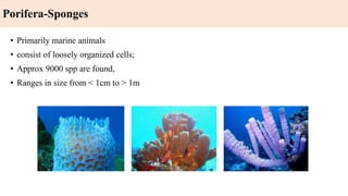
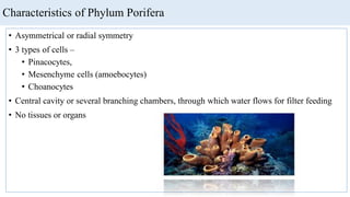
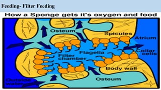
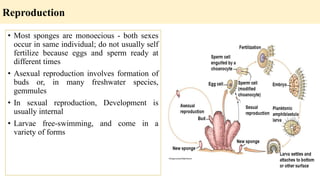
Phylum Porifera
Sponges
Lecture 3
Porifera-Sponges
• Primarily marine animals
• consist of loosely organized cells;
• Approx 9000 spp are found,
• Ranges in size from < 1cm to > 1m
Characteristics of Phylum Porifera
• Asymmetrical or radial symmetry
• 3 types of cells –
• Pinacocytes,
• Mesenchyme cells (amoebocytes)
• Choanocytes
• Central cavity or several branching chambers, through which water flows for filter feeding
• No tissues or organs
Cell types and Body wall
Sponge cells are specialized for particular functions (division of labor)
a. Pinacocytes
• These cells are the “skin cells”of sponges. They line the exterior of the sponge body wall.
They are thin, leathery and tightly packed together.
• may be slightly contractile and help sponge change shape.
• Some pinacocytes specialized into porocytes, which regulate water circulation
b. Jelly like layer under pinacocytes is termed mesohyl . Mesenchyme cells are amoeboid,
and move about in the mesohyl. Specialized for reproduction, transporting and storing
food, secreting skeletal elements ( spicules )
c. Beneath mesenchyme, lining inner chambers are choanocytes - collar cells . Flagellated
cells with ring of microvilli surrounding flagella. Microfilaments connect microvilli,
forming a net that helps filter edible particles
Microscopic View of Poriferan Wall
Skeleton
• Sponges are supported by skeleton that may consist of spicules - needlelike spikes
• Spicules are formed by amoeboid cells
• Made of CaCO3 or silica
• May take on a variety of shapes
• Alternatively, skeleton may be made of spongin, a fibrous protein made of collagen - dried
beaten and washed to produce commercial sponges
Spicules and their types
Water currents and body forms
• Sponges lives depend on the
water currents that coenocytes
create
• Water brings food and O2,
removes wastes
• Methods of food filtration and
circulation reflect body forms in
the phylum
Body forms
Three types of body forms:
a. Ascon body form
• Simplest and least common.
• Vaselike form ostia are outer openings of
porocytes and lead directly to chamber called
spongocoel
• Choanocytes line spongocoel and their flagellar
movements draw water into the spongocoel thru
the ostia
• Water exits sponge thru osculum, single large
opening at the top of the sponge
b. Sycon body form
• Sponge wall appears folded
• Water enters through dermal pores, which are
openings of incurrent canals
• Pores in body walls open to radial canals, and
radial canals lead to spongocoel
• Choanocytes line radial canals and beating of
flagella moves water from ostia, through
incurrent and radial canals, to spongocoel and
out the osculum.
Body forms
c. Leucon body forms
• An extensively branched canal system.
• Water enters the ostium and moves through
branched incurrent canals incurrent canals lead
to choanocyte lined chambers.
• Canals leading away from the chambers are
called excurrent canals
• Proliferation of chambers and canals has resulted
in absence of spongocoel.
• Often there are multiple exit points for water
leaving sponge
Body forms
Maintenance functions
1. Sponges feed on particles that range in size from .1 to 50 um.
a. Bacteria
b. Microscopic algae
c. Protists
d. Other suspended particles
2. Important in reducing coastal turbidity as 1 leucon sponge, 1 cm in diameter and 10 cm
high, filters 20 liters of water/day
3. A few sponges are carnivorous - catch small crustaceans (deep water) with spicule-covered
filaments.
4. Feeding methods-choanocytes filter small suspended particles.
a. Water passes thru collar near base and moves into spongocoel at open end of collar
b. Suspended food is trapped on collar and moved along microvilli to base of collar,
where it is incorporated into a food vacuole
c. Lysozymal enzymes and pH changes digest particle in vacuole
Feeding- Filter Feeding
5. Partly digested food passed to amoeboid cells, that distribute it
6. Pinacocytes lining incurrent canals may phagocytize larger food particles. Sponges may
also absorb nutrients in sea water thru active transport
7. Sponges get rid of waste thru diffusion, since all cells are in close contact with water
8. Sponges have no nerve cells for communication/coordination, but somehow choanocytes
can cease activities more or less simultaneously, ceasing water circulation
9. Chemical messages sent by amoeboid cells is one possible method
Maintenance functions
Reproduction
• Most sponges are monoecious - both sexes
occur in same individual; do not usually self
fertilize because eggs and sperm ready at
different times
• Asexual reproduction involves formation of
buds or, in many freshwater species,
gemmules
• In sexual reproduction, Development is
usually internal
• Larvae free-swimming, and come in a
variety of forms
Classes of Porifera
a. Hexactinellida
• Glass sponges, siliceous lattice, syconoid forms
• Spicules – hexaxons
• Lattice – siliceous w/ sieve plate over osculum
• Basal spicules w/ tufts for soft sediment
• Structure and habitat, Individualized cup, urns or vase
shape
• Body wall- w/out pinacoderm, syncitium externally and
internally
• Deep water (200 meters-abyss) / Cosmopolitan w/ more
in Antarctic
b. Class Calcarea (Calcispongeae)
• Spicules - Mon-, tri-, or tetraxon shapes
• Calcium carbonate, No spongin
• Small (<10 cm) Occupy shallow water
• Cosmopolitan
• All 3 body types
c. Class Demospongiae
• Most common form,
• leuconoid forms Spicules - Tri- or tetraxon Along with spongin
• Brightly colored, Shape reflects habitat & resources available
• Encrusting on vertical surfaces or in crevices
• Tubular (w/ branching) on limited substrates (conserves space)
• Shallow to deep water Algal symbionts- non-motile zooxanthella or cyanobacters in mesohyl or
amebocytes
d. Class Sclerospongiae
• Leuconoid forms
• Found in grottos or coral tunnels Internal siliceous spicules & spngin
• External calcareous portion
Classes of Porifera
Calcispongiae Domospongiae Sclerospongiae

Lecture 3 (1) porifera sponges

  • 1.
  • 2.
    Porifera-Sponges • Primarily marineanimals • consist of loosely organized cells; • Approx 9000 spp are found, • Ranges in size from < 1cm to > 1m
  • 3.
    Characteristics of PhylumPorifera • Asymmetrical or radial symmetry • 3 types of cells – • Pinacocytes, • Mesenchyme cells (amoebocytes) • Choanocytes • Central cavity or several branching chambers, through which water flows for filter feeding • No tissues or organs
  • 4.
    Cell types andBody wall Sponge cells are specialized for particular functions (division of labor) a. Pinacocytes • These cells are the “skin cells”of sponges. They line the exterior of the sponge body wall. They are thin, leathery and tightly packed together. • may be slightly contractile and help sponge change shape. • Some pinacocytes specialized into porocytes, which regulate water circulation b. Jelly like layer under pinacocytes is termed mesohyl . Mesenchyme cells are amoeboid, and move about in the mesohyl. Specialized for reproduction, transporting and storing food, secreting skeletal elements ( spicules ) c. Beneath mesenchyme, lining inner chambers are choanocytes - collar cells . Flagellated cells with ring of microvilli surrounding flagella. Microfilaments connect microvilli, forming a net that helps filter edible particles
  • 5.
    Microscopic View ofPoriferan Wall
  • 6.
    Skeleton • Sponges aresupported by skeleton that may consist of spicules - needlelike spikes • Spicules are formed by amoeboid cells • Made of CaCO3 or silica • May take on a variety of shapes • Alternatively, skeleton may be made of spongin, a fibrous protein made of collagen - dried beaten and washed to produce commercial sponges Spicules and their types
  • 7.
    Water currents andbody forms • Sponges lives depend on the water currents that coenocytes create • Water brings food and O2, removes wastes • Methods of food filtration and circulation reflect body forms in the phylum
  • 8.
    Body forms Three typesof body forms: a. Ascon body form • Simplest and least common. • Vaselike form ostia are outer openings of porocytes and lead directly to chamber called spongocoel • Choanocytes line spongocoel and their flagellar movements draw water into the spongocoel thru the ostia • Water exits sponge thru osculum, single large opening at the top of the sponge
  • 9.
    b. Sycon bodyform • Sponge wall appears folded • Water enters through dermal pores, which are openings of incurrent canals • Pores in body walls open to radial canals, and radial canals lead to spongocoel • Choanocytes line radial canals and beating of flagella moves water from ostia, through incurrent and radial canals, to spongocoel and out the osculum. Body forms
  • 10.
    c. Leucon bodyforms • An extensively branched canal system. • Water enters the ostium and moves through branched incurrent canals incurrent canals lead to choanocyte lined chambers. • Canals leading away from the chambers are called excurrent canals • Proliferation of chambers and canals has resulted in absence of spongocoel. • Often there are multiple exit points for water leaving sponge Body forms
  • 11.
    Maintenance functions 1. Spongesfeed on particles that range in size from .1 to 50 um. a. Bacteria b. Microscopic algae c. Protists d. Other suspended particles 2. Important in reducing coastal turbidity as 1 leucon sponge, 1 cm in diameter and 10 cm high, filters 20 liters of water/day 3. A few sponges are carnivorous - catch small crustaceans (deep water) with spicule-covered filaments. 4. Feeding methods-choanocytes filter small suspended particles. a. Water passes thru collar near base and moves into spongocoel at open end of collar b. Suspended food is trapped on collar and moved along microvilli to base of collar, where it is incorporated into a food vacuole c. Lysozymal enzymes and pH changes digest particle in vacuole
  • 12.
  • 13.
    5. Partly digestedfood passed to amoeboid cells, that distribute it 6. Pinacocytes lining incurrent canals may phagocytize larger food particles. Sponges may also absorb nutrients in sea water thru active transport 7. Sponges get rid of waste thru diffusion, since all cells are in close contact with water 8. Sponges have no nerve cells for communication/coordination, but somehow choanocytes can cease activities more or less simultaneously, ceasing water circulation 9. Chemical messages sent by amoeboid cells is one possible method Maintenance functions
  • 14.
    Reproduction • Most spongesare monoecious - both sexes occur in same individual; do not usually self fertilize because eggs and sperm ready at different times • Asexual reproduction involves formation of buds or, in many freshwater species, gemmules • In sexual reproduction, Development is usually internal • Larvae free-swimming, and come in a variety of forms
  • 15.
    Classes of Porifera a.Hexactinellida • Glass sponges, siliceous lattice, syconoid forms • Spicules – hexaxons • Lattice – siliceous w/ sieve plate over osculum • Basal spicules w/ tufts for soft sediment • Structure and habitat, Individualized cup, urns or vase shape • Body wall- w/out pinacoderm, syncitium externally and internally • Deep water (200 meters-abyss) / Cosmopolitan w/ more in Antarctic
  • 16.
    b. Class Calcarea(Calcispongeae) • Spicules - Mon-, tri-, or tetraxon shapes • Calcium carbonate, No spongin • Small (<10 cm) Occupy shallow water • Cosmopolitan • All 3 body types c. Class Demospongiae • Most common form, • leuconoid forms Spicules - Tri- or tetraxon Along with spongin • Brightly colored, Shape reflects habitat & resources available • Encrusting on vertical surfaces or in crevices • Tubular (w/ branching) on limited substrates (conserves space) • Shallow to deep water Algal symbionts- non-motile zooxanthella or cyanobacters in mesohyl or amebocytes d. Class Sclerospongiae • Leuconoid forms • Found in grottos or coral tunnels Internal siliceous spicules & spngin • External calcareous portion Classes of Porifera
  • 17.