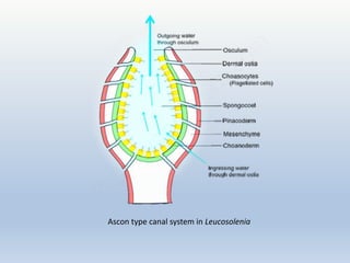
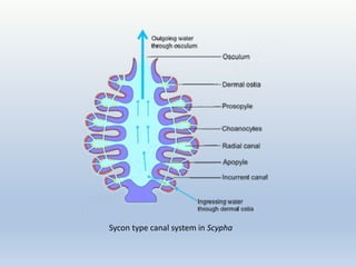
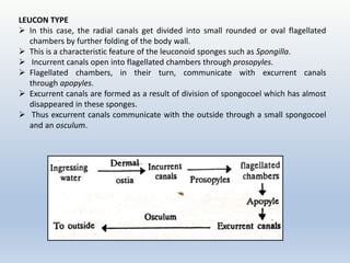
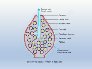
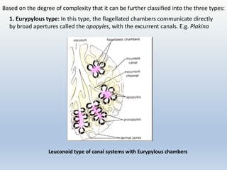
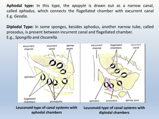
Canal system in
sponges
Asst.Prof.Devdhe Geetanjali Rajendra
M.Sc.Zoology NET,SET
Department of Zoology
New Arts,Commerce and Science College,Ahmednagar(Autonomous)
Sponges,are pore bearing,multicellular,diploblastic animals that belong to
phylum Porifera
Body of all sponges is perforated by large number of pores called ostia
through which water enters Inside body and flows through a system of criss-
crossing canals known as canal system
Three main types of canal systems in the order of increasing complexity are
Asconoid, Syconoid and Leuconoid type.
Significance of canal system:
Canal system helps the sponges in
(i) Nutrition
(ii) Respiration
(iii) Excretion
(iv) Reproduction
ASCON TYPE
Simplest type of canal system
Found in asconoid sponges e.g. Leucosolenia and developmental stages of
Syconoid sponges
Ostia are present on the surface of body and lead directly into the
spongocoel which is lined by flagellated choanocyte cells.
Spongocoel opens to the outside through a narrow circular opening,
the osculum located at the distal free end of the sponge body.
Water enters through ostia into spongocoel and goes out of body through
the osculum.
Ingressing water
Through Ostia
Spongocoel
Through Osculum
Outside
Ascon type canal system in Leucosolenia
SYCON TYPE
Characteristic of syconoid sponges, e.g. Scypha and Grantia.
Body wall is secondarily folded to form incurrent and radial canals, which open into
the spongocoel by an opening called apopyle.
Ostia open into the incurrent canals, which lead into adjacent radial canals through
minute openings called prospyles.
Radial canals are the flagellated chambers that open into central spongocoel by
internal openings called apopyles.
Spongocoel is a narrow, without flagellated cells but is lined by pinacocytes and
opens to exterior through the osculum.
In more complex sycon type e.g. Grantia, the incurrent canals travel along an
irregular course through the tissue and connect to the radial canals, thus forming large
sub-dermal spaces.
Sycon type canal system in Scypha
LEUCON TYPE
 In this case, the radial canals get divided into small rounded or oval flagellated
chambers by further folding of the body wall.
 This is a characteristic feature of the leuconoid sponges such as Spongilla.
 Incurrent canals open into flagellated chambers through prosopyles.
 Flagellated chambers, in their turn, communicate with excurrent canals
through apopyles.
 Excurrent canals are formed as a result of division of spongocoel which has almost
disappeared in these sponges.
 Thus excurrent canals communicate with the outside through a small spongocoel
and an osculum.
Leucon type canal system in Spongilla
1. Eurypylous type: In this type, the flagellated chambers communicate directly
by broad apertures called the apopyles, with the excurrent canals. E.g. Plakina
Based on the degree of complexity that it can be further classified into the three types:
Leuconoid type of canal systems with Eurypylous chambers
Aphodal type: In this type, the apopyle is drawn out as a narrow canal,
called aphodus, which connects the flagellated chamber with excurrent canal
E.g. Geodia.
Diplodal Type: In some sponges, besides aphodus, another narrow tube, called
prosodus, is present between incurrent canal and flagellated chamber.
E.g., Spongilla and Oscarella.
Leuconoid type of canal systems with
aphodal chambers
Leuconoid type of canal systems with
diplodal chambers
Skeleton in sponges
Skeleton-Embedded in mesenchyme
Skeletal structures of sponges are spicules and spongin fibres.
Separate spicules/spongin fibres/both
 Spicules are formed by calcium carbonate or silica in the form
of needle like pieces.
 Spongin fibres are composed of a silk-like scleroprotein.
Function- Supports and protects the soft body parts
Forms basis for sponge classification
Spicules:
Crystalline
Consist of spines or rays that radiate from the centre
Secreted by mesenchymal amoebocytes called scleroblasts
Possess a core of organic material around which is deposited
either calcium carbonate or silica
Calcareous e.g. Class calcarea
Siliceous e.g class hexactinellida
Formation of spicules
The spicules are secreted by specialized mesenchyme cells known
as scleroblasts. Scleroblast secreting a calcareous spicule is
called calcoblast while that producing a siliceous spicule is called silicoblast
Monaxon spicule or each ray of triradiate spicule is secreted by two sclerocytes
i.e. a thickener cell and a founder cell formed by division of binucleate scleroblast
CaCO3 particle deposition betn 2 nuclei- grows and draws apart the nuclei and
sclerocytes- thickener adds more CaCO3 -after spicule is fully formed, two cells
wander in mesenchyme
6 rayed triaxon-secreted within multinucleate mass formed by repeated nuclear
division of a silicoblast
Spongin is an organic, horny, elastic substance
,resembles silk in chemical composition,insoluble in
water and chemically resistant to protein digesting
enzymes.
Spongin fibers are fine threads consisting of a soft
granular axial core externally surrounded by
concentric layers of spongin.
May occur as cement connecting siliceous spicules
or in the form of branched fibres embedding spicules
 Sometimes spicules are totally absent ,only
spongin fibres are present e.g keratosa
Development of spongin
Secreted by flask shaped mesenchymal
spongioblasts
e.g. Demospongiae
The spongioblasts arrange themselves in rows
and develop a vacuole within which spongin
material is collected
 Later, spongin secreted by each spongioblast
cell fuses with the neighbouring cells to form long
fibres
Spongin fibres form a mesh work to provide
firmness to the sponge body

Canal system in sponges.pptx

  • 1.
    Canal system in sponges Asst.Prof.DevdheGeetanjali Rajendra M.Sc.Zoology NET,SET Department of Zoology New Arts,Commerce and Science College,Ahmednagar(Autonomous)
  • 2.
    Sponges,are pore bearing,multicellular,diploblasticanimals that belong to phylum Porifera Body of all sponges is perforated by large number of pores called ostia through which water enters Inside body and flows through a system of criss- crossing canals known as canal system Three main types of canal systems in the order of increasing complexity are Asconoid, Syconoid and Leuconoid type. Significance of canal system: Canal system helps the sponges in (i) Nutrition (ii) Respiration (iii) Excretion (iv) Reproduction
  • 4.
    ASCON TYPE Simplest typeof canal system Found in asconoid sponges e.g. Leucosolenia and developmental stages of Syconoid sponges Ostia are present on the surface of body and lead directly into the spongocoel which is lined by flagellated choanocyte cells. Spongocoel opens to the outside through a narrow circular opening, the osculum located at the distal free end of the sponge body. Water enters through ostia into spongocoel and goes out of body through the osculum. Ingressing water Through Ostia Spongocoel Through Osculum Outside
  • 5.
    Ascon type canalsystem in Leucosolenia
  • 6.
    SYCON TYPE Characteristic ofsyconoid sponges, e.g. Scypha and Grantia. Body wall is secondarily folded to form incurrent and radial canals, which open into the spongocoel by an opening called apopyle. Ostia open into the incurrent canals, which lead into adjacent radial canals through minute openings called prospyles. Radial canals are the flagellated chambers that open into central spongocoel by internal openings called apopyles. Spongocoel is a narrow, without flagellated cells but is lined by pinacocytes and opens to exterior through the osculum. In more complex sycon type e.g. Grantia, the incurrent canals travel along an irregular course through the tissue and connect to the radial canals, thus forming large sub-dermal spaces.
  • 7.
    Sycon type canalsystem in Scypha
  • 8.
    LEUCON TYPE  Inthis case, the radial canals get divided into small rounded or oval flagellated chambers by further folding of the body wall.  This is a characteristic feature of the leuconoid sponges such as Spongilla.  Incurrent canals open into flagellated chambers through prosopyles.  Flagellated chambers, in their turn, communicate with excurrent canals through apopyles.  Excurrent canals are formed as a result of division of spongocoel which has almost disappeared in these sponges.  Thus excurrent canals communicate with the outside through a small spongocoel and an osculum.
  • 9.
    Leucon type canalsystem in Spongilla
  • 10.
    1. Eurypylous type:In this type, the flagellated chambers communicate directly by broad apertures called the apopyles, with the excurrent canals. E.g. Plakina Based on the degree of complexity that it can be further classified into the three types: Leuconoid type of canal systems with Eurypylous chambers
  • 11.
    Aphodal type: Inthis type, the apopyle is drawn out as a narrow canal, called aphodus, which connects the flagellated chamber with excurrent canal E.g. Geodia. Diplodal Type: In some sponges, besides aphodus, another narrow tube, called prosodus, is present between incurrent canal and flagellated chamber. E.g., Spongilla and Oscarella. Leuconoid type of canal systems with aphodal chambers Leuconoid type of canal systems with diplodal chambers
  • 12.
    Skeleton in sponges Skeleton-Embeddedin mesenchyme Skeletal structures of sponges are spicules and spongin fibres. Separate spicules/spongin fibres/both  Spicules are formed by calcium carbonate or silica in the form of needle like pieces.  Spongin fibres are composed of a silk-like scleroprotein. Function- Supports and protects the soft body parts Forms basis for sponge classification
  • 13.
    Spicules: Crystalline Consist of spinesor rays that radiate from the centre Secreted by mesenchymal amoebocytes called scleroblasts Possess a core of organic material around which is deposited either calcium carbonate or silica Calcareous e.g. Class calcarea Siliceous e.g class hexactinellida
  • 16.
    Formation of spicules Thespicules are secreted by specialized mesenchyme cells known as scleroblasts. Scleroblast secreting a calcareous spicule is called calcoblast while that producing a siliceous spicule is called silicoblast Monaxon spicule or each ray of triradiate spicule is secreted by two sclerocytes i.e. a thickener cell and a founder cell formed by division of binucleate scleroblast CaCO3 particle deposition betn 2 nuclei- grows and draws apart the nuclei and sclerocytes- thickener adds more CaCO3 -after spicule is fully formed, two cells wander in mesenchyme 6 rayed triaxon-secreted within multinucleate mass formed by repeated nuclear division of a silicoblast
  • 19.
    Spongin is anorganic, horny, elastic substance ,resembles silk in chemical composition,insoluble in water and chemically resistant to protein digesting enzymes. Spongin fibers are fine threads consisting of a soft granular axial core externally surrounded by concentric layers of spongin. May occur as cement connecting siliceous spicules or in the form of branched fibres embedding spicules  Sometimes spicules are totally absent ,only spongin fibres are present e.g keratosa
  • 20.
    Development of spongin Secretedby flask shaped mesenchymal spongioblasts e.g. Demospongiae The spongioblasts arrange themselves in rows and develop a vacuole within which spongin material is collected  Later, spongin secreted by each spongioblast cell fuses with the neighbouring cells to form long fibres Spongin fibres form a mesh work to provide firmness to the sponge body

Editor's Notes

  • #3 (i) Nutrition: The sponges are holozoic and bring various kinds of microscopic or­ganisms as food such as bacteria, diatoms, protozoans and other organic particles with the water current inside the body. The se­lected food are digested by choanocytes of the flagellated chambers and serves the pur­pose of nutrition. (ii) Respiration: continuous flow of water brings oxygen inside, and exchange of gases takes place between the dissolved oxy­gen of the flowing water and the cells of the sponge along the course of water flow. (iii) Excretion: The outgoing flow of water current removes the various kinds of meta­bolic waste materials such as ammonia, urea, uric acid and other nitrogenous excretory products. (iv) Reproduction: The incoming water current brings sperms which are captured by the choanocytes and help in fertilization. (i) Nutrition: The sponges are holozoic and bring various kinds of microscopic or­ganisms as food such as bacteria, diatoms, protozoans and other organic particles with the water current inside the body. The se­lected food are digested by choanocytes of the flagellated chambers and serves the pur­pose of nutrition. (ii) Respiration: continuous flow of water brings oxygen inside, and exchange of gases takes place between the dissolved oxy­gen of the flowing water and the cells of the sponge along the course of water flow. (iii) Excretion: The outgoing flow of water current removes the various kinds of meta­bolic waste materials such as ammonia, urea, uric acid and other nitrogenous excretory products. (iv) Reproduction: The incoming water current brings sperms which are captured by the choanocytes and help in fertilization. (i) Nutrition: The sponges are holozoic and bring various kinds of microscopic or­ganisms as food such as bacteria, diatoms, protozoans and other organic particles with the water current inside the body. The se­lected food are digested by choanocytes of the flagellated chambers and serves the pur­pose of nutrition. (ii) Respiration: continuous flow of water brings oxygen inside, and exchange of gases takes place between the dissolved oxy­gen of the flowing water and the cells of the sponge along the course of water flow. (iii) Excretion: The outgoing flow of water current removes the various kinds of meta­bolic waste materials such as ammonia, urea, uric acid and other nitrogenous excretory products. (iv) Reproduction: The incoming water current brings sperms which are captured by the choanocytes and help in fertilization.