Sponges,are pore bearing,multicellular,diploblastic animals that belong to phylum Porifera
Body of all sponges is perforated by large number of pores called ostia through which water enters Inside body and flows through a system of criss-crossing canals known as canal system
Three main types of canal systems in the order of increasing complexity are Asconoid, Syconoid and Leuconoid type.
are worm-like parasites. The clinically relevant groups are separated according to their general external shape and the host organ they inhabit. There are both hermaphroditic and bisexual species.
The definitive classification is based on the external and internal morphology of egg, larval, and adult stages.
Helminth is a general term meaning worm. The helminths are invertebrates characterized by elongated, flat or round bodies.
In flatworms or platyhelminths (platy from the Greek root meaning “flat”) include flukes and tapeworms.
Roundworms are nematodes (nemato from the Greek root meaning “thread”).
looking after the eggs or young until they are independent to defend from predators is known as parental care.
Amphibians show great diversity in Parental care.
ORIGIN OF CHORDATES
Animal kingdom is basically divided into two sub kingdoms:
Non-chordata- including animals without notochord.
Chordata- This comprising animals having notochord or chorda dorsalis.
Chordates were evolved sometime 500 million years ago during Cambrian period (invertebrates were also began to evolve in this period) .
Chamberlain (1900) pointed out that all modern chordates possess glomerular kidneys that are designed to remove excess water from body.
It is believed that Chordates have originated from invertebrates.
It is difficult to determine from which invertebrate group the chordates were developed.
Chordate ancestors were soft bodied animals. Hence they were not preserved as Fossils.
However, early fossils of chordates have all been recovered from marine sediments and even modern protochordates are all marine forms.
Also glomerular kidneys are also found in some marine forms such as myxinoids and sharks. That makes the marine origin of chordates more believable.
Chordates evolved from some deuterostome ancestor (echinoderms, hemichordates, pogonophorans etc.) as they have similarities in embryonic development, type of coelom and larval stages.
Many theories infers origin of chordates, hemichordates and echinoderms from a common ancestor.
paramecium is a microscopic organism. it is an protozoan that comes under ciliates. they are even visible under naked eyes. Paramecium are unicellular organism they lives in aquatic environment. they are used as live feed for fishes.
DENTITION IN MAMMALS
The study of arrangement structure and number of types of teeth collectively is called as dentition. Teeth are present in the foetal as well as in adults of mammals, based on the presence of teeth Mammals are two types.
Edentata : In some animals teeth are absent hence called as edentate. e.g., Echidna or spiny ant-eater (Tachyglossus) the teeth are absent in all stages of life.
Dentata : Teeth are present in all mammals though a secon¬dary toothless condition is found in some mammals. Modern turtles and birds lack teeth. The adult platypus (Ornithorhynchus) bears epidermal teeth but no true teeth are present. In platypus embryonic teeth are replaced by horny epidermal teeth in adult.
Classification According to the Shape and Size of the Teeth:
Homodont:
Homodont or Isodont type of teeth is a condition where the teeth are all alike in their shape and size in the toothed whales e.g., Pinnipedians. Fishes, amphibians, reptiles and in the extinct toothed birds.
Heterodont
Heterodont condition is the usual feature in mammals, i.e. the teeth are distinguished according to their shape, size and function. The function is also different at different parts of the tooth row.
According to the Mode of Attachment of Teeth:
Thecodont : The teeth are lodged in bony sockets or alveoli of the jaw bone and capillaries and nerves enter the pulp cavity through the open tips of the hollow roots e.g., mammals, crocodiles and in some fishes.
Acrodont: The teeth are fused to the surface of the underlying jawbone. They have no roots and are attached to the edge of the jawbone by fibrous membrane e.g., fishes, amphibians and some reptiles.
Pleurodont:
The teeth are attached to the inner-side of the jawbone. The tooth touches the bone only with the outer surface of its root. In acrodont and pleurodont types of dentition, there are no roots, and nerves and blood vessels do not enter the pulp cavity at the base, e.g., Necturus (Amphibia) and some reptiles.
According to the Succession or Replace¬ment of Teeth:
Porifera is a phylum of primitive invertebrate animals comprising the sponges and having a cellular grade of construction without true tissue or organ formation but with the body permeated by canals and chambers through which a current of water flows and passes in its course through one or more cavities lined with choanocytes.
In this Presentation, Phylum Porifera, Sponge is described. After watching this you will learn the characteristics, Cell Types, Body Wall, Skeletons, Water Currents, Body Forms, Maintenance of Functions, Reproduction, example and taxonomy of Phylum Porifera. It is part of BS Zoology Course Animal diversity
Indian Dental Academy: will be one of the most relevant and exciting training center with best faculty and flexible training programs for dental professionals who wish to advance in their dental practice,Offers certified courses in Dental implants,Orthodontics,Endodontics,Cosmetic Dentistry, Prosthetic Dentistry, Periodontics and General Dentistry.
Physiology of Respiration in InvertebratesPRANJAL SHARMA
In physiology, respiration is the movement of oxygen from the outside environment to the cells within tissues, and the removal of carbon dioxide in the opposite direction. In these slides you will get to know about Physiology of Respiration in Invertibrates.
This pdf is about the Schizophrenia.
For more details visit on YouTube; @SELF-EXPLANATORY;
https://www.youtube.com/channel/UCAiarMZDNhe1A3Rnpr_WkzA/videos
Thanks...!
Observation of Io’s Resurfacing via Plume Deposition Using Ground-based Adapt...Sérgio Sacani
Since volcanic activity was first discovered on Io from Voyager images in 1979, changes
on Io’s surface have been monitored from both spacecraft and ground-based telescopes.
Here, we present the highest spatial resolution images of Io ever obtained from a groundbased telescope. These images, acquired by the SHARK-VIS instrument on the Large
Binocular Telescope, show evidence of a major resurfacing event on Io’s trailing hemisphere. When compared to the most recent spacecraft images, the SHARK-VIS images
show that a plume deposit from a powerful eruption at Pillan Patera has covered part
of the long-lived Pele plume deposit. Although this type of resurfacing event may be common on Io, few have been detected due to the rarity of spacecraft visits and the previously low spatial resolution available from Earth-based telescopes. The SHARK-VIS instrument ushers in a new era of high resolution imaging of Io’s surface using adaptive
optics at visible wavelengths.
Professional air quality monitoring systems provide immediate, on-site data for analysis, compliance, and decision-making.
Monitor common gases, weather parameters, particulates.
The increased availability of biomedical data, particularly in the public domain, offers the opportunity to better understand human health and to develop effective therapeutics for a wide range of unmet medical needs. However, data scientists remain stymied by the fact that data remain hard to find and to productively reuse because data and their metadata i) are wholly inaccessible, ii) are in non-standard or incompatible representations, iii) do not conform to community standards, and iv) have unclear or highly restricted terms and conditions that preclude legitimate reuse. These limitations require a rethink on data can be made machine and AI-ready - the key motivation behind the FAIR Guiding Principles. Concurrently, while recent efforts have explored the use of deep learning to fuse disparate data into predictive models for a wide range of biomedical applications, these models often fail even when the correct answer is already known, and fail to explain individual predictions in terms that data scientists can appreciate. These limitations suggest that new methods to produce practical artificial intelligence are still needed.
In this talk, I will discuss our work in (1) building an integrative knowledge infrastructure to prepare FAIR and "AI-ready" data and services along with (2) neurosymbolic AI methods to improve the quality of predictions and to generate plausible explanations. Attention is given to standards, platforms, and methods to wrangle knowledge into simple, but effective semantic and latent representations, and to make these available into standards-compliant and discoverable interfaces that can be used in model building, validation, and explanation. Our work, and those of others in the field, creates a baseline for building trustworthy and easy to deploy AI models in biomedicine.
Bio
Dr. Michel Dumontier is the Distinguished Professor of Data Science at Maastricht University, founder and executive director of the Institute of Data Science, and co-founder of the FAIR (Findable, Accessible, Interoperable and Reusable) data principles. His research explores socio-technological approaches for responsible discovery science, which includes collaborative multi-modal knowledge graphs, privacy-preserving distributed data mining, and AI methods for drug discovery and personalized medicine. His work is supported through the Dutch National Research Agenda, the Netherlands Organisation for Scientific Research, Horizon Europe, the European Open Science Cloud, the US National Institutes of Health, and a Marie-Curie Innovative Training Network. He is the editor-in-chief for the journal Data Science and is internationally recognized for his contributions in bioinformatics, biomedical informatics, and semantic technologies including ontologies and linked data.
(May 29th, 2024) Advancements in Intravital Microscopy- Insights for Preclini...Scintica Instrumentation
Intravital microscopy (IVM) is a powerful tool utilized to study cellular behavior over time and space in vivo. Much of our understanding of cell biology has been accomplished using various in vitro and ex vivo methods; however, these studies do not necessarily reflect the natural dynamics of biological processes. Unlike traditional cell culture or fixed tissue imaging, IVM allows for the ultra-fast high-resolution imaging of cellular processes over time and space and were studied in its natural environment. Real-time visualization of biological processes in the context of an intact organism helps maintain physiological relevance and provide insights into the progression of disease, response to treatments or developmental processes.
In this webinar we give an overview of advanced applications of the IVM system in preclinical research. IVIM technology is a provider of all-in-one intravital microscopy systems and solutions optimized for in vivo imaging of live animal models at sub-micron resolution. The system’s unique features and user-friendly software enables researchers to probe fast dynamic biological processes such as immune cell tracking, cell-cell interaction as well as vascularization and tumor metastasis with exceptional detail. This webinar will also give an overview of IVM being utilized in drug development, offering a view into the intricate interaction between drugs/nanoparticles and tissues in vivo and allows for the evaluation of therapeutic intervention in a variety of tissues and organs. This interdisciplinary collaboration continues to drive the advancements of novel therapeutic strategies.
Nutraceutical market, scope and growth: Herbal drug technologyLokesh Patil
As consumer awareness of health and wellness rises, the nutraceutical market—which includes goods like functional meals, drinks, and dietary supplements that provide health advantages beyond basic nutrition—is growing significantly. As healthcare expenses rise, the population ages, and people want natural and preventative health solutions more and more, this industry is increasing quickly. Further driving market expansion are product formulation innovations and the use of cutting-edge technology for customized nutrition. With its worldwide reach, the nutraceutical industry is expected to keep growing and provide significant chances for research and investment in a number of categories, including vitamins, minerals, probiotics, and herbal supplements.
2. INTERNAL STRUCTURE OF SYCON
Sycon is a diploblastic cells. These are highly contractile. They cover the entire outer body
surface of the sponge. Pinacocytes covering the outer body surface from the dermal epithelium
and which cover paragastric cavity and form the gastral epithelium.
The body wall is made by two layers
1) Derma layer-
2) Gastral layer. In between them mesenchyme is present.
3) Mesenchyme-
A) Dermal layer : This layer contains pinacocytes and porocytes.
1. Pinacocytes : These are simple flat, polygonal external characters.
The colony contains groups of cylinders, which are branched. Each cylinder grows three inches
in length. All the branched cylinders are connected to a base. At the apex of the cylinder an
opening is present called osculum. Around this opening monaxon spicules are arranged in a
circle, called oscular fringe.
2. Potocytes :These are tubular cells distributed among the pinacocytes. They form the
openings on the dermal layer.
B) Gastral layer : it shows choanocytes and epithelial cells.
1.Choanocytes : These are round cells. They show big nucleus A long flagellum rises from each
cell. At base of the flagellum a protoplasmic collar is present. The action of flagellum brings in
water. This cell is useful in digestion, respiration and other functions.
3. C) Mesenchyme : It is present between dermal and gastral layers. It contains amoebocytes. They are many types.
1) Scleroblasts - secrete skeleton. Scleroblasts are of three types :
i) Calcoblasts - secrete calcareous spicules.
ii) Silicoblasts -secrete silicious spicules
iii') Spongioblasts -secrete spongin fibres.
2) Chromocytes--pigment and give colour to the body.
3) Thesocytes - reserve food material.
4) Archeocytes - give rise to sex cells.
5) Myocytes - These are highly contractile cells. They are arranged circularly around the osculum arid other openings. They
guard and regulate the apertures.
6) Gland cells : They are attached to the surface of the sponge. They produce slime.
4. Canal System in Sponges
Introduction-
The water circulatory system of sponges also called as canal system is the characteristic feature of the
phylum Porifera. Canal system is also known as aquiferous system. The canal system of sponges helps in
food acquisition, respiratory gas exchange and also in excretion.
The water current flows through a certain system of spaces where by the food is captured from the
incoming water and the excretory material is sent out into the outgoing water.
5. canal system is composed by following components:
(a) Incurrent canal – It opens externally to the outside by a small pore known as
incurrent pore or ostia, but internally it ends blindly. but opens internally by
minute pores or prosopyle. Prosopyle It is a smaller canal or passage-way
connecting incurrent canal with radial canal.The incurrent canals are lined by
flat squamous cells
(b) Radial canal or excurrent canal- It is closed externally but opens internally by
minute pores or apopyles into a central cavity gastral cavity or spongocoel, which
cannot be compared in any way with the stomach or intestine of other animals.
The radial canals are lined by collar cells opening at the surface and are provided
with flagella . The lashing movements of flagellum procure the food particles and
push them into the cell-mouth. Thus, this is food-capturing arrangement of sponges
Spongocoel cavity is lined by a thin gastric epithelium. It opens to the outside by an
aperture, called osculum.
6. Type of canal System
1. Ascon type-
This canal system is the simples of all the three.
The asconoid type of sponges is pierced by a large number of minute openings called as incurrent
pores or ostia.
These pores are intracellular spaces within the tube like cells called porocytes.
These pores extend radially into mesenchyme and open directly into the spongocoel.
The spongocoel is the single largest spacious cavity in the body of the sponge.
The spongocoel is lined by the flattened collar cells or choanocytes.
Spongocoel opens outside through a narrow circular opening called as osculum.
The surrounding sea water enters the canal system through the ostia.
The flow of the water is maintained by the beating of the flagella of the collar cells. The rate of
water flow is slow as the large spongocoel contains much water which cannot be pumped out
through a single osculum.
EX. – Leucosolenia simple sponges.
The course of water current through the canal system can be represented as follows:-
Entry of water-dermal ostia – Spongocoel- Osculum - Outside
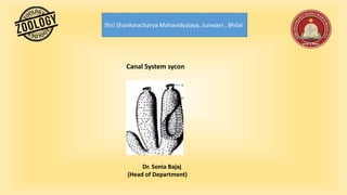
7. 2. Sycon type-
This type canal system is more complex compared to the ascon type.
This canal system can be derived from asconoid type by horizontal folding of its walls.
The radial canals and the incurrent canals paralleling and alternating with each other.
Incurrent pores also known as dermal ostia are found on the outer surface of the body.
The incurrent canals are non-flagellated as they are lined by pinacocytes and not
choanocytes.
The incurrent canals leas into adjacent radial canals through the minute openings called
prosopyles.
Radial canals are flagellated as they are lined by choanocytes.
These canals open into the central spongocoel by internal ostia or apopyles.
In sycon type of canal system, spongocoel is a narrow, non-flagellated cavity lined by
pinacocytes. It opens to the exterior though an excurrent opening called osculum which is
similar to that of the ascon type of canal system.
EX. Scypha.
The course of water current through the canal system can be represented as follows:
Entry of water-dermal ostia- incurrent canal - Prosopyles - Radial canals -
Apopyles – Spongocoel- Osculum - Outside.
8. 3. Leucon type
This type of canal system results due to further folding of body wall of the sycon type of
canal system.
In this type the radial symmetry is lost due to the complexity of the canal system and this
result in an irregular symmetry.
The flagellated chambers are small compared to that of the asconoid and syconoid type.
These chambers are lined by choanocytes and are spherical in shape.
The incurrent canals open into flagellated chambers through prosopyles. These
flagellated chambers in turn communicate with the excurrent canals through apopyles.
The excurrent canals develop as a result of shrinkage and division of spongocoel.
Here the spongocoel is much reduced. This excurrent canal finally communicates with
the outside through the osculum.
EX. Spongilla
The course of water current through the canal system can be represented as follows:
Entry of water -dermal ostia- incurrent canal- Prosopyles -Flagellated
chambers - Apopyles - excurrent canals -Osculum- Outside
9. Leucon type of canal system can be divided into 3 sub-types:
Eurypylous type: This is the simplest and the most primitive type of leuconoid canal system.
In this type the flagellated chambers directly communicate with the excurrent canal through
broad apertures called the apopyles. Ex: Plakina
Aphodal type: In this type of canal system the apopyles are drawn out as a narrow canal called
aphodas. This connects the flagellated chambers with the excurrent canals.Ex: Geodia
Diplodal type: in some of the sponges, along with aphodas another narrow tube called prosodus
is present between incurrent canal and flagellated chamber. This arrangement gives rise to
diplodal type of canal system. Ex: Spongilla
4. Rhagon type
This type of canal system is found in the larva of Demospongiae called rhagon .
It has a broad base are called hypophare and is conical in shape.
Due to excessive growth of mesenchyme sub-dermal spaces are formed in its body wall.
The ostia open in these spaces which lead into incurrent canals. The incurrent canals open by
prosopyles into flagellated canals which are lined with choanocytes.
The flagellated canals open by apopyles into excurrent canals which lead into paragastric cavity.
The paragastric cavity opens to the outside by the osculum which is present at the apex.
EX. Spongilla
Entry of water -dermal ostia- Subdermal pore- incurrent canal- Prosopyles - Flagellated
chambers - Apopyles - excurrent canals - spongocoel-Osculum- Outside.
10. Points Ascon Type Sycon Type Leucon Type Rhagon Type
Wall simple Evaginated to
produce radial and
incurrent canals.
Irregular simple
Mesenchyme Simple and thin.
completely traversed
by posocytes.
Thickened and not
completely
traversed by
posocytes.
Highly elaborated ,traversed
by incurrent canals .
Considerably
thickened ,traversed
by incurrent canals .
Choanocytes Lined the
spongocoel.
Limited to the radial
canals.
Limited to the flagellated
chamber. flagellated
chambers open by narrow
apopyles.
Limited to the
flagellated chamber
which open by wide
apopyles.
Spongocoel Spacious Spacious Usually obliterated Spacious
11. Mechanism of current production
To produce an incurrent or cxcurrent condition there are two factors which are essential:
(i) For entering water through ostia into the body there must be a pressure within it less than that in the incurrent canals.
(ii) For escaping water through osculum there must be a pressure within chambers higher than that in the excurrent canals.
But as the pressure in the incurrent and excurrent canals is the same, there must be a difference of pressure within the
chamber itself and the lower pressure must be towards the periphery. Such a distribution of pressure is set up when each
flagellum causes a flow of water towards the Centre of the chamber.
12. Functions of the Canal System
1. The canal system serves the purpose of nutrition. Smaller food-particles e.g. diatoms, protozoa and particles of
organic debris are ingested into the cells protoplasm and digested. The digestion is intracellular.
2. Streaming currents of water have dissolved air, therefore, gaseous exchange or respiration takes place in the
cells. Oxygen is taken in by simple process of diffusion and carbon-dioxide is given out. The respiration is also
intracellular.
3. The function of the canal system is also excretory. Currents of water, which pass outside the osculum remove the
carbonic acid and other nitrogenous waste substances, which are the excretory products of the body.
4. The purpose of the canal system is also to increase the surface area of the animal in water. This is a characteristic
point by which increase of volume is allowed by keeping the ratio of the surface to the volume.