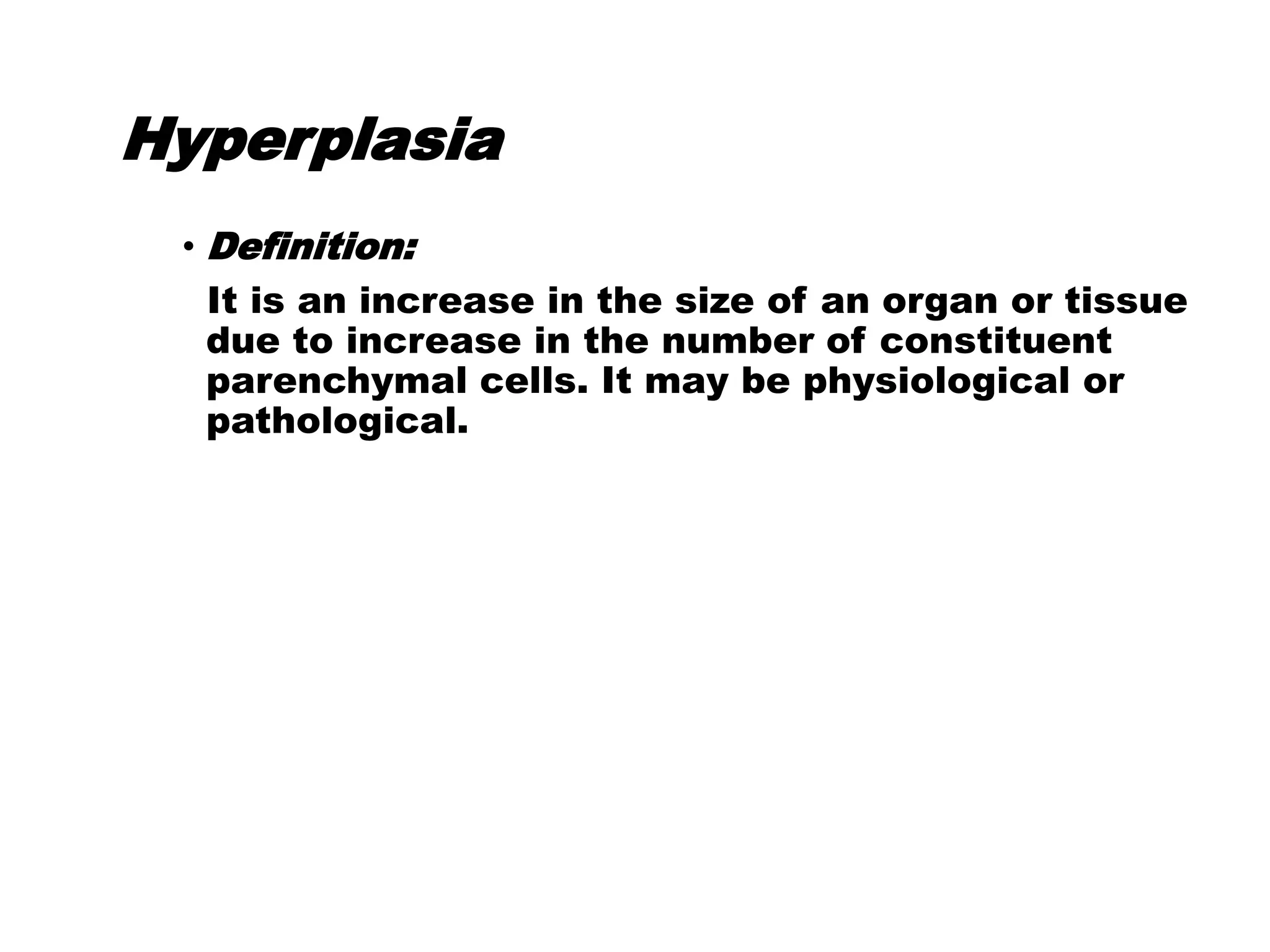
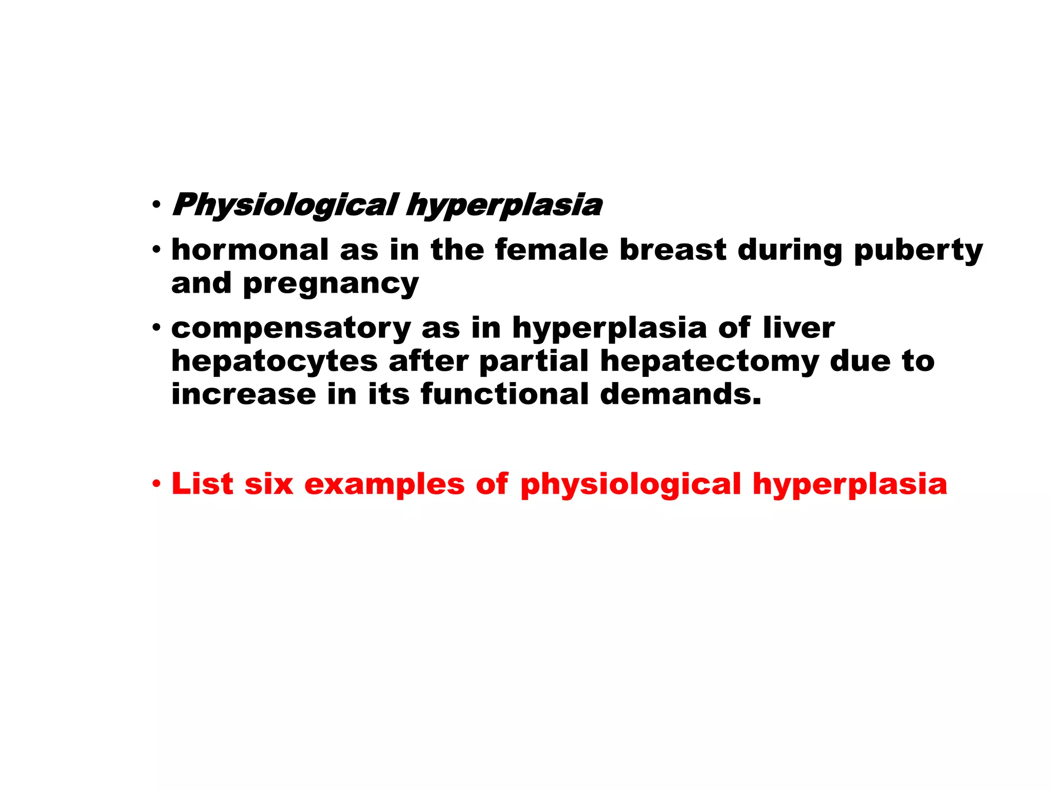
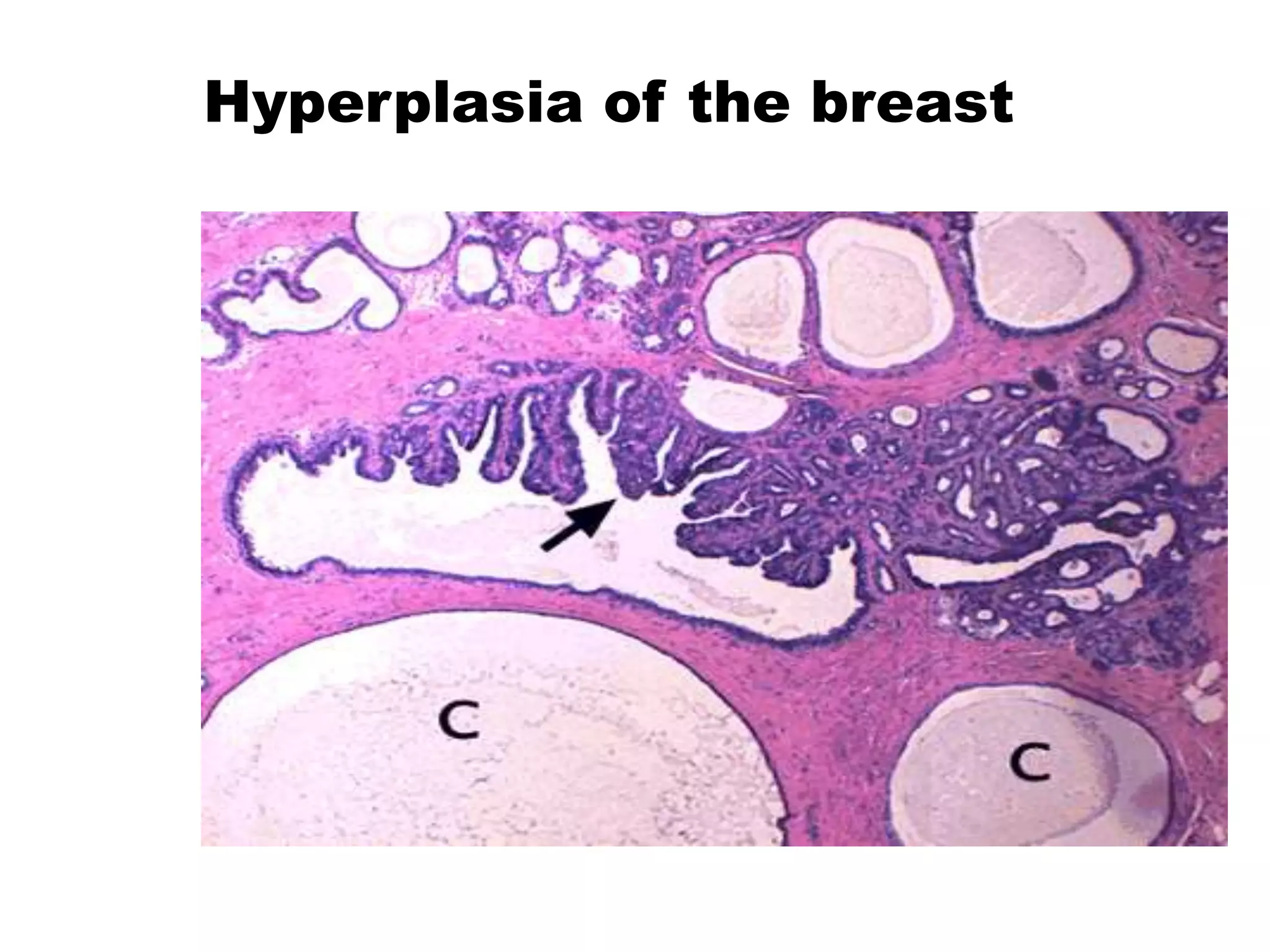
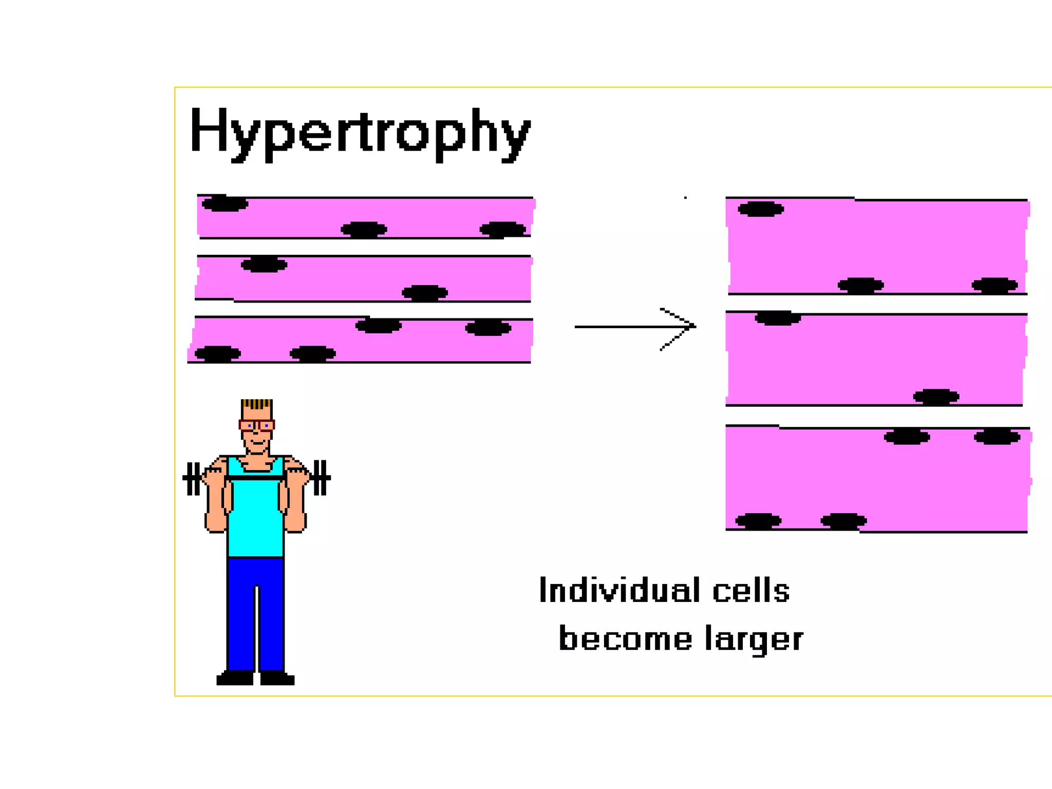
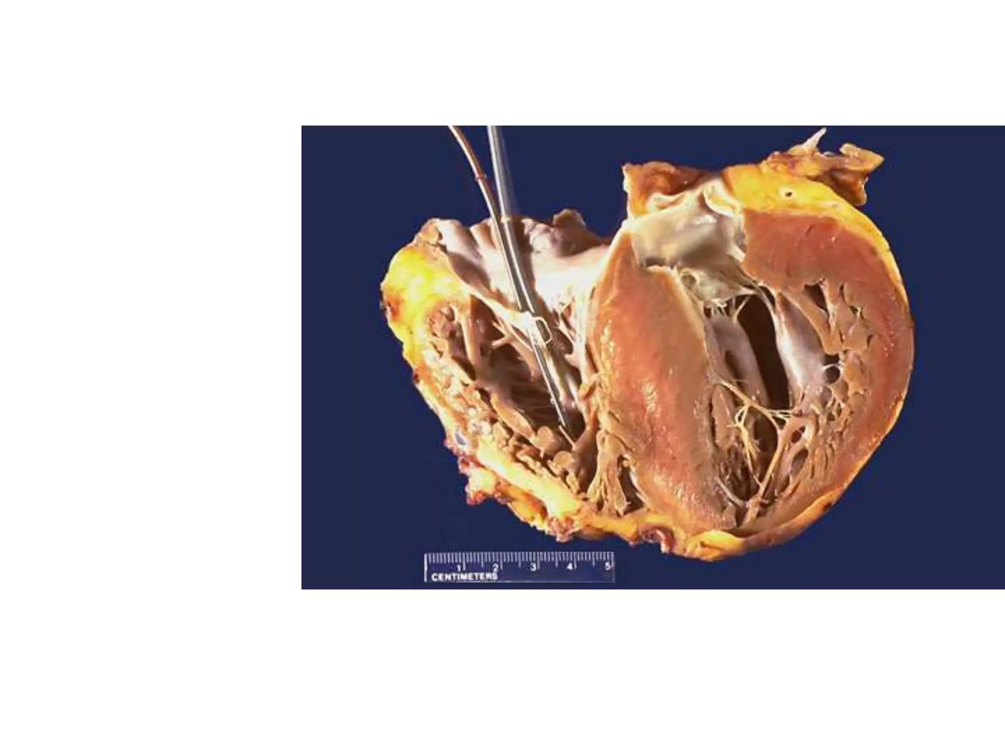
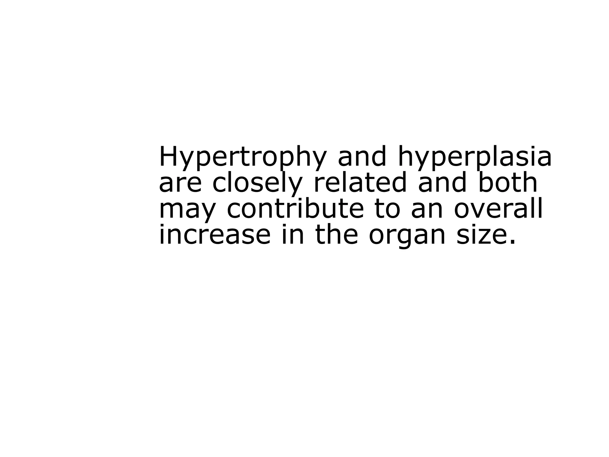
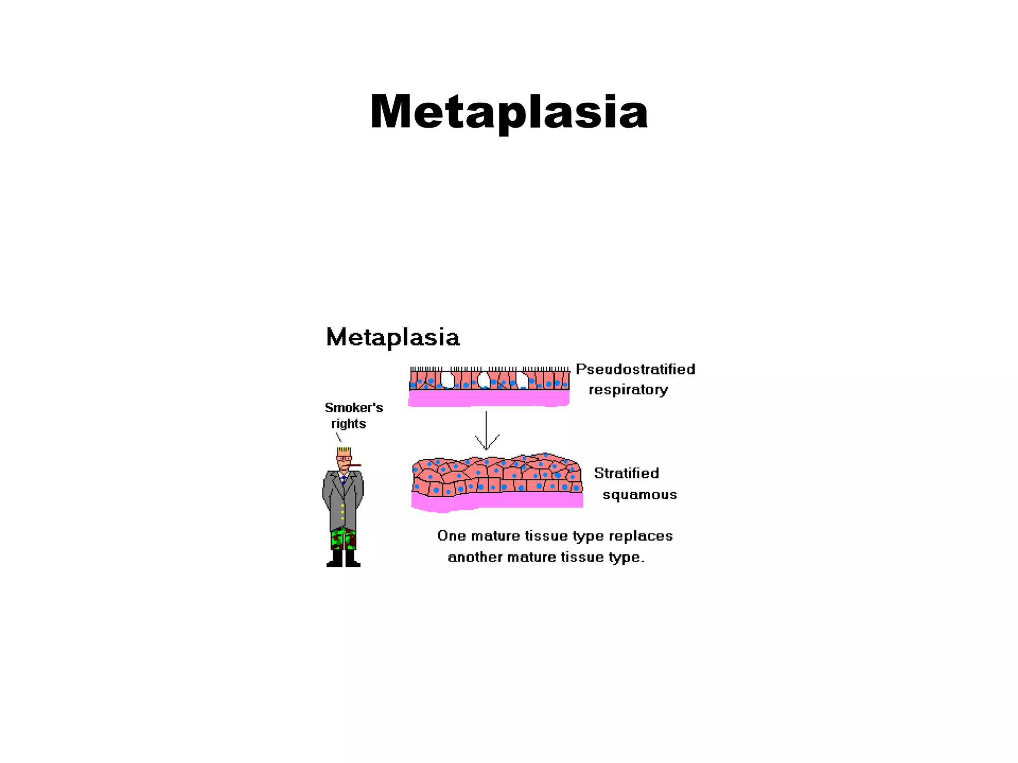
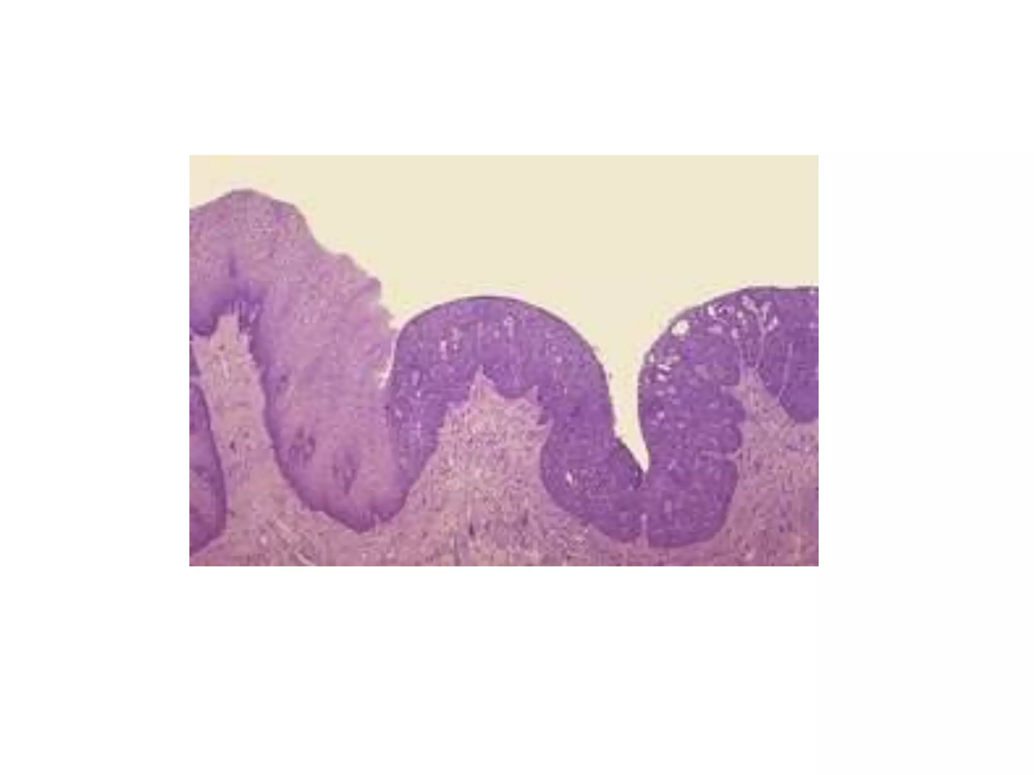
Cellular Adaptations of
Growth and
Differentiation
In response to excessive physiologic
stresses or pathologic stimuli cell
may adapt, where a new altered
steady state is achieved preserving
cell viability.
Adaptation
• 1- Physiological adaptations
They represent response of cells to normal stimulation
by hormones or endogenous chemical substances
e.g.
a. enlargement of the breast and induction of lactation
by pregnancy.
b. replacement of labile tissues, e.g. skin, bone marrow
c. cyclic as endometrium.
•2-Pathological adaptations
•adaptations occuring as a result of
certain stimuli which are not lethal,
•i.e. a state intermediate between
normal unstressed cells and injured
overstressed ones.
Adaptive changes in cell growth
and differentiation include:
a- hyperplasia
b- hypertrophy
c- atrophy
d- metaplasia
Hyperplasia
• Definition:
It is an increase in the size of an organ or tissue
due to increase in the number of constituent
parenchymal cells. It may be physiological or
pathological.
• Physiological hyperplasia
• hormonal as in the female breast during puberty
and pregnancy
• compensatory as in hyperplasia of liver
hepatocytes after partial hepatectomy due to
increase in its functional demands.
• List six examples of physiological hyperplasia
Hyperplasia of the breast
•Pathological hyperplasia
•Increased level of circulating
hormones produce hyperplasia in the
target organs
e.g. a- hyperplasia of the
endometrium
due to hyperestrinism
b-senile prostatic hyperplasia
due to increase in the level of
androgen and estrogen.
However, hyperplasia due to
certain stimuli stops with
removal of these stimuli and may
serve a useful function.
List six examples of pathological hyperplasia
Hypertrophy
• Definition
• It is an increase in the size of an organ or
tissue due to enlargement of individual
cells without increase in the number of
cells.
• It usually occurs in organs in which
proliferation and mitosis are restricted
e.g. skeletal muscle and heart muscles.
• It may be physiological or pathological.
• 1- Physiological hypertrophy
e.g. pregnant uterus, and muscles of athletes.
List six examples of physiological hypertrophy
• 2- Pathological hypertrophy
(adaptive hypertrophy)
e.g. a- hypertrophy of the stomach in pyloric
stenosis
b- cardiac muscle hypertrophy in
chronically hypertensive patients.
List 10 examples of physiological hypertrophy
Hypertrophy and hyperplasia
are closely related and both
may contribute to an overall
increase in the organ size.
Atrophy
Definition
• Reduction of the size of an organ
after reaching its normal adult size.
• As a result of decrease in both
the number and size of the cells.
• Often associated with fibrosis.
• May be physiological or pathological
•Physiological atrophy
•Involution:
•A form of physiologic atrophy.
•Return to normal size after
hyperplasia and hypertrophy, if the
causative stimuli are removed and
equilibrium returns back..
Eg
•uterus after labour.
•atrophy of the ovary after menopause
• senile atrophy of geriatrics.
• Pathological atrophy
• It depends on the cause. It may be localized or
generalized
• Eg.
a- Ischemic atrophy: due to decrease of blood
supply e.g. atherosclerosis
Renal artery stenosis
• b- Pressure atrophy:
due to long continued pressure on a tissue leading to
decrease in its blood supply with atrophy of its cells
• e.g. amyloidosis of the liver in which the extracellular
deposition of
amyloid substance leads to pressure atrophy of
hepatocytes.
Amyloidosis liver
•c-Neuropathic atrophy:
•e.g. when a motor nerve supplying a
muscle is affected as in poliomyelitis
•d- Decreased work load:
•e.g. immobilized limb
•e- Starvation: leading to generalized
atrophy.
The atrophic cells in all cases, are
still surviving cells, but with
diminished function.
Metaplasia
Definition
•It is a reversible change in which an
adult cell type (epithelial or
mesenchymal) is replaced by
another adult cell type of the same
tissue.
It may represent an adaptive
substitution of cells more
sensitive to stress by other cell
types better able to withstand
the adverse environment.
Metaplasia is thought to arise by
genetic 'deprogramming" of
epithelial stem cell or of
undifferentiated mesenchymal
cells in connective tissue.
•Epithelial squamous metaplasia
e.g. a- uterine endocervical glandular
epithelium due to chronic irritation
b-transitional epithelium of
urinary bladder, ureter or renal pelvis
due to chronic irritation by bilharziasis
or stones
c-columnar epithelium of gall
bladder due to chronic irritation by
stones or inflammation
d-pseudostratified ciliated
columnar respiratory epithelium due to
chronic irritation by habitual cigarette
smoking or in vitamin A deficiency.
Squamous metaplasia
Respiratory epithelium
Metaplasia
• The adaptive metaplastic epithelium is
better able to survive, but important
protective mechanisms (such as mucus
secretion and ciliary clearance of
particulate matter in the respiratory
epithelium) are lost.
• If the influences that induce metaplastic
transformation of the epithelium are
persistent, they may lead to cancer
transformation in the metaplastic
epithelium, e.g. squamous metaplasia of
transitional epithelium of the urinary bladder
by bilharziasis will lead to squamous cell
carcinoma.
• Mesenchymal cell metaplasia:
Myxomatous change of fibrous tissue.
Transformation of fibroblasts to osteoblasts or
chondroblasts to produce bone or cartilage in soft
tissues in foci of injury.
• Hypoplasia
It is the decrease in size of an organ due to
incomplete development in embryonic or fetal life
e.g. kidney and uterus.
• Agenesis
• Complete absence of an organ or a part of an organ
e.g. solitary kidney, in such case the other kidney is
absent.
• Dysplasia
• A term used to describe disorderly but non-
neoplastic proliferation.
• It is a loss in the uniformity of individual cells as
well as in their architectural orientation
• Dysplastic changes reveal pleomorphism,
hyperchromatism, abundant mitoses, and loss of
normal orientation. Dysplastic changes are mostly
seen in epithelial cells especially in the cervix uteri.
When dysplastic changes are mild they
are often reversible by removal of the
inciting cause (e.g. chronic irritation).
On the other hand, when dysplastic
changes are marked and involve the
entire thickness of the epithelium, the
lesion in this case is considered as pre-
invasive neoplasm and is referred to as
"carcinoma in situ".
LEC 6 cellular adaptation.ppt

LEC 6 cellular adaptation.ppt

  • 1.
    Cellular Adaptations of Growthand Differentiation
  • 2.
    In response toexcessive physiologic stresses or pathologic stimuli cell may adapt, where a new altered steady state is achieved preserving cell viability. Adaptation
  • 3.
    • 1- Physiologicaladaptations They represent response of cells to normal stimulation by hormones or endogenous chemical substances e.g. a. enlargement of the breast and induction of lactation by pregnancy. b. replacement of labile tissues, e.g. skin, bone marrow c. cyclic as endometrium.
  • 4.
    •2-Pathological adaptations •adaptations occuringas a result of certain stimuli which are not lethal, •i.e. a state intermediate between normal unstressed cells and injured overstressed ones.
  • 5.
    Adaptive changes incell growth and differentiation include: a- hyperplasia b- hypertrophy c- atrophy d- metaplasia
  • 6.
    Hyperplasia • Definition: It isan increase in the size of an organ or tissue due to increase in the number of constituent parenchymal cells. It may be physiological or pathological.
  • 7.
    • Physiological hyperplasia •hormonal as in the female breast during puberty and pregnancy • compensatory as in hyperplasia of liver hepatocytes after partial hepatectomy due to increase in its functional demands. • List six examples of physiological hyperplasia
  • 8.
  • 9.
    •Pathological hyperplasia •Increased levelof circulating hormones produce hyperplasia in the target organs e.g. a- hyperplasia of the endometrium due to hyperestrinism b-senile prostatic hyperplasia due to increase in the level of androgen and estrogen.
  • 10.
    However, hyperplasia dueto certain stimuli stops with removal of these stimuli and may serve a useful function. List six examples of pathological hyperplasia
  • 12.
    Hypertrophy • Definition • Itis an increase in the size of an organ or tissue due to enlargement of individual cells without increase in the number of cells. • It usually occurs in organs in which proliferation and mitosis are restricted e.g. skeletal muscle and heart muscles. • It may be physiological or pathological.
  • 13.
    • 1- Physiologicalhypertrophy e.g. pregnant uterus, and muscles of athletes. List six examples of physiological hypertrophy • 2- Pathological hypertrophy (adaptive hypertrophy) e.g. a- hypertrophy of the stomach in pyloric stenosis b- cardiac muscle hypertrophy in chronically hypertensive patients. List 10 examples of physiological hypertrophy
  • 16.
    Hypertrophy and hyperplasia areclosely related and both may contribute to an overall increase in the organ size.
  • 17.
    Atrophy Definition • Reduction ofthe size of an organ after reaching its normal adult size. • As a result of decrease in both the number and size of the cells. • Often associated with fibrosis. • May be physiological or pathological
  • 18.
    •Physiological atrophy •Involution: •A formof physiologic atrophy. •Return to normal size after hyperplasia and hypertrophy, if the causative stimuli are removed and equilibrium returns back.. Eg •uterus after labour. •atrophy of the ovary after menopause • senile atrophy of geriatrics.
  • 19.
    • Pathological atrophy •It depends on the cause. It may be localized or generalized • Eg. a- Ischemic atrophy: due to decrease of blood supply e.g. atherosclerosis
  • 20.
  • 21.
    • b- Pressureatrophy: due to long continued pressure on a tissue leading to decrease in its blood supply with atrophy of its cells • e.g. amyloidosis of the liver in which the extracellular deposition of amyloid substance leads to pressure atrophy of hepatocytes.
  • 22.
  • 23.
    •c-Neuropathic atrophy: •e.g. whena motor nerve supplying a muscle is affected as in poliomyelitis •d- Decreased work load: •e.g. immobilized limb •e- Starvation: leading to generalized atrophy. The atrophic cells in all cases, are still surviving cells, but with diminished function.
  • 24.
    Metaplasia Definition •It is areversible change in which an adult cell type (epithelial or mesenchymal) is replaced by another adult cell type of the same tissue.
  • 25.
    It may representan adaptive substitution of cells more sensitive to stress by other cell types better able to withstand the adverse environment. Metaplasia is thought to arise by genetic 'deprogramming" of epithelial stem cell or of undifferentiated mesenchymal cells in connective tissue.
  • 26.
    •Epithelial squamous metaplasia e.g.a- uterine endocervical glandular epithelium due to chronic irritation b-transitional epithelium of urinary bladder, ureter or renal pelvis due to chronic irritation by bilharziasis or stones c-columnar epithelium of gall bladder due to chronic irritation by stones or inflammation d-pseudostratified ciliated columnar respiratory epithelium due to chronic irritation by habitual cigarette smoking or in vitamin A deficiency.
  • 27.
  • 28.
  • 29.
    • The adaptivemetaplastic epithelium is better able to survive, but important protective mechanisms (such as mucus secretion and ciliary clearance of particulate matter in the respiratory epithelium) are lost. • If the influences that induce metaplastic transformation of the epithelium are persistent, they may lead to cancer transformation in the metaplastic epithelium, e.g. squamous metaplasia of transitional epithelium of the urinary bladder by bilharziasis will lead to squamous cell carcinoma.
  • 30.
    • Mesenchymal cellmetaplasia: Myxomatous change of fibrous tissue. Transformation of fibroblasts to osteoblasts or chondroblasts to produce bone or cartilage in soft tissues in foci of injury.
  • 31.
    • Hypoplasia It isthe decrease in size of an organ due to incomplete development in embryonic or fetal life e.g. kidney and uterus.
  • 33.
    • Agenesis • Completeabsence of an organ or a part of an organ e.g. solitary kidney, in such case the other kidney is absent.
  • 34.
    • Dysplasia • Aterm used to describe disorderly but non- neoplastic proliferation. • It is a loss in the uniformity of individual cells as well as in their architectural orientation
  • 35.
    • Dysplastic changesreveal pleomorphism, hyperchromatism, abundant mitoses, and loss of normal orientation. Dysplastic changes are mostly seen in epithelial cells especially in the cervix uteri.
  • 36.
    When dysplastic changesare mild they are often reversible by removal of the inciting cause (e.g. chronic irritation). On the other hand, when dysplastic changes are marked and involve the entire thickness of the epithelium, the lesion in this case is considered as pre- invasive neoplasm and is referred to as "carcinoma in situ".