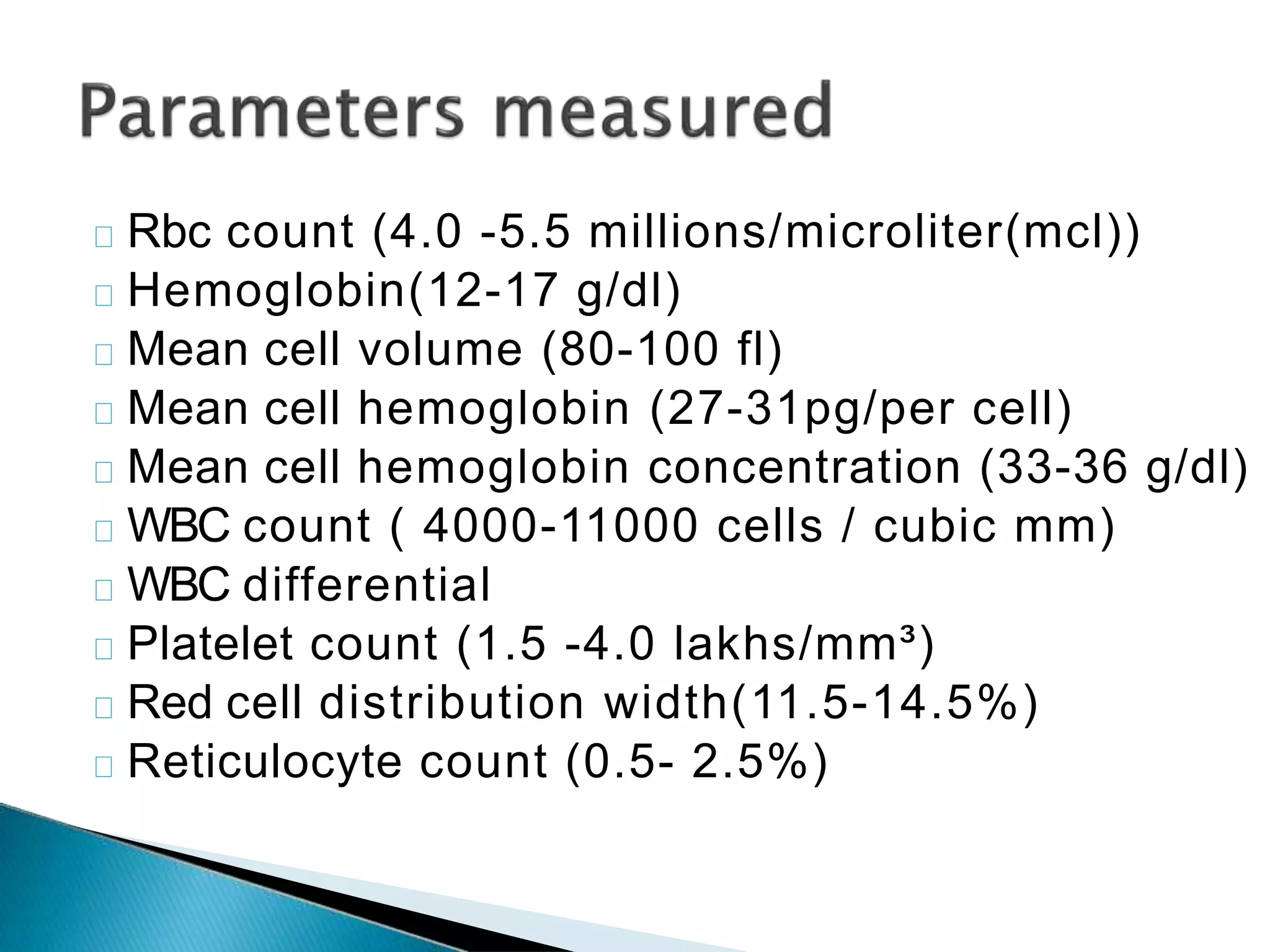
Presented by – Raju
Moderator –Dr Charu Batra
AUTOMATION
IN HEMATOLOGY
IIIIIIIIIIt is process of replacement of task performed
by humans by computerized methods
Until recently hematological tests were
performed only by manual methods.
Both manual and automated laboratory
techniques have advantages and
disadvantages and it is unlikely that one will
completely replace the other.
Semi-automated instruments
Require some steps, as dilution of blood
samples
Often measure only a small number of
variables
Fully automated instruments
Require only that an appropriate blood sample
is presented to the instrument.
They can measure 8-20 variables including
some new parameters which do not have any
equivalent in manual methods.
AUTOMATED HEMATOLOGY ANALYZERS ARE OF TWO TYPES-
Speed with efficient handling of a large
number of samples
Accuracy and precision in quantitative blood
tests
Ability to perform multiple tests on asingle
platform
Significant reduction of labor requirements
Invaluable for accurate determination of red
cell indices
Flags : flagging of laboratory test result
demands labor –intensive manual
examination of a blood smear
Comments on red cell morphology cannot be
generated . Abnormal red cell shapes (such as
fragmented cells) cannot be recognized.
Erroneously increased or decreased results
due to interfering factors
Expensive with high running costs
Electrical impedance
Light scatter
Fluorescence
Light absorption
Electrical conductivity
A stream of cells in suspension passes
through a small aperture across which
an electrical current is applied. Each
cell that passes alters the electrical
impedance and can thus be counted
and sized.
An anticoagulated whole blood sample
is aspirated into the systems , divided
into two portions—
One diluton is passed to red cell aperture bath, and the other is delivered
to the WBC aperture bath .
Particles b/w to 2-20 fl are counted as platelets , while those b/w 36-360
fl are counted as RBC’s .
Each cell flows in a single line through a flow
cell . A laser device is focused on the flow cell
as the laser light beam strikes a cell it is
scattered in various directions.One detector
captures the forward scatter light that is
proportional to cell size and other captures
the side scatter which is equal to cell
granularity & complexity
Light absorption :Concentration of
hemoglobin is measured by absorption
spectrophotometry , after conversion of
hemoglobin to cynmethemoglobin.
Some analyzers use peroxidase cytochemistry
to classify leukocytes
Electrical conductivity : Some analyzers use
conductivity of high frequency current to
determine physical and chemical composition
of leucocytes for their classification .
Rbc count (4.0 -5.5 millions/microliter(mcl))
Hemoglobin(12-17 g/dl)
Mean cell volume (80-100 fl)
Mean cell hemoglobin (27-31pg/per cell)
Mean cell hemoglobin concentration (33-36 g/dl)
WBC count ( 4000-11000 cells / cubic mm)
WBC differential
Platelet count (1.5 -4.0 lakhs/mm³)
Red cell distribution width(11.5-14.5%)
Reticulocyte count (0.5- 2.5%)
Reticulocyte hemoglobin content
Mean platelet volume
Platelet distribution width
Reticulated platelets
◦ Hb is measured automatically by a modification of
the manual (cyanide) method(cyanamethemoglobin
method).
◦ To reduce toxicity of Cyanide some systems replace
it by a non-toxic material Na- lauryl sulphate.
The RBCs are counted automatically by two
methods
◦ Aperture impedance: where cells are counted as
they pass in a stream through an aperture.
◦ Or by light scattering technology
The precision of an electronic counting for
RBCs is much better than the manual count,
and it is available in a fraction of time.
This made the use of RBCindices of more
clinical relevance.
Red cell count and volume are directly
measured by aperture impedance or light
scatter analysis . In a red cell histogram , cell
numbers are plotted on Y axis,white cell
volume on X-axis
Analyzer counts those cells with volume of
b/w 36fl & 360 fl
MCV is used for anemia classification in
microcytic, macrocytic and normocytic types
Mean cell hemoglobin =hemoglobin (g/l)/red
cell count (10/ml)
Mean cell hemoglobin concentartion (g/dl)=
hemoglobin(g/dl)/hemotocrit(%)
Hemocrit (%)=mean cell volume(fl)*red cell
count (10 /ml)
RBC count
Mean cell volume
Red cell distribution width
Hemoglobin
Reticulocyte count
WBC count
Differential WBC count
Platelet count
Mean platelet volume
Automated instruments produce volume
distribution histograms which allow the
presence of more than one population of cells
to be appreciated.
Most instrument produce a quantitative
measurement of variation in cell volume, an
equivalent of the microscopic assessment of
the degree of anisocytosis.
This is known as the RDW.
The total WBCcount is determined in whole
blood in which red cells have been lysed.
Fully automated multichannel instruments
perform WBCcounting by either
◦ Impedance
◦ Or light scattering
◦ Or both
Automated differential counters which are
available now generally use flow cytometry
incorporated into a full blood counter rather
than being standard alone differential
counters
Automated counters provide a three-part or
five- to seven-part differential count.
3-part differential usually cont
◦ Granulocytes or large cells
◦ Lymphocytes or small cells
◦ Monocytes(mononuclear cells) or (middle cells)
5-part classify cells to
◦ Neutrophils
◦ Eosinophils
◦ Basophils
◦ Lymphocytes
◦ Monocytes
A sixth category designated “large unstained
cells” include cells larger than normal and lack
the peroxidase activity this include
◦ Atypical lymphocytes
◦ Various other abnormal cells.
Other counters identifies 7 categories
including
◦ Large immature cells(composed of blasts and
immature granulocytes)
◦ Atypical lymphocytes(including blast cells).
The accuracy of automated counters is less
impressive than their precision.
In general automated differential counters are
favorable to the manual in 2 conditions
◦ Exam of normal blood samples
◦ Flagging of abnormal samples
Platelets can be counted in whole blood
using the same tech. Of electrical or electro-
optical detection as are employed for RBCs.
Other parameters include
◦ MPV (7-10 fl)
◦ PDW
An automated retic count can be performed
using the fact that various fluoro-chromes
combine with the RNA of the reticulocytes.
Fluorescent cells can then be enumerated
using a flowcytometer.
An automated retic counter also permits the
assessment of retic maturity since the more
immature reticulocytes have more
RNAfluoresce more strongly than the mature
retics found normally in PB.
They are precise but care should be taken so
that they are also accurate.
Some problems which could be faced:
◦ Two cells passing through the orifice at the same
time, counted as one cell.
◦ RBCagglutination(clump of cells)
◦ Counting bubbles as cells
◦ Dust particles counted as cell.
Flow cytometry
FLUORESCENCE
A fluorescence absorb light energy and limits excess
energy in the form of photon light. Fluorescence is
the property of molecules to absorb light at one
wavelength and emit light at a longer wavelength.
LIGHT SCATTER
Light is scattered when the incident light is deflected
by a particle traversing through a beam of light .This
depends on physical properties of the cell.
DATA ANALYSIS
The data collected and stored in the computer can be
displayed in various formats . A parameter means
forward scatter , or side scatter , or emitted fluorescence
from a particle as it passes through a laser beam.
GATING
A gates is boundary that can be set to restrict the
analysis to a specific population within the sample.
SORTING
When a cell passes through the laser beam , it is
sent to waste . Sorting consists of collecting cells
of interest for further analysis.
Raju automation Msc MLT PATHOLOGY

Raju automation Msc MLT PATHOLOGY

  • 1.
    Presented by –Raju Moderator –Dr Charu Batra AUTOMATION IN HEMATOLOGY
  • 2.
    IIIIIIIIIIt is processof replacement of task performed by humans by computerized methods Until recently hematological tests were performed only by manual methods. Both manual and automated laboratory techniques have advantages and disadvantages and it is unlikely that one will completely replace the other.
  • 3.
    Semi-automated instruments Require somesteps, as dilution of blood samples Often measure only a small number of variables Fully automated instruments Require only that an appropriate blood sample is presented to the instrument. They can measure 8-20 variables including some new parameters which do not have any equivalent in manual methods. AUTOMATED HEMATOLOGY ANALYZERS ARE OF TWO TYPES-
  • 4.
    Speed with efficienthandling of a large number of samples Accuracy and precision in quantitative blood tests Ability to perform multiple tests on asingle platform Significant reduction of labor requirements Invaluable for accurate determination of red cell indices
  • 5.
    Flags : flaggingof laboratory test result demands labor –intensive manual examination of a blood smear Comments on red cell morphology cannot be generated . Abnormal red cell shapes (such as fragmented cells) cannot be recognized. Erroneously increased or decreased results due to interfering factors Expensive with high running costs
  • 6.
  • 7.
    A stream ofcells in suspension passes through a small aperture across which an electrical current is applied. Each cell that passes alters the electrical impedance and can thus be counted and sized. An anticoagulated whole blood sample is aspirated into the systems , divided into two portions— One diluton is passed to red cell aperture bath, and the other is delivered to the WBC aperture bath . Particles b/w to 2-20 fl are counted as platelets , while those b/w 36-360 fl are counted as RBC’s .
  • 8.
    Each cell flowsin a single line through a flow cell . A laser device is focused on the flow cell as the laser light beam strikes a cell it is scattered in various directions.One detector captures the forward scatter light that is proportional to cell size and other captures the side scatter which is equal to cell granularity & complexity
  • 9.
    Light absorption :Concentrationof hemoglobin is measured by absorption spectrophotometry , after conversion of hemoglobin to cynmethemoglobin. Some analyzers use peroxidase cytochemistry to classify leukocytes Electrical conductivity : Some analyzers use conductivity of high frequency current to determine physical and chemical composition of leucocytes for their classification .
  • 10.
    Rbc count (4.0-5.5 millions/microliter(mcl)) Hemoglobin(12-17 g/dl) Mean cell volume (80-100 fl) Mean cell hemoglobin (27-31pg/per cell) Mean cell hemoglobin concentration (33-36 g/dl) WBC count ( 4000-11000 cells / cubic mm) WBC differential Platelet count (1.5 -4.0 lakhs/mm³) Red cell distribution width(11.5-14.5%) Reticulocyte count (0.5- 2.5%)
  • 11.
    Reticulocyte hemoglobin content Meanplatelet volume Platelet distribution width Reticulated platelets
  • 12.
    ◦ Hb ismeasured automatically by a modification of the manual (cyanide) method(cyanamethemoglobin method). ◦ To reduce toxicity of Cyanide some systems replace it by a non-toxic material Na- lauryl sulphate.
  • 13.
    The RBCs arecounted automatically by two methods ◦ Aperture impedance: where cells are counted as they pass in a stream through an aperture. ◦ Or by light scattering technology The precision of an electronic counting for RBCs is much better than the manual count, and it is available in a fraction of time. This made the use of RBCindices of more clinical relevance.
  • 14.
    Red cell countand volume are directly measured by aperture impedance or light scatter analysis . In a red cell histogram , cell numbers are plotted on Y axis,white cell volume on X-axis Analyzer counts those cells with volume of b/w 36fl & 360 fl MCV is used for anemia classification in microcytic, macrocytic and normocytic types
  • 15.
    Mean cell hemoglobin=hemoglobin (g/l)/red cell count (10/ml) Mean cell hemoglobin concentartion (g/dl)= hemoglobin(g/dl)/hemotocrit(%) Hemocrit (%)=mean cell volume(fl)*red cell count (10 /ml)
  • 16.
    RBC count Mean cellvolume Red cell distribution width Hemoglobin Reticulocyte count WBC count Differential WBC count Platelet count Mean platelet volume
  • 17.
    Automated instruments producevolume distribution histograms which allow the presence of more than one population of cells to be appreciated. Most instrument produce a quantitative measurement of variation in cell volume, an equivalent of the microscopic assessment of the degree of anisocytosis. This is known as the RDW.
  • 18.
    The total WBCcountis determined in whole blood in which red cells have been lysed. Fully automated multichannel instruments perform WBCcounting by either ◦ Impedance ◦ Or light scattering ◦ Or both
  • 19.
    Automated differential counterswhich are available now generally use flow cytometry incorporated into a full blood counter rather than being standard alone differential counters Automated counters provide a three-part or five- to seven-part differential count.
  • 20.
    3-part differential usuallycont ◦ Granulocytes or large cells ◦ Lymphocytes or small cells ◦ Monocytes(mononuclear cells) or (middle cells) 5-part classify cells to ◦ Neutrophils ◦ Eosinophils ◦ Basophils ◦ Lymphocytes ◦ Monocytes
  • 21.
    A sixth categorydesignated “large unstained cells” include cells larger than normal and lack the peroxidase activity this include ◦ Atypical lymphocytes ◦ Various other abnormal cells. Other counters identifies 7 categories including ◦ Large immature cells(composed of blasts and immature granulocytes) ◦ Atypical lymphocytes(including blast cells).
  • 22.
    The accuracy ofautomated counters is less impressive than their precision. In general automated differential counters are favorable to the manual in 2 conditions ◦ Exam of normal blood samples ◦ Flagging of abnormal samples
  • 23.
    Platelets can becounted in whole blood using the same tech. Of electrical or electro- optical detection as are employed for RBCs. Other parameters include ◦ MPV (7-10 fl) ◦ PDW
  • 24.
    An automated reticcount can be performed using the fact that various fluoro-chromes combine with the RNA of the reticulocytes. Fluorescent cells can then be enumerated using a flowcytometer. An automated retic counter also permits the assessment of retic maturity since the more immature reticulocytes have more RNAfluoresce more strongly than the mature retics found normally in PB.
  • 25.
    They are precisebut care should be taken so that they are also accurate. Some problems which could be faced: ◦ Two cells passing through the orifice at the same time, counted as one cell. ◦ RBCagglutination(clump of cells) ◦ Counting bubbles as cells ◦ Dust particles counted as cell.
  • 26.
  • 27.
    FLUORESCENCE A fluorescence absorblight energy and limits excess energy in the form of photon light. Fluorescence is the property of molecules to absorb light at one wavelength and emit light at a longer wavelength. LIGHT SCATTER Light is scattered when the incident light is deflected by a particle traversing through a beam of light .This depends on physical properties of the cell.
  • 28.
    DATA ANALYSIS The datacollected and stored in the computer can be displayed in various formats . A parameter means forward scatter , or side scatter , or emitted fluorescence from a particle as it passes through a laser beam. GATING A gates is boundary that can be set to restrict the analysis to a specific population within the sample. SORTING When a cell passes through the laser beam , it is sent to waste . Sorting consists of collecting cells of interest for further analysis.