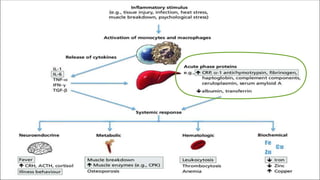
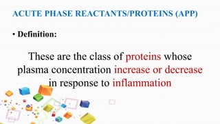
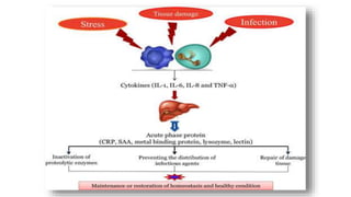
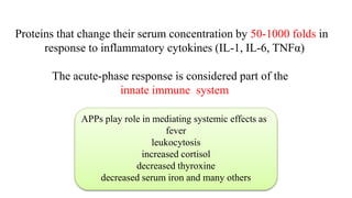
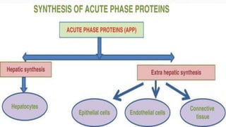
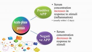
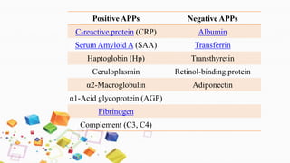
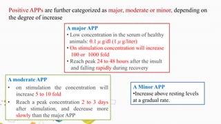
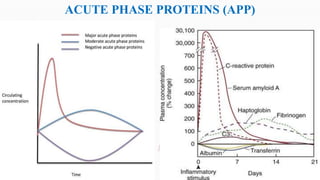
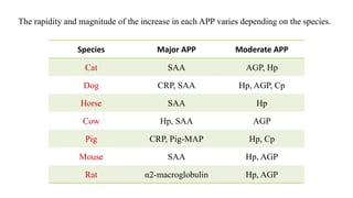
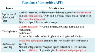
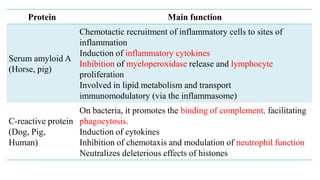
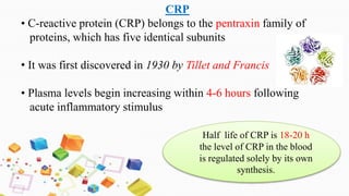
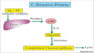
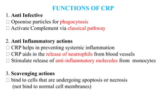
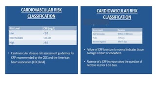
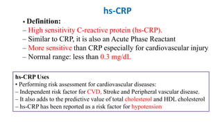
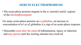
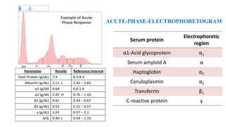
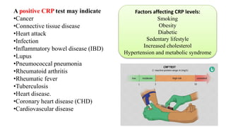
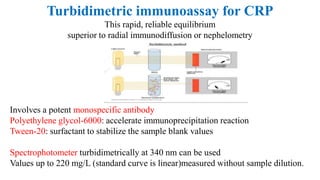
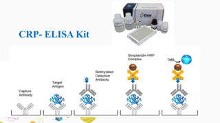
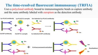
ACUTE PHASE
PROTEINS (APP)
ACUTE PHASE REACTANTS/PROTEINS (APP)
• Definition:
These are the class of proteins whose
plasma concentration increase or decrease
in response to inflammation
Proteins that change their serum concentration by 50-1000 folds in
response to inflammatory cytokines (IL-1, IL-6, TNFα)
The acute-phase response is considered part of the
innate immune system
APPs play role in mediating systemic effects as
fever
leukocytosis
increased cortisol
decreased thyroxine
decreased serum iron and many others
Positive
APP
• Serum
concentration
increases in
response to stimuli
(inflammation)
• (usually within 1-2 days)
Negati
ve APP
• Serum
concentration
decreases in
response to
stimuli
Positive APPs Negative APPs
C-reactive protein (CRP) Albumin
Serum Amyloid A (SAA) Transferrin
Haptoglobin (Hp) Transthyretin
Ceruloplasmin Retinol-binding protein
α2-Macroglobulin Adiponectin
α1-Acid glycoprotein (AGP)
Fibrinogen
Complement (C3, C4)
A major APP
• Low concentration in the serum of healthy
animals: 0.1 μ g/dl (1 μ g/liter)
• On stimulation concentration will increase
100 or 1000 fold
• Reach peak 24 to 48 hours after the insult
and falling rapidly during recovery
A moderate APP
• on stimulation the concentration will
increase 5 to 10 fold
• Reach a peak concentration 2 to 3 days
after stimulation, and decrease more
slowly than the major APP
Positive APPs are further categorized as major, moderate or minor, depending on
the degree of increase
A Minor APP
•Increase above resting levels
at a gradual rate.
ACUTE PHASE PROTEINS (APP)
The rapidity and magnitude of the increase in each acute phase protein varies depending on the
species.
Species Major APP Moderate APP
Cat SAA AGP, Hp
Dog CRP, SAA Hp, AGP, Cp
Horse SAA Hp
Cow Hp, SAA AGP
Pig CRP, Pig-MAP Hp, Cp
Mouse SAA Hp, AGP
Rat α2-macroglobulin Hp, AGP
The rapidity and magnitude of the increase in each APP varies depending on the species.
Protein Main function
Alpha-1-acid
glycoprotein
(cat)
Antiinflammatory and immunomodulatory agent: has antineutrophil
and anticomplement activity and increases macrophage secretion of
IL-1 receptor antagonist.
Binds to lipophilic and acidic drugs.
Ceruloplasmin
Copper transport (for wound healing, collagen formation and
maturation)
Antioxidant
Reduces the number of neutrophils attaching to endothelium
Haptoglobin
(Cow, Pig)
Binds free hemoglobin (limiting Hb iron availability for bacterial
growth)
Natural antagonist for receptor-ligand activation of the immune
system. Inhibition of granulocyte chemotaxis and phagocytosis
Functions of the positive APPs
Protein Main function
Serum amyloid A
(Horse, pig)
Chemotactic recruitment of inflammatory cells to sites of
inflammation
Induction of inflammatory cytokines
Inhibition of myeloperoxidase release and lymphocyte
proliferation
Involved in lipid metabolism and transport
immunomodulatory (via the inflammasome)
C-reactive protein
(Dog, Pig,
Human)
On bacteria, it promotes the binding of complement, facilitating
phagocytosis.
Induction of cytokines
Inhibition of chemotaxis and modulation of neutrophil function
Neutralizes deleterious effects of histones
Negative acute phase proteins
• Decrease in plasma concentration by greater than 25% in
response to inflammation
• This reduction can occur rapidly (within 24 hours) or may
decrease gradually over a period of days
The mechanism by which their concentrations decrease is likely
 including decreased production by the liver in response to
inflammatory cytokines
possibly increased loss or increased proteolysis.
Protein Main function
Albumin Reduced production of albumin allows greater increase in the
amount of amino acids available for positive APP production
Albumin concentration falls gradually and more noticeable in
chronic inflammatory disease
Transferrin Usually measured to assess iron status
Ovotransferrin is the avian analog, but it is a positive acute phase
protein
Adiponectin Produced in adipose tissue, and promotes energy usage through
increasing sensitivity to insulin, has anti-inflammatory properties.
Decreased concentrations may be seen in obese animals or animals
with diabetes mellitus
Functions of the negative APPs
C-reactive protein (CRP)
• A major acute-phase protein produced by the liver in response
to tissue damage
• CRP was the first acute phase protein to be recognized
• CRP is a beta-globulin and has a molecular weight of 115-140 kD
• CRP Gene- Chromosome 1, Short arm
• Cytokines especially, IL-6 and IL-1 regulate CRP at transcritional level
• Normal range: Less than 10mg/L
CRP
• C-reactive protein (CRP) belongs to the pentraxin family of
proteins, which has five identical subunits
• It was first discovered in 1930 by Tillet and Francis
• Plasma levels begin increasing within 4-6 hours following
acute inflammatory stimulus
Half life of CRP is 18-20 h
the level of CRP in the blood
is regulated solely by its own
synthesis.
C- REACTIVE PROTEIN
•Its name derives from its pattern recognition activity:
C-reactive protein binds to the C-polysaccharide cell-wall
component found on a variety of bacteria and fungi
• This binding activates the complement system, resulting in increased
clearance of the pathogen either by complement-mediated lysis or
by a complement mediated phagocytosis
• It has highest affinity for,
– Phosphocholine on bacteria
– Mixture of sphingomyeline and phosphatidylcholine in
eukaryotic membranes
FUNCTIONS OF CRP
1. Anti Infective
Opsonise particles for phagocytosis
Activate Complement via classical pathway
2. Anti Inflammatory actions
CRP helps in preventing systemic inflammation
CRP aids in the release of neutrophils from blood vessels
Stimulate release of anti-inflammatory molecules from monocytes
3. Scavenging actions
bind to cells that are undergoing apoptosis or necrosis
(not bind to normal cell membranes)
• CRP begins to rise within 4-6 h
of stimulus, peaks within 36-50
hours, and returns to normal 3-7
days following resolution
•ESR shows a much slower
response, taking up to a week to
peak, and up to several weeks to
return to normal.
CRP and ESR patterns of response
• CRP is more sensitive than ESR to
subtle changes in the acute phase
response
• There are distinct ranges of normal
and abnormal in CRP reference
ranges, without variations for age and
gender
• CRP is not affected by conditions
such as pregnancy, intercurrent drug
use, anaemia and plasma protein
variations.
CRP is a better measure of acute phase response
10
CLINICAL IMPORTANCE OF CRP
Provides fast and adequate information of the actual clinical situation
Useful for monitoring the effect of treatment
Early detection of postoperative complications or intercurrent infections
• Elevated CRP levels:
– Osteoarthritis
– Proinflammatory or prothrombotic effects (Cerebral vein or sinus thrombosis)
_An extremely elevated suggestive of a possible bacterial infection
_Elevated levels were seen in majority of kidney or heart transplant
• Mild elevation of CRP levels:
– SLE
– Scleroderma
– Sjogren syndrome
– Dermatomyositis/Polymyositis
Pathophysiology
There is considerable species variation in the pathophysiology of CRP
Dog: (infectious diseases -babesiosis, leishmaniasis, leptospirosis, parvoviruses,
trypanosomiasis, infection with Bordetella bronchiseptica, Ehrlichia canis , and
Escherichia coli sepsis and in hematological and neoplastic diseases of the dog )
Pig: CRP concentration increases following aseptic Inflammation and in
experimental infection with Actinobacillus pleuropneumoniae
Bovine: respiratory syncytial virus, multiple myeloma, mastitis, lymphatic
neoplasia.
hs-CRP
• Definition:
– High sensitivity C-reactive protein (hs-CRP).
– Similar to CRP, it is also an Acute Phase Reactant
– More sensitive than CRP especially for cardiovascular injury
– Normal range: less than 0.3 mg/dL
hs-CRP Uses
• Performing risk assessment for cardiovascular diseases:
– Independent risk factor for CVD, Stroke and Peripheral vascular disease.
– It also adds to the predictive value of total cholesterol and HDL cholesterol
– hs-CRP has been reported as a risk factor for hypotension
SERUM ELECTROPHORESIS
• The acute-phase proteins migrate in the α- (mostly) and β- regions
of the electrophoretogram
•As many acute-phase proteins are α globulins, an increase in
concentration of α1 or α2 globulins is a sign of an acute-phase response
• Detectable soon after the onset of inflammation, injury, or infection
and may persist until the inciting stimulus has resolved
Acute-phase-electrophoretogram
ACUTE-PHASE-ELECTROPHORETOGRAM
Serum protein
Electrophoretic
region
α1-Acid glycoprotein α1
Serum amyloid A α
Haptoglobin α2
Ceruloplasmin α2
Transferrin β1
C-reactive protein γ
•CRP is prominent among the acute-phase proteins
•It is a blood test marker for inflammation in the body
Sample considerations
Storage: stable at -10º C for 3 months
Anticoagulant: Do not use citrate tube as levels are significantly
lowered
CRP : a direct and quantitative measure of the acute-
phase reactions
C-REACTIVE PROTEIN
Tests
Slide/Rapid latex agglutination tests
Turbidimetric immunoassay: used in humans and has been adapted
for automated biochemical analyzers. However, there is variation in
cross-activity with different antihuman CRP antibodies. Hemolysis
will interfere with immunoturbidimetric testing.
ELISA: a commercially available kit for canine CRP
Time-resolved fluorometry (TRFIA): recently developed for CRP
assays in canine whole blood, saliva and effusions
• Rapid latex agglutination test: Principle
•Based on the reaction between patient serum
containing CRP as the antigen & the
corresponding antibody coated to the treated
surface of latex particle.
INTERPRETATION
• A negative reaction is indicated by a
uniform milky suspension with no
agglutination as observed with the CRP
Negative Control.
• A positive reaction is indicated by any
observable agglutination in the reaction
mixture.
A positive CRP test may indicate
•Cancer
•Connective tissue disease
•Heart attack
•Infection
•Inflammatory bowel disease (IBD)
•Lupus
•Pneumococcal pneumonia
•Rheumatoid arthritis
•Rheumatic fever
•Tuberculosis
•Heart disease.
•Coronary heart disease (CHD)
•Cardiovascular disease
Factors affecting CRP levels:
Smoking
Obesity
Diabetic
Sedentary lifestyle
Increased cholesterol
Hypertension and metabolic syndrome
Turbidimetric immunoassay for CRP
This rapid, reliable equilibrium
superior to radial immunodiffusion or nephelometry
Involves a potent monospecific antibody
Polyethylene glycol-6000: accelerate immunoprecipitation reaction
Tween-20: surfactant to stabilize the sample blank values
Spectrophotometer turbidimetrically at 340 nm can be used
Values up to 220 mg/L (standard curve is linear)measured without sample dilution.
CRP- ELISA Kit
The time-resolved fluorescent immunoassay (TRFIA)
Uses a polyclonal antibody bound to immunomagnetic beads as capture antibody
and the same antibody labeled with europium as the detection antibody
Ceruloplasmin
Sample considerations
Anticoagulant: concentrations are higher with heparin and lower with EDTA
Tests
There are problems with assays - lack of commercially available reference
materials to standardize ceruloplasmin concentrations.
Therefore, different arbitrary units based on increased absorbance per unit time
have been used (oxidase units UI/L).
Haptoglobin
Sample considerations
Storage: Values decrease in serum stored at -20ºC. Storage at -70°C
Anticoagulants: concentration is increased with heparin
Interferences: Dogs with high endogenous steroids or on exogenous steroid therapy
will have increase Hp concentrations. Canine Hp concentrations in health or
disease are significantly higher than other species.
Tests
Spectrophotometric assays
Hemoglobin-haptoglobin complexes that alter the absorbance characteristic of
Hb in proprotion of the concentration of Hb in serum
Peroxidase acitivity at an acidic pH detected & quantified
Immunoassays
Nephelometric assay: rate of precipitation of the Ag-Ab complex is measured.
Alpha-1 acid glycoprotein
Tests:
Estimation by precipitation of majority of serum proteins by perchloric acid
and quantification of the remaining soluble proteins.
Single radial immunodiffusion on agarose gel impregnated with anti-species
AGP rabbit serum. Dog and cat specific assays have been developed.
Immunoturbidimetric assays have been developed for canine and feline
AGP measurement
Acute phase proteins

Acute phase proteins

  • 1.
  • 4.
    ACUTE PHASE REACTANTS/PROTEINS(APP) • Definition: These are the class of proteins whose plasma concentration increase or decrease in response to inflammation
  • 6.
    Proteins that changetheir serum concentration by 50-1000 folds in response to inflammatory cytokines (IL-1, IL-6, TNFα) The acute-phase response is considered part of the innate immune system APPs play role in mediating systemic effects as fever leukocytosis increased cortisol decreased thyroxine decreased serum iron and many others
  • 8.
    Positive APP • Serum concentration increases in responseto stimuli (inflammation) • (usually within 1-2 days) Negati ve APP • Serum concentration decreases in response to stimuli
  • 9.
    Positive APPs NegativeAPPs C-reactive protein (CRP) Albumin Serum Amyloid A (SAA) Transferrin Haptoglobin (Hp) Transthyretin Ceruloplasmin Retinol-binding protein α2-Macroglobulin Adiponectin α1-Acid glycoprotein (AGP) Fibrinogen Complement (C3, C4)
  • 10.
    A major APP •Low concentration in the serum of healthy animals: 0.1 μ g/dl (1 μ g/liter) • On stimulation concentration will increase 100 or 1000 fold • Reach peak 24 to 48 hours after the insult and falling rapidly during recovery A moderate APP • on stimulation the concentration will increase 5 to 10 fold • Reach a peak concentration 2 to 3 days after stimulation, and decrease more slowly than the major APP Positive APPs are further categorized as major, moderate or minor, depending on the degree of increase A Minor APP •Increase above resting levels at a gradual rate.
  • 11.
  • 12.
    The rapidity andmagnitude of the increase in each acute phase protein varies depending on the species. Species Major APP Moderate APP Cat SAA AGP, Hp Dog CRP, SAA Hp, AGP, Cp Horse SAA Hp Cow Hp, SAA AGP Pig CRP, Pig-MAP Hp, Cp Mouse SAA Hp, AGP Rat α2-macroglobulin Hp, AGP The rapidity and magnitude of the increase in each APP varies depending on the species.
  • 13.
    Protein Main function Alpha-1-acid glycoprotein (cat) Antiinflammatoryand immunomodulatory agent: has antineutrophil and anticomplement activity and increases macrophage secretion of IL-1 receptor antagonist. Binds to lipophilic and acidic drugs. Ceruloplasmin Copper transport (for wound healing, collagen formation and maturation) Antioxidant Reduces the number of neutrophils attaching to endothelium Haptoglobin (Cow, Pig) Binds free hemoglobin (limiting Hb iron availability for bacterial growth) Natural antagonist for receptor-ligand activation of the immune system. Inhibition of granulocyte chemotaxis and phagocytosis Functions of the positive APPs
  • 14.
    Protein Main function Serumamyloid A (Horse, pig) Chemotactic recruitment of inflammatory cells to sites of inflammation Induction of inflammatory cytokines Inhibition of myeloperoxidase release and lymphocyte proliferation Involved in lipid metabolism and transport immunomodulatory (via the inflammasome) C-reactive protein (Dog, Pig, Human) On bacteria, it promotes the binding of complement, facilitating phagocytosis. Induction of cytokines Inhibition of chemotaxis and modulation of neutrophil function Neutralizes deleterious effects of histones
  • 15.
    Negative acute phaseproteins • Decrease in plasma concentration by greater than 25% in response to inflammation • This reduction can occur rapidly (within 24 hours) or may decrease gradually over a period of days The mechanism by which their concentrations decrease is likely  including decreased production by the liver in response to inflammatory cytokines possibly increased loss or increased proteolysis.
  • 16.
    Protein Main function AlbuminReduced production of albumin allows greater increase in the amount of amino acids available for positive APP production Albumin concentration falls gradually and more noticeable in chronic inflammatory disease Transferrin Usually measured to assess iron status Ovotransferrin is the avian analog, but it is a positive acute phase protein Adiponectin Produced in adipose tissue, and promotes energy usage through increasing sensitivity to insulin, has anti-inflammatory properties. Decreased concentrations may be seen in obese animals or animals with diabetes mellitus Functions of the negative APPs
  • 18.
    C-reactive protein (CRP) •A major acute-phase protein produced by the liver in response to tissue damage • CRP was the first acute phase protein to be recognized • CRP is a beta-globulin and has a molecular weight of 115-140 kD • CRP Gene- Chromosome 1, Short arm • Cytokines especially, IL-6 and IL-1 regulate CRP at transcritional level • Normal range: Less than 10mg/L
  • 19.
    CRP • C-reactive protein(CRP) belongs to the pentraxin family of proteins, which has five identical subunits • It was first discovered in 1930 by Tillet and Francis • Plasma levels begin increasing within 4-6 hours following acute inflammatory stimulus Half life of CRP is 18-20 h the level of CRP in the blood is regulated solely by its own synthesis.
  • 20.
    C- REACTIVE PROTEIN •Itsname derives from its pattern recognition activity: C-reactive protein binds to the C-polysaccharide cell-wall component found on a variety of bacteria and fungi • This binding activates the complement system, resulting in increased clearance of the pathogen either by complement-mediated lysis or by a complement mediated phagocytosis • It has highest affinity for, – Phosphocholine on bacteria – Mixture of sphingomyeline and phosphatidylcholine in eukaryotic membranes
  • 22.
    FUNCTIONS OF CRP 1.Anti Infective Opsonise particles for phagocytosis Activate Complement via classical pathway 2. Anti Inflammatory actions CRP helps in preventing systemic inflammation CRP aids in the release of neutrophils from blood vessels Stimulate release of anti-inflammatory molecules from monocytes 3. Scavenging actions bind to cells that are undergoing apoptosis or necrosis (not bind to normal cell membranes)
  • 23.
    • CRP beginsto rise within 4-6 h of stimulus, peaks within 36-50 hours, and returns to normal 3-7 days following resolution •ESR shows a much slower response, taking up to a week to peak, and up to several weeks to return to normal. CRP and ESR patterns of response
  • 24.
    • CRP ismore sensitive than ESR to subtle changes in the acute phase response • There are distinct ranges of normal and abnormal in CRP reference ranges, without variations for age and gender • CRP is not affected by conditions such as pregnancy, intercurrent drug use, anaemia and plasma protein variations. CRP is a better measure of acute phase response 10
  • 25.
    CLINICAL IMPORTANCE OFCRP Provides fast and adequate information of the actual clinical situation Useful for monitoring the effect of treatment Early detection of postoperative complications or intercurrent infections • Elevated CRP levels: – Osteoarthritis – Proinflammatory or prothrombotic effects (Cerebral vein or sinus thrombosis) _An extremely elevated suggestive of a possible bacterial infection _Elevated levels were seen in majority of kidney or heart transplant • Mild elevation of CRP levels: – SLE – Scleroderma – Sjogren syndrome – Dermatomyositis/Polymyositis
  • 26.
    Pathophysiology There is considerablespecies variation in the pathophysiology of CRP Dog: (infectious diseases -babesiosis, leishmaniasis, leptospirosis, parvoviruses, trypanosomiasis, infection with Bordetella bronchiseptica, Ehrlichia canis , and Escherichia coli sepsis and in hematological and neoplastic diseases of the dog ) Pig: CRP concentration increases following aseptic Inflammation and in experimental infection with Actinobacillus pleuropneumoniae Bovine: respiratory syncytial virus, multiple myeloma, mastitis, lymphatic neoplasia.
  • 28.
    hs-CRP • Definition: – Highsensitivity C-reactive protein (hs-CRP). – Similar to CRP, it is also an Acute Phase Reactant – More sensitive than CRP especially for cardiovascular injury – Normal range: less than 0.3 mg/dL hs-CRP Uses • Performing risk assessment for cardiovascular diseases: – Independent risk factor for CVD, Stroke and Peripheral vascular disease. – It also adds to the predictive value of total cholesterol and HDL cholesterol – hs-CRP has been reported as a risk factor for hypotension
  • 30.
    SERUM ELECTROPHORESIS • Theacute-phase proteins migrate in the α- (mostly) and β- regions of the electrophoretogram •As many acute-phase proteins are α globulins, an increase in concentration of α1 or α2 globulins is a sign of an acute-phase response • Detectable soon after the onset of inflammation, injury, or infection and may persist until the inciting stimulus has resolved
  • 31.
    Acute-phase-electrophoretogram ACUTE-PHASE-ELECTROPHORETOGRAM Serum protein Electrophoretic region α1-Acid glycoproteinα1 Serum amyloid A α Haptoglobin α2 Ceruloplasmin α2 Transferrin β1 C-reactive protein γ
  • 32.
    •CRP is prominentamong the acute-phase proteins •It is a blood test marker for inflammation in the body Sample considerations Storage: stable at -10º C for 3 months Anticoagulant: Do not use citrate tube as levels are significantly lowered CRP : a direct and quantitative measure of the acute- phase reactions
  • 33.
    C-REACTIVE PROTEIN Tests Slide/Rapid latexagglutination tests Turbidimetric immunoassay: used in humans and has been adapted for automated biochemical analyzers. However, there is variation in cross-activity with different antihuman CRP antibodies. Hemolysis will interfere with immunoturbidimetric testing. ELISA: a commercially available kit for canine CRP Time-resolved fluorometry (TRFIA): recently developed for CRP assays in canine whole blood, saliva and effusions
  • 35.
    • Rapid latexagglutination test: Principle •Based on the reaction between patient serum containing CRP as the antigen & the corresponding antibody coated to the treated surface of latex particle. INTERPRETATION • A negative reaction is indicated by a uniform milky suspension with no agglutination as observed with the CRP Negative Control. • A positive reaction is indicated by any observable agglutination in the reaction mixture.
  • 36.
    A positive CRPtest may indicate •Cancer •Connective tissue disease •Heart attack •Infection •Inflammatory bowel disease (IBD) •Lupus •Pneumococcal pneumonia •Rheumatoid arthritis •Rheumatic fever •Tuberculosis •Heart disease. •Coronary heart disease (CHD) •Cardiovascular disease Factors affecting CRP levels: Smoking Obesity Diabetic Sedentary lifestyle Increased cholesterol Hypertension and metabolic syndrome
  • 37.
    Turbidimetric immunoassay forCRP This rapid, reliable equilibrium superior to radial immunodiffusion or nephelometry Involves a potent monospecific antibody Polyethylene glycol-6000: accelerate immunoprecipitation reaction Tween-20: surfactant to stabilize the sample blank values Spectrophotometer turbidimetrically at 340 nm can be used Values up to 220 mg/L (standard curve is linear)measured without sample dilution.
  • 38.
  • 39.
    The time-resolved fluorescentimmunoassay (TRFIA) Uses a polyclonal antibody bound to immunomagnetic beads as capture antibody and the same antibody labeled with europium as the detection antibody
  • 40.
    Ceruloplasmin Sample considerations Anticoagulant: concentrationsare higher with heparin and lower with EDTA Tests There are problems with assays - lack of commercially available reference materials to standardize ceruloplasmin concentrations. Therefore, different arbitrary units based on increased absorbance per unit time have been used (oxidase units UI/L).
  • 41.
    Haptoglobin Sample considerations Storage: Valuesdecrease in serum stored at -20ºC. Storage at -70°C Anticoagulants: concentration is increased with heparin Interferences: Dogs with high endogenous steroids or on exogenous steroid therapy will have increase Hp concentrations. Canine Hp concentrations in health or disease are significantly higher than other species. Tests Spectrophotometric assays Hemoglobin-haptoglobin complexes that alter the absorbance characteristic of Hb in proprotion of the concentration of Hb in serum Peroxidase acitivity at an acidic pH detected & quantified Immunoassays Nephelometric assay: rate of precipitation of the Ag-Ab complex is measured.
  • 42.
    Alpha-1 acid glycoprotein Tests: Estimationby precipitation of majority of serum proteins by perchloric acid and quantification of the remaining soluble proteins. Single radial immunodiffusion on agarose gel impregnated with anti-species AGP rabbit serum. Dog and cat specific assays have been developed. Immunoturbidimetric assays have been developed for canine and feline AGP measurement

Editor's Notes

  • #3 IL-1,IL-6, TNF ALPHA acts on LIVER- APP- activation of complement opsonisation Bone marrow- neutrophil mobilization-phagocytosis Hypothalamus- increased body temperature- decrease bacterial and viral infection Fat, Muscle- protein and energy mobilization to increase body temperature-decrease bacterial and viral infection All together restoration of homeostasis and promotion of healing
  • #4  Among the chemical mediators released in response to tissue damage are various serum proteins called acute-phase proteins. The concentrations of these proteins increase dramatically in tissue-damaging infections
  • #5 Following injury, trauma or infection of a tissue, a complex series of reactions occur in an effort to prevent ongoing tissue damage, and activate the repair processes. This cumulative homeostatic process is known as inflammation, and the early of reactions are known as the acute phase response (APR)
  • #6 Th cells,B cells , macrophages and blood monocytes. These cells release pro-inflammatory cytokines such as IL-1 and TNF
  • #7 Acute-phase proteins are defense to pathological damage and restoration of homeostasis. There are more moderate increases in the level of other proteins such as ferritin, transferrin recptor which may affect assessment of iron status in the presence of Inflammation.
  • #9 Increased production of positive acute phase proteins is a sensitive indicator of inflammation which can occur prior to the development of an inflammatory leukogram
  • #13 MAP-major acute phase protein cP -ceruloplasmin
  • #14 DISCUSS ABOUT c REACTIVE PROTEIN IN DETAIL
  • #20 Relatively long half life due to stable pentraxin sructure. being composed of five subunits ( 20 kDa)
  • #21 It was named because it reacts with the somatic C polysaccharide of Streptococcus pneumonae • CRP can recognize self ligands, – Damaged cell membrane – Several phospholopids – Small nuclear ribonucleoprotein components – Apoptotic Cells
  • #22 Following bacterial infection, CRP binds to pathogen and activates the classical complement pathway leading to the opsonization of the bacteria. Binding of CRP to pathogen also interacts with specific receptors on phagocytes, induces anti-inflammatory cytokine production, and modulates neutrophil function
  • #23 CRP will not bind to normal cell membrane, it binds to cell that undergoes apoptosis possibly because it recognises particular receptors that appear on the surface of dying cells. This in turn binds and activates complement
  • #24 Both CRP and ESR have characteristic patterns of response CRP generally rised beyond 10mg/l with a concomitant elevation of ESR.
  • #26 Helpful tool in determining the response to Antibiotic therapy and duration of treatment
  • #31 Measurement of specific acute-phase proteins is a more sensitive test of the acute-phase response than electrophoresis  
  • #32 there is a tall peak in the α2 region indicating an acute phase An an increase in α-globulins may only be observed  when acute-phase proteins normally found in high concentrations (milligram or gram quantities, e.g. haptoglobin) are increased in serum. Acute-phase proteins found in smaller amounts (nanogram or picogram quantities, e.g. serum amyloid A) will not result in an increase in α-globulins, even when markedly increased in serum.
  • #33 CRP -produced in the liver and its level is measured by testing the blood (e-g. in the differentiation between a bacterial and a viral infection)
  • #37 There are several ways to reduce CRP Non-pharmacological methods 1- Aerobic exercise 2- Smoking cessation 3-Weight loss and diet Drug therapy Lipitor (atorvastatin) Mevacor (lovastatin) Pravachol (pravastatin) Zocor(simvastatin)