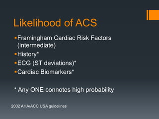
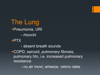
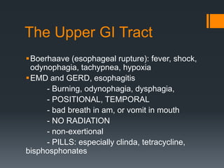
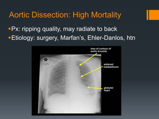
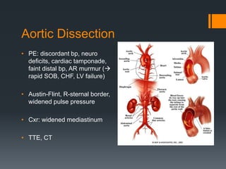
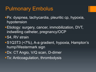
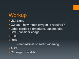
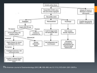
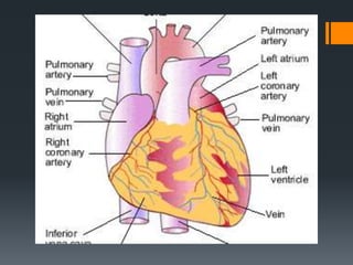
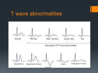
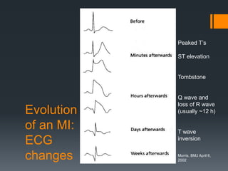
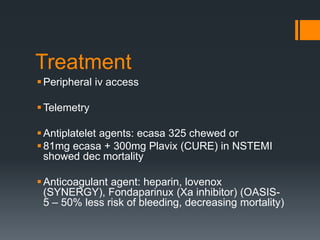
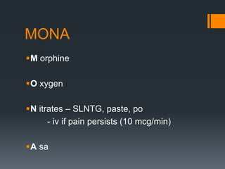
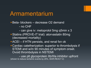
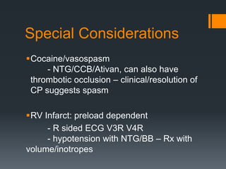
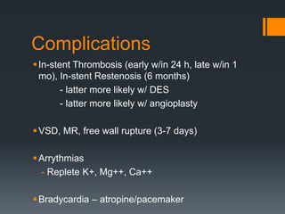
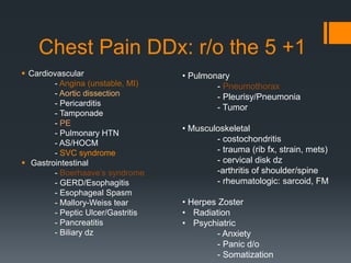
Chest Pain
It’s all in the story
Everyone has one
Very common
Acute management. Mostly benign, AND
rarely **life-threatening **
Therefore concentrate on the work-up and
treatment of those
Roadmap
Context/Background/Epidemiology
DDx
Presentation/History = Workup
AA/Dissection, PTX, PE, SVC Syndrome
ACS
Acute Management
Epidemiology
Up to 30% cardiac catheterizations are
negative for CAD*
3.15 million to 1.8 billion $/yr
Low mortality and high morbidity, i.e. the
cause and effect of your hard work
Nevens, F, Janssens, J, Piessens, J, et al. Prospective study on prevalence of esophageal chest pain in patients referred
on an elective basis to a cardiac unit for suspected myocardial ischemia. Dig Dis Sci 1991;36:229–235.
Achem, S, DeVault, K. Recent developments in chest pain of undetermined origin. Curr Gastroenterol Rep 2000;2:201–
209.
DDx
mainly cardiac, pulmonary, gastrointestinal,
and psychiatric
Must r/o ACS (angina, NSTEMI, STEMI),
PE, tamponade, Boerhaave’s, aortic
dissection, tension PTX, SVC syndrome
Mind your PQRSTs
Provoke, palliation, position
Prior occurrence
Quality
Radiation
Severity
Surgery (PE, CABG)
Timing
The Heart
QUALITY: pressure, squeezing, burning
RADIATION to jaw, shoulder, arm
Related to EXERTION?
palpitations, diaphoresis
early am (sympathetic stimulation)
PND (at night)
POSITION
HYPOXIA
The Heart
LIMITED FLOW: vasoconstriction, anemia,
hypotention (coronary vasospasm,
arrhythmia, valvular disease, hypertensive
urgency/emergency, cocaine)
Pericarditis, pericardial effusion
CHF
Likelihood of ACS
Framingham Cardiac Risk Factors
(intermediate)
History*
ECG (ST deviations)*
Cardiac Biomarkers*
* Any ONE connotes high probability
2002 AHA/ACC USA guidelines
The Lung
Pneumonia, URI
- rhonchi
PTX
- absent breath sounds
COPD, sarcoid, pulmonary fibrosis,
pulmonary htn, i.e. increased pulmonary
resistance
- no air movt, wheeze, velcro rales
The Upper GI Tract
Boerhaave (esophageal rupture): fever, shock,
odynophagia, tachypnea, hypoxia
EMD and GERD, esophagitis
- Burning, odynophagia, dysphagia,
- POSITIONAL, TEMPORAL
- bad breath in am, or vomit in mouth
- NO RADIATION
- non-exertional
- PILLS: especially clinda, tetracycline,
bisphosphonates
GERD, esophagitis
• Can account for >20-65% of UCP
• Diagnostic and Therapeutic: Antacids
- PPI
- H2-blockers
- sucralfate
Psychiatric:
dx of exclusion
Panic disorder
Anxiety
Somatization
Don’t Forget
AAA/Aortic Dissection
PE
PTX
SVC syndrome
Aortic Dissection: High Mortality
Px: ripping quality, may radiate to back
Etiology: surgery, Marfan’s, Ehler-Danlos, htn
Aortic Dissection
• PE: discordant bp, neuro
deficits, cardiac tamponade,
faint distal bp, AR murmur (
rapid SOB, CHF, LV failure)
• Austin-Flint, R-sternal border,
widened pulse pressure
• Cxr: widened mediastinum
• TTE, CT
Pulmonary Embolus
Px: dyspnea, tachycardia, pleuritic cp, hypoxia,
hypotension
Etiology: surgery, cancer, immobilization, DVT,
indwelling catheter, pregnancy/OCP
S4, RV strain
S1Q3T3 (<7%), A-a gradient, hypoxia, Hampton’s
hump/Westermark sign
Dx: CT Angio, V/Q scan, D-dimer
Tx: Anticoagulation, thrombolysis
Pulmonary
Embolus
Pneumothorax
 Px: Dyspnea, pleuritic cp
 Etiology: spontaneous, recent
instrumentation or intubation
 PE: tracheal deviation, absent
breath sounds
 CXR
 Minor: observe and o2
 Major: CHEST TUBE >>
needle decompression (mid
clavicular, 2nd ICS)
SVC syndrome*
Low-pressure, and thin-walled
Neighbors: trachea, R bronchus, Aorta,
sternum
Head, neck, upper thorax and arms
Etiology: R sided mass – 80% cancer (80%
mostly NSCLCa, 15% NHL), infection (Histo,
TB, syphilis), vasculitides (AA, Takayasu,
mediastinitis).
SVC syndrome*
Px: cough, head fullness, SOB, facial swelling,
unilateral arm swelling, dysphagia, dysphonia,
stridor, orthopnea, lightheadedness  worsened
by lying down, bending over
SVC Syndrome*
Tx: elevation of head, o2
Steroids, intubation, lasix (cerebral or
laryngeal edema)
consider XRT
Thrombolytics, anticoagulation (cerebral
hemorrhage)
PTA (perc transluminal angio)
Other Diagnoses
Malignancy
Musculoskeletal (cough), compression
fracture, rheum diseases, costochondritis,
fibromyalgia
Zoster
Post-radiation
Workup
vital signs
O2 sat – how much oxygen is required?
Labs: cardiac biomarkers, lactate, cbc,
BMP, consider coags
ECG
CXR
- mediastinal or aortic widening
ABG
CT angio: if stable
The American Journal of Gastroenterology (2001) 96, 958–968; doi:10.1111/j.1572-0241.2001.03678.x
Back to the
ECG findings
Always look at old ECG
ECG findings
LAD – V1-V4, aka “septal, precordial leads”
- Diagonal artery
- supplies LV
LCX – I, L, V5 and V6, aka “lateral leads”
- Obtuse marginal (OM) artery
- reciprocal changes in II, III, F
RCA – II, III, avF, aka “posterior leads”
- posterior descending artery (PDA)
- supplies SA node, and RV  arrythmias
- preload dependent
Conduction Pathways
ECG findings
ST Elevation (injury)
– DDx: MI, Pericarditis, early repolarization,
vasospasm, LV aneurysm
New LBBB: QRS >120ms, notched R in lateral
leads (no q’s), rS in septal (V1) leads, L axis
deviation
ST Depressions (ischemia)
T wave abnormalities: flattening, inversions,
biphasic
T wave abnormalities
Evolution
of an MI:
ECG
changes
Peaked T’s
ST elevation
Tombstone
Q wave and
loss of R wave
(usually ~12 h)
T wave
inversion
Morris, BMJ April 6,
2002
ECG findings: RV infarct
V1-V3  turn over
and upside down or
ST-depression, R: S
is >1, R may be
broad
V7-V9: place leads
V4-V6 horizontally
at level of V6 (post
axillary, mid
scapula)
ECG findings: RV infarct
Cardiac Biomarkers
AST: first test, not specific
 CK-MB: 6-12 hours (artifactual if skelatal
muscle is present). Disappears quickly but good
for extension of infarction
 troponin: 12 hours, persists up to 7 days. Can
correlate to size of infarct but can take up to 3
days to result
LDH: 72 hours, not specific
Cardiac Biomarkers
Adapted from ACC/AHA Guidelines 2005
Treatment
Peripheral iv access
Telemetry
Antiplatelet agents: ecasa 325 chewed or
81mg ecasa + 300mg Plavix (CURE) in NSTEMI
showed dec mortality
Anticoagulant agent: heparin, lovenox
(SYNERGY), Fondaparinux (Xa inhibitor) (OASIS-
5 – 50% less risk of bleeding, decreasing mortality)
MONA
M orphine
O xygen
N itrates – SLNTG, paste, po
- iv if pain persists (10 mcg/min)
A sa
Armamentarium
Beta- blockers – decrease O2 demand
- no CHF
- can give iv: metoprolol 5mg q5min x 3
Statins (PROVE-IT trial): atorvastatin 80mg
(decreased mortality)
ACEI – if HTN persists, and renal fxn ok
Cardiac catetherization: superior to thrombolysis if
STEMI and w/in 90 minutes of symptom onset.
Ovoid thrombolysis in NSTEMI
- use plt glycoprotein IIb/IIIa inhibitor upfront
(shown to reduce ischemic events by 25%, ISAR-REACT 2)
Special Considerations
Cocaine/vasospasm
- NTG/CCB/Ativan, can also have
thrombotic occlusion – clinical/resolution of
CP suggests spasm
RV Infarct: preload dependent
- R sided ECG V3R V4R
- hypotension with NTG/BB – Rx with
volume/inotropes
Complications
In-stent Thrombosis (early w/in 24 h, late w/in 1
mo), In-stent Restenosis (6 months)
- latter more likely w/ DES
- latter more likely w/ angioplasty
VSD, MR, free wall rupture (3-7 days)
Arrythmias
- Replete K+, Mg++, Ca++
Bradycardia – atropine/pacemaker
Chest Pain DDx: r/o the 5 +1
 Cardiovascular
- Angina (unstable, MI)
- Aortic dissection
- Pericarditis
- Tamponade
- PE
- Pulmonary HTN
- AS/HOCM
- SVC syndrome
 Gastrointestinal
- Boerhaave’s syndrome
- GERD/Esophagitis
- Esophageal Spasm
- Mallory-Weiss tear
- Peptic Ulcer/Gastritis
- Pancreatitis
- Biliary dz
• Pulmonary
- Pneumothorax
- Pleurisy/Pneumonia
- Tumor
• Musculoskeletal
- costochondritis
- trauma (rib fx, strain, mets)
- cervical disk dz
-arthritis of shoulder/spine
- rheumatologic: sarcoid, FM
• Herpes Zoster
• Radiation
• Psychiatric
- Anxiety
- Panic d/o
- Somatization
Conclusions
Chest Pain does not necessarily = Acute
Coronary Syndrome
Recognize DDx and clinical manifestations
PE, Aortic Dissection, PTX, Boerhaave’s, (SVC
syndrome)
 Angina/ACS – myocardial 02 supply
insufficient to meet demand; recognize
“secondary angina” and treat correctable
causes
Shock: contractility agents (dobutamine), IVF,
reperfusion,
Call for help if needed

Chest pain

  • 1.
  • 2.
    Everyone has one Verycommon Acute management. Mostly benign, AND rarely **life-threatening ** Therefore concentrate on the work-up and treatment of those
  • 3.
  • 4.
    Epidemiology Up to 30%cardiac catheterizations are negative for CAD* 3.15 million to 1.8 billion $/yr Low mortality and high morbidity, i.e. the cause and effect of your hard work Nevens, F, Janssens, J, Piessens, J, et al. Prospective study on prevalence of esophageal chest pain in patients referred on an elective basis to a cardiac unit for suspected myocardial ischemia. Dig Dis Sci 1991;36:229–235. Achem, S, DeVault, K. Recent developments in chest pain of undetermined origin. Curr Gastroenterol Rep 2000;2:201– 209.
  • 5.
    DDx mainly cardiac, pulmonary,gastrointestinal, and psychiatric Must r/o ACS (angina, NSTEMI, STEMI), PE, tamponade, Boerhaave’s, aortic dissection, tension PTX, SVC syndrome
  • 6.
    Mind your PQRSTs Provoke,palliation, position Prior occurrence Quality Radiation Severity Surgery (PE, CABG) Timing
  • 7.
    The Heart QUALITY: pressure,squeezing, burning RADIATION to jaw, shoulder, arm Related to EXERTION? palpitations, diaphoresis early am (sympathetic stimulation) PND (at night) POSITION HYPOXIA
  • 8.
    The Heart LIMITED FLOW:vasoconstriction, anemia, hypotention (coronary vasospasm, arrhythmia, valvular disease, hypertensive urgency/emergency, cocaine) Pericarditis, pericardial effusion CHF
  • 9.
    Likelihood of ACS FraminghamCardiac Risk Factors (intermediate) History* ECG (ST deviations)* Cardiac Biomarkers* * Any ONE connotes high probability 2002 AHA/ACC USA guidelines
  • 10.
    The Lung Pneumonia, URI -rhonchi PTX - absent breath sounds COPD, sarcoid, pulmonary fibrosis, pulmonary htn, i.e. increased pulmonary resistance - no air movt, wheeze, velcro rales
  • 11.
    The Upper GITract Boerhaave (esophageal rupture): fever, shock, odynophagia, tachypnea, hypoxia EMD and GERD, esophagitis - Burning, odynophagia, dysphagia, - POSITIONAL, TEMPORAL - bad breath in am, or vomit in mouth - NO RADIATION - non-exertional - PILLS: especially clinda, tetracycline, bisphosphonates
  • 12.
    GERD, esophagitis • Canaccount for >20-65% of UCP • Diagnostic and Therapeutic: Antacids - PPI - H2-blockers - sucralfate
  • 13.
    Psychiatric: dx of exclusion Panicdisorder Anxiety Somatization
  • 14.
  • 15.
    Aortic Dissection: HighMortality Px: ripping quality, may radiate to back Etiology: surgery, Marfan’s, Ehler-Danlos, htn
  • 16.
    Aortic Dissection • PE:discordant bp, neuro deficits, cardiac tamponade, faint distal bp, AR murmur ( rapid SOB, CHF, LV failure) • Austin-Flint, R-sternal border, widened pulse pressure • Cxr: widened mediastinum • TTE, CT
  • 17.
    Pulmonary Embolus Px: dyspnea,tachycardia, pleuritic cp, hypoxia, hypotension Etiology: surgery, cancer, immobilization, DVT, indwelling catheter, pregnancy/OCP S4, RV strain S1Q3T3 (<7%), A-a gradient, hypoxia, Hampton’s hump/Westermark sign Dx: CT Angio, V/Q scan, D-dimer Tx: Anticoagulation, thrombolysis
  • 18.
  • 19.
    Pneumothorax  Px: Dyspnea,pleuritic cp  Etiology: spontaneous, recent instrumentation or intubation  PE: tracheal deviation, absent breath sounds  CXR  Minor: observe and o2  Major: CHEST TUBE >> needle decompression (mid clavicular, 2nd ICS)
  • 20.
    SVC syndrome* Low-pressure, andthin-walled Neighbors: trachea, R bronchus, Aorta, sternum Head, neck, upper thorax and arms Etiology: R sided mass – 80% cancer (80% mostly NSCLCa, 15% NHL), infection (Histo, TB, syphilis), vasculitides (AA, Takayasu, mediastinitis).
  • 21.
    SVC syndrome* Px: cough,head fullness, SOB, facial swelling, unilateral arm swelling, dysphagia, dysphonia, stridor, orthopnea, lightheadedness  worsened by lying down, bending over
  • 22.
    SVC Syndrome* Tx: elevationof head, o2 Steroids, intubation, lasix (cerebral or laryngeal edema) consider XRT Thrombolytics, anticoagulation (cerebral hemorrhage) PTA (perc transluminal angio)
  • 23.
    Other Diagnoses Malignancy Musculoskeletal (cough),compression fracture, rheum diseases, costochondritis, fibromyalgia Zoster Post-radiation
  • 24.
    Workup vital signs O2 sat– how much oxygen is required? Labs: cardiac biomarkers, lactate, cbc, BMP, consider coags ECG CXR - mediastinal or aortic widening ABG CT angio: if stable
  • 25.
    The American Journalof Gastroenterology (2001) 96, 958–968; doi:10.1111/j.1572-0241.2001.03678.x
  • 26.
  • 27.
  • 30.
    ECG findings LAD –V1-V4, aka “septal, precordial leads” - Diagonal artery - supplies LV LCX – I, L, V5 and V6, aka “lateral leads” - Obtuse marginal (OM) artery - reciprocal changes in II, III, F RCA – II, III, avF, aka “posterior leads” - posterior descending artery (PDA) - supplies SA node, and RV  arrythmias - preload dependent
  • 31.
  • 32.
    ECG findings ST Elevation(injury) – DDx: MI, Pericarditis, early repolarization, vasospasm, LV aneurysm New LBBB: QRS >120ms, notched R in lateral leads (no q’s), rS in septal (V1) leads, L axis deviation ST Depressions (ischemia) T wave abnormalities: flattening, inversions, biphasic
  • 33.
  • 34.
    Evolution of an MI: ECG changes PeakedT’s ST elevation Tombstone Q wave and loss of R wave (usually ~12 h) T wave inversion Morris, BMJ April 6, 2002
  • 35.
    ECG findings: RVinfarct V1-V3  turn over and upside down or ST-depression, R: S is >1, R may be broad V7-V9: place leads V4-V6 horizontally at level of V6 (post axillary, mid scapula)
  • 36.
  • 37.
    Cardiac Biomarkers AST: firsttest, not specific  CK-MB: 6-12 hours (artifactual if skelatal muscle is present). Disappears quickly but good for extension of infarction  troponin: 12 hours, persists up to 7 days. Can correlate to size of infarct but can take up to 3 days to result LDH: 72 hours, not specific
  • 38.
    Cardiac Biomarkers Adapted fromACC/AHA Guidelines 2005
  • 39.
    Treatment Peripheral iv access Telemetry Antiplateletagents: ecasa 325 chewed or 81mg ecasa + 300mg Plavix (CURE) in NSTEMI showed dec mortality Anticoagulant agent: heparin, lovenox (SYNERGY), Fondaparinux (Xa inhibitor) (OASIS- 5 – 50% less risk of bleeding, decreasing mortality)
  • 40.
    MONA M orphine O xygen Nitrates – SLNTG, paste, po - iv if pain persists (10 mcg/min) A sa
  • 41.
    Armamentarium Beta- blockers –decrease O2 demand - no CHF - can give iv: metoprolol 5mg q5min x 3 Statins (PROVE-IT trial): atorvastatin 80mg (decreased mortality) ACEI – if HTN persists, and renal fxn ok Cardiac catetherization: superior to thrombolysis if STEMI and w/in 90 minutes of symptom onset. Ovoid thrombolysis in NSTEMI - use plt glycoprotein IIb/IIIa inhibitor upfront (shown to reduce ischemic events by 25%, ISAR-REACT 2)
  • 42.
    Special Considerations Cocaine/vasospasm - NTG/CCB/Ativan,can also have thrombotic occlusion – clinical/resolution of CP suggests spasm RV Infarct: preload dependent - R sided ECG V3R V4R - hypotension with NTG/BB – Rx with volume/inotropes
  • 43.
    Complications In-stent Thrombosis (earlyw/in 24 h, late w/in 1 mo), In-stent Restenosis (6 months) - latter more likely w/ DES - latter more likely w/ angioplasty VSD, MR, free wall rupture (3-7 days) Arrythmias - Replete K+, Mg++, Ca++ Bradycardia – atropine/pacemaker
  • 44.
    Chest Pain DDx:r/o the 5 +1  Cardiovascular - Angina (unstable, MI) - Aortic dissection - Pericarditis - Tamponade - PE - Pulmonary HTN - AS/HOCM - SVC syndrome  Gastrointestinal - Boerhaave’s syndrome - GERD/Esophagitis - Esophageal Spasm - Mallory-Weiss tear - Peptic Ulcer/Gastritis - Pancreatitis - Biliary dz • Pulmonary - Pneumothorax - Pleurisy/Pneumonia - Tumor • Musculoskeletal - costochondritis - trauma (rib fx, strain, mets) - cervical disk dz -arthritis of shoulder/spine - rheumatologic: sarcoid, FM • Herpes Zoster • Radiation • Psychiatric - Anxiety - Panic d/o - Somatization
  • 45.
    Conclusions Chest Pain doesnot necessarily = Acute Coronary Syndrome Recognize DDx and clinical manifestations PE, Aortic Dissection, PTX, Boerhaave’s, (SVC syndrome)  Angina/ACS – myocardial 02 supply insufficient to meet demand; recognize “secondary angina” and treat correctable causes Shock: contractility agents (dobutamine), IVF, reperfusion, Call for help if needed

Editor's Notes

  • #19 TTE: underfilled LA and “D-sign” from large RV
  • #31 T wave abnormalities – hyperacute: hyper K, early MI – inversions: DDx: ischemia, pericardial dz, myocarditis, CNS, drugs (TCA, phenothiazine) • Q waves –  pathologic: >.04 wide, 25% of total qrs height –  precordial R wave progression (should increase V1-V5/6) • Special – – – – Posterior MI: early transition (tall R V1 or V2, ST depression) RV infarct: ST elevation V3R V4R new LBBB less than 50% of acute MI have clear ECG findings on 1st ECG, 10% of pt with MI may not show ECG changes
  • #33 T wave abnormalities – hyperacute: hyper K, early MI – inversions: DDx: ischemia, pericardial dz, myocarditis, CNS, drugs (TCA, phenothiazine) • Q waves –  pathologic: >.04 wide, 25% of total qrs height –  precordial R wave progression (should increase V1-V5/6) • Special – – – – Posterior MI: early transition (tall R V1 or V2, ST depression) RV infarct: ST elevation V3R V4R new LBBB less than 50% of acute MI have clear ECG findings on 1st ECG, 10% of pt with MI may not show ECG changes
  • #36 T wave abnormalities – hyperacute: hyper K, early MI – inversions: DDx: ischemia, pericardial dz, myocarditis, CNS, drugs (TCA, phenothiazine) • Q waves –  pathologic: >.04 wide, 25% of total qrs height –  precordial R wave progression (should increase V1-V5/6) • Special – – – – Posterior MI: early transition (tall R V1 or V2, ST depression) RV infarct: ST elevation V3R V4R new LBBB less than 50% of acute MI have clear ECG findings on 1st ECG, 10% of pt with MI may not show ECG changes
  • #37 T wave abnormalities – hyperacute: hyper K, early MI – inversions: DDx: ischemia, pericardial dz, myocarditis, CNS, drugs (TCA, phenothiazine) • Q waves –  pathologic: >.04 wide, 25% of total qrs height –  precordial R wave progression (should increase V1-V5/6) • Special – – – – Posterior MI: early transition (tall R V1 or V2, ST depression) RV infarct: ST elevation V3R V4R new LBBB less than 50% of acute MI have clear ECG findings on 1st ECG, 10% of pt with MI may not show ECG changes