SlideShare a Scribd company logo
1 of 53
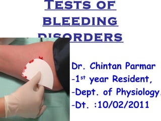
Tests of 
bleeding 
disorders 
Dr. Chintan Parmar 
-1st year Resident, 
-Dept. of Physiology. 
-Dt. :10/02/2011
OUTLINE 
• Normal Hemostasis 
• Coagulation Factors 
• Coagulation Pathway 
• Bleeding Disorders 
• Laboratory Tests
Normal Hemostasis 
• Trauma, disease or surgery – Disruption of 
vascular endothelial lining 
• Primary – Platelet Plug Formation – within 
seconds – capillaries, small arterioles & 
venules 
- Platelet adhesion, granule release & 
aggregation 
- Role of vWF ( Von Willebrand Factor ) 
- Role of fibrinogen
Normal Hemostasis 
• Secondary – plasma coagulation 
system – fibrin formation – several 
minutes – larger vessels 
• Interlinking – Activated platelets 
accelerate plasma coagulation & 
products of plasma coagulation 
reaction such as thrombin induce 
platelet activation.
5 
Normal Hemostasis 
BV Injury 
Platelet 
Activation 
Plt-Fusion 
Blood Vessel 
Constriction 
Coagulation 
Activation 
Stable Hemostatic Plug 
Thromibn, 
Fibrin 
Reduced 
Blood flow 
Tissue 
Factor, 
blood 
Primary hemostatic plug 
Neural
Coagulation Factors 
• Factor I : Fibrinogen 
• Factor II : Prothrombin 
• Factor III : Thromboplastin ( Tissue Factor ) 
• Factor IV : Calcium 
• Factor V : Labile factor or proaccelerin 
• Factor VII : Stable factor or proconvertin 
• Factor VIII : Antihemophilic globulin (AHA) 
• Factor IX : Christmas factor (AHB) 
• Factor X : Stuart-Prower factor 
• Factor XI : Plasma thromboplastin antecedent (AHC) 
• Factor XII : Hageman factor 
• Factor XIII : Fibrin-stabilizing factor
Bleeding Disorders 
• Increased fragility of vessels 
– Aging 
– Medications (steroids) 
• Platelet defect (deficiency) 
– Low platelet count 
– Medication (aspirin, clopidogrel) 
– Congenital platelet defect 
• Deranged clotting 
– Factor deficiency 
– Medication (coumarin) 
– Coagulation inhibitor
Coagulation Factor 
Disorders 
• Acquired 
– Vitamin K deficiency 
– Parenchymal disease of liver 
• Hereditary deficiencies 
– Factor VIII : hemophilia A 
– Factor IX : hemophilia B 
– Factor XI : hemophilia C 
–Von Willebrand’s Disease
Laboratory tests 
1. Tests of the Vascular Platelet Phase of 
Hemostasis: 
• BBlleeeeddiinngg TTiimmee ((BBTT) 
2. Tests of the Coagulation Cascade: 
• CClloottttiinngg TTiimmee (( CCTT) oorr CCooaagguullaattiioonn ttiimmee 
• AAccttiivvaatteedd PPaarrttiiaall TThhrroommbbooppllaassttiinn TTiimmee 
((AAPPTTTT).. 
• PPrrootthhrroommbbiinn TTiimmee ((PPTT) 
• TThhrroommbbiinn TTiimmee ((TTTT)
Laboratory tests 
3. OTHERS: 
- Platelet Count 
- Capillary fragility test 
- Clot Solubility Test 
- Clot Retraction Test 
- Thromboplastin Generation Test 
- Factors, fibrinogen, anticoagulants assay 
- Platelet function tests (aggregation) 
- Tests for Liver function
1. Bleeding Time 
• It is the time taken from the puncture of the 
blood vessel to the stoppage of bleeding. 
• The bleeding time test is a useful tool to test 
for platelet plug formation and capillary 
integrity. 
• BT is more imp. than CT. 
• CT concerns the blood only i.e. how firm 
the the clot is formed, whereas BT involves 
the interaction of blood with the injured 
tissues.
Duke Method 
• With the Duke method, the patient is 
pricked with a special needle or lancet, 
preferably on the earlobe or fingertip, 
after having been swabbed with alcohol. 
• The prick is about 3-4 mm deep. The 
patient then wipes the blood every 30 
seconds with a filter paper. 
• The test ends when bleeding stops. The 
usual time is about 2-6 minutes.
Ivy method 
٠ Clean the anterior surface of the forearm with spirit. 
• The blood pressure cuff is placed on the upper arm 
and inflated to 40 mmHg. 
• A lancet or scalpel blade is used to make a shallow 
incision that is 1 millimeter deep on the anterior of 
the forearm. 
• The time from when the incision is made until all 
bleeding has stopped is measured and is called the 
bleeding time. Every 30 seconds, filter paper or a 
paper towel is used to draw off the blood. 
• Normal BT by this method is 3-6 minutes.
Bleeding Time
Bleeding Time 
• A prolonged bleeding time may be a result from 
decreased number of thrombocytes or impaired 
blood vessels. 
• Bleeding time is affected by platelet function, 
certain vascular disorders and von Willebrand 
Disease, not by other coagulation factors. 
• Diseases that cause prolonged bleeding time include 
thrombocytopenia, disseminated intravascular 
coagulation (DIC). 
• Aspirin and other cyclooxygenase inhibitors can 
prolong bleeding time significantly.
Bleeding Time 
• People with von Willebrand disease usually 
experience increased bleeding time. 
• Von Willebrand factor is a platelet agglutination 
protein, but this is not considered an effective 
diagnostic test for this condition. 
• It is also prolonged in hypofibrinogenemia. 
• Many experts regard the bleeding time as useless, in 
that it does not predict surgical bleeding. 
• Role in post Myocardial Infraction patients.
2. Clotting Time 
• It is the time taken from the puncture of the blood 
vessel to the formation of a fibrin thread. 
• A. Capillary Glass Tube Method : Here the blood is 
collected in capillary tube & total time is noted to 
form FIBRIN THREADS on breaking tube every 30 
seconds. N : 3-8 minutes 
• B. Lee & White method : Here venous blood is 
collected in 8 mm diameter glass tube, rocked in a 
water bath at 37°C & time is noted from the time of 
vene puncture till the blood stops flowing. N : 6-12 
minutes.
Clotting Time 
• Mechanism Involved is INTRINSIC Pathway. 
• CT depends on presence of all clotting factors. 
• It gets prolonged in : 
- 1. Deficiency of clotting factors – Hemophilia. 
- 2. Vitamin K Deficiency – Factor II, VII, IX & X. 
- 3. Anticoagulant overdose. 
• BT & CT is measured before surgery & liver or 
bone marrow biopsy. 
• PURPURA : BT increased, CT normal. 
• HEMOPHILIA : BT normal, CT increased.
Clotting Time
3. Capillary Fragility Test 
• A tourniquet test (also known as a 
Rumpel-Leede Capillary-Fragility Test or 
Hess capillary fragility test) determines 
capillary fragility. 
• It is a clinical diagnostic method to 
determine a patient's haemorrhagic 
tendency. 
• It assesses fragility of capillary walls and 
is used to identify thrombocytopenia.
Capillary Fragility Test 
• The test is defined by the WHO as 
one of the necessary requisites for 
diagnosis of Dengue fever. 
• A blood pressure cuff is applied and 
inflated to a point between the 
systolic and diastolic blood pressures 
for five minutes. 
• The test is positive if there are 10 or 
more petechiae per square inch. 
• In DHF the test usually gives a 
definite positive result with 20 
petechiae or more.
4. Prothombin Time 
• The prothrombin time was discovered by Dr. Armand Quick 
and colleagues in 1935 and a second method was published 
by Dr.Paul Owren also called the "p and p" or "prothrombin 
and proconvertin" method. 
• It aided in the identification of the anticoagulants dicumarol 
and warfarin and was used subsequently as a measure of 
activity for warfarin when used therapeutically. 
• The INR was introduced in the early 1980s when it turned 
out that there was a large degree of variation between the 
various prothrombin time assays, a discrepancy mainly due 
to problems with the purity of the thromboplastin (tissue 
factor) concentrate. 
• The INR became widely accepted worldwide, especially 
after endorsement by the World Health Organisation.
Prothombin Time 
• The prothrombin time (PT) and its derived 
measures of prothrombin ratio (PR) and 
international normalised ratio (INR) are measures 
of the extrinsic pathway of coagulation. 
• They are used to determine the clotting tendency of 
blood, in the measure of warfarin dosage, liver 
damage and vitamin K status. 
• PT measures factors I, II, V, VII and X. 
• The reference range for prothrombin time is usually 
around 11-16 seconds; the normal range for the INR 
is 0.8–1.2.
Prothombin Time 
• Method 
• The prothrombin time is most commonly measured using 
blood plasma. Blood is drawn into a test tube containing 
liquid citrate, which acts as an anticoagulant by binding the 
calcium in a sample. 
• The blood is mixed, then centrifuged to separate blood cells 
from plasma. 
• The plasma is analyzed by an automated instrument at 
37°C. An excess of calcium is added to reverse the effects 
of citrate , which enables the blood to clot again. 
• Tissue factor (also known as factor III) is added, and the 
time the sample takes to clot is measured. 
• The prothrombin ratio is the prothrombin time for a patient, 
divided by the result for control plasma.
Prothombin Time
Prothombin Time 
• International normalised ratio 
• The result (in seconds) for a prothrombin time performed on 
a normal individual will vary depending on what type of 
analytical system it is performed. 
• This is due to the differences between different batches of 
manufacturer's tissue factor used in the reagent to perform 
the test. 
• The INR was devised to standardize the results. Each 
manufacturer assigns an ISI value (International Sensitivity 
Index) for any tissue factor they manufacture. The ISI value 
indicates how a particular batch of tissue factor compares to 
an internationally standardized sample. The ISI is usually 
between 1.0 and 2.0.
Prothombin Time
Prothombin Time 
• Interpretation 
• The prothrombin time is the time it takes plasma to clot 
after addition of tissue factor obtained from animals. This 
measures the quality of the extrinsic pathway as well as the 
common pathway of coagulation. 
• The speed of the extrinsic pathway is greatly affected by 
levels of factor VII in the body. Factor VII has a short half-life 
and its synthesis requires vitamin K. 
• The prothrombin time can be prolonged as a result of 
deficiencies in vitamin K, which can be caused by warfarin, 
malabsorption, newborns. 
• In addition, poor factor VII synthesis due to liver disease or 
increased consumption in disseminated intravascular 
coagulation may prolong the PT.
Prothombin Time
5. APTT 
• The partial thromboplastin time (PTT) or activated 
partial thromboplastin time (aPTT or APTT) is a 
performance indicator measuring the efficacy of both 
the intrinsic and the common coagulation pathways. 
• Apart from detecting abnormalities in blood clotting, it 
is also used to monitor the treatment effects with 
heparin, a major anticoagulant. 
• Kaolin cephalin clotting time (KccT) is a historic 
name for the activated partial thromboplastin time. 
• The aPTT was first described in 1953 by researchers at 
the University of North Carolina at Chapel Hill.
APTT 
• Method 
• Collection of blood samples is done in vacu-tubes with 
oxalate or citrate to arrest coagulation by binding 
calcium. 
• In order to activate the intrinsic pathway, phospholipid, 
an activator such as silica, celite, kaolin, ellagic acid 
and calcium to reverse the anticoagulant effect of the 
oxalate are mixed into the plasma sample. 
• The time is measured until a thrombus (clot) forms. 
• The test is termed "partial" due to the absence of tissue 
factor from the reaction mixture.
APTT 
• After 
centrifugation, the 
Plasma contains all 
the intrinsic 
coagulation factors 
except calcium 
(removed during 
anticoagulation) 
and the platelets 
(removed during 
centrifugation).
APTT 
• Interpretation 
• Normal value : 25 – 39 seconds. 
• Normal PTT times require the presence of the following 
coagulation factors: I, II, V, VIII, IX, X, XI & XII. 
• Deficiencies in factors VII or XIII will not be detected with 
the PTT test. 
• Prolonged APTT may indicate: 
- use of heparin 
- coagulation factor deficiency (e.g. hemophilia A , B ) 
- antiphospholipid antibody especially lupus anticoagulant, 
which increases propensity to thrombosis.
APTT 
• To distinguish the causes, mixing tests are 
performed, in which the patient's plasma is mixed 
initially at a 50 : 50 dilution with normal plasma. 
• If the abnormality does not disappear, the sample is 
said to contain an "inhibitor" , either heparin, 
antiphospholipid antibodies or coagulation factor 
specific inhibitors. 
• If it does correct, a factor deficiency is more likely. 
• Deficiencies of factors VIII, IX, XI and XII and 
rarely von Willebrand factor, if causing a low factor 
VIII level, may lead to a prolonged aPTT, that 
corrects on mixing studies.
APTT
APTT
APTT
6. Thrombin Time 
• The Thrombin Time (TT) , is a blood test which measures 
the time it takes for a clot to form in the plasma from a 
blood sample in anticoagulant which had added an excess of 
thrombin. 
• This test is repeated with pooled plasma from normal 
patients. 
• The difference in time between the test and the 'normal' 
indicates an abnormality in the conversion of fibrinogen (a 
soluble protein) to fibrin, an insoluble protein. 
• This test is also known as the Thrombin Clotting Time 
(TCT).
Thrombin Time 
• Thrombin time compares a patient's rate of clot 
formation to that of a sample of normal pooled 
plasma. 
• Thrombin is added to the samples of plasma. If the 
plasma does not clot immediately, a quantitative 
(fibrinogen deficiency) or qualitative (dysfunctional 
fibrinogen) defect is present. 
• If a patient is receiving heparin, a substance derived 
from snake venom called reptilase is used instead of 
thrombin. 
• Reptilase has a similar action to thrombin but unlike 
thrombin, it is not inhibited by heparin.
Thrombin Time 
• The thrombin time is used to diagnose bleeding 
disorders and to assess the effectiveness of fibrinolytic 
therapy. 
• The time between the addition of the thrombin and the 
clot formation is recorded as the thrombin clotting time 
• Normal values for thrombin time are 10 to 15 seconds. 
• If reptilase is used, the reptilase time should be between 
15 and 20 seconds. 
• Thrombin time can be prolonged by : heparin, FDPs, 
factor XIII deficiency and fibrinogen deficiency / 
abnormality.
Thrombin Time
7. Platelet Count 
• Platelet Count can be determined by improved 
Neubauer’s counting chamber with RBC 
pipette & 1% ammonium oxalate. 
• They can be seen as tiny diameter, well 
seprated, highly refractile rounded bodies with 
silvery appearance. 
• N : 1.5 – 4 lacs / cumm. 
• Leishman stain : in clumps, blue cytoplasm, 
reddish purple granules, no nucleus.
Platelet Count
Platelet Count 
• Surgical bleeding usually does 
not occur until the platelet count 
is less than 50,000. 
• Spontaneous bleeding does not 
occur until the platelet count is 
less than 10,000-20,000.
Platelet Count 
• THOMBOCYTOPENIA : 
• Bone marrow depression – Aplastic anemia 
• Hypersplenism 
• Viral infections 
• Drugs : Aspirin, Heparin, Chemotherapy 
• ITP 
• TTP 
• HUS 
• DIC
8. Thromboplastin Generation Test 
• This test measures generation of 
thromboplastin – intrinsic mechanism 
• N : 12 sec or less 
• Prolonged TGT : deficiency of factor V, VIII, 
IX & X. 
• Hemophilia : PT normal, TGT prolonged 
• Pure factor VII def. : PT prolonged, TGT 
normal 
• Factor X def. : Both PT & TGT prolonged
9. Clot Solubility Test 
• This test detects def. of factor XIII. 
• The test is based on the solubility of non 
crosslinked fibrin. 
• Test plasma is clotted in either calcium chloride 
or thrombin, then suspended at 37°C in either 
urea, 1% monochloroacetic acid or 2% acetic 
acid. 
• If the clot dissolves in one of the solvents, this 
indicates either low levels or total absence of 
factor XIII. 
• Confirmed by different Assays.
Clot Solubility Test 
• Low Factor XIII Levels are seen in : 
- Individuals with inherited F XIII deficiency - umbilical 
stump bleeding 
- F XIII inhibitors – Isoniazid , sodium valproate , 
phenytoin , penicillin or procainamide. 
- Henoch-Schoenlein purpura ( HSP ) 
- In patients on and following cardiopulmonary bypass 
- Chronic inflammatory bowel disease 
- Severe GvHD of the gut 
- Levels fall in pregnancy. Severe inherited F XIII 
deficiency is associated with recurrent miscarriage 
- Excessive activation, as seen in DIC. 
• LATE BLEEDERS
10. Clot Retraction Test 
• This test measures time needed for contraction 
of an undisturbed clot. 
• It indicates function & number of platelets. 
• N : begins within 2 hrs, completed within 24 
hrs. 
• Clot Retraction retarded in thrombocytopenia. 
• Clot is soft & small in thromboasthenia – 
functional disturbance of platelets.
Summary 
• CT, APTT increased – Hemophilia A or B, 
factor deficiency or inhibitors – factor assays 
• BT, APTT increased – vWD – VIII, vW factor 
assay ; platelet aggregation test 
• PT, APTT increased – def. factor II, V, X ; def 
vit. K ; liver disease ; warfarin – factor assays 
• PT increased – def. factor VII – factor VII 
assay 
• BT, CT, PT, TT, APTT increased – 
fibrinogenemia – Physio-chemical methods
Summary 
• Clot solubility test positive – def. factor XIII – 
factor XIII assay 
• BT increased, tourniquet / clot retraction test 
positive – thromboasthenia – platelet function tests 
• BT increased, tourniquet / clot retraction test 
positive, platelet count reduced – thrombocytopenia, 
platelet function defects - platelet function tests, 
platelet morphology 
• BT, CT, PT, TT, APTT increased ; platelet count 
reduced – liver disease, DIC - LFTs
• Take home message - All Bleeding 
stops…. Eventually

More Related Content

What's hot

Platelet Function Tests
Platelet Function TestsPlatelet Function Tests
Platelet Function TestsAhmed Makboul
 
Clotting time - Coagulation of whole blood
Clotting time - Coagulation of whole bloodClotting time - Coagulation of whole blood
Clotting time - Coagulation of whole bloodSHRUTHI VASAN
 
Coagulation tests
Coagulation testsCoagulation tests
Coagulation teststemis cola
 
Coagulation profile and its uses
Coagulation profile and its usesCoagulation profile and its uses
Coagulation profile and its usesAppy Akshay Agarwal
 
Blood coagulation mechanism (Intrinsic Coagulation pathway and Extrinsic Coag...
Blood coagulation mechanism (Intrinsic Coagulation pathway and Extrinsic Coag...Blood coagulation mechanism (Intrinsic Coagulation pathway and Extrinsic Coag...
Blood coagulation mechanism (Intrinsic Coagulation pathway and Extrinsic Coag...Shaista Jabeen
 
PLATELET COUNT by Dr. Pandian M .pptx
PLATELET COUNT by Dr. Pandian M .pptxPLATELET COUNT by Dr. Pandian M .pptx
PLATELET COUNT by Dr. Pandian M .pptxPandian M
 
Platelet function tests.pptx 2.pptx final
Platelet function tests.pptx 2.pptx finalPlatelet function tests.pptx 2.pptx final
Platelet function tests.pptx 2.pptx finalAnupam Singh
 
Bleeding disorders
Bleeding disordersBleeding disorders
Bleeding disordersManisha Meda
 
Erythrocyte sedimentation rate
Erythrocyte sedimentation rateErythrocyte sedimentation rate
Erythrocyte sedimentation rateHajra Mehdi
 
Exfoliative cytology
Exfoliative cytologyExfoliative cytology
Exfoliative cytologyShamin Joshi
 
Pleural fluid examination
Pleural fluid examinationPleural fluid examination
Pleural fluid examinationNasir Nazeer
 
Investigation of bleeding disorder || bleeding disorder
Investigation of bleeding disorder ||  bleeding disorderInvestigation of bleeding disorder ||  bleeding disorder
Investigation of bleeding disorder || bleeding disorderparveen singh
 
Bleeding & clotting disorders
Bleeding & clotting disordersBleeding & clotting disorders
Bleeding & clotting disordersshabeel pn
 

What's hot (20)

Approach to bleeding disorders
Approach to bleeding disordersApproach to bleeding disorders
Approach to bleeding disorders
 
Platelet Function Tests
Platelet Function TestsPlatelet Function Tests
Platelet Function Tests
 
Bleeding disorders
Bleeding disordersBleeding disorders
Bleeding disorders
 
Clotting time - Coagulation of whole blood
Clotting time - Coagulation of whole bloodClotting time - Coagulation of whole blood
Clotting time - Coagulation of whole blood
 
Coagulation tests
Coagulation testsCoagulation tests
Coagulation tests
 
Coagulation profile and its uses
Coagulation profile and its usesCoagulation profile and its uses
Coagulation profile and its uses
 
Blood coagulation mechanism (Intrinsic Coagulation pathway and Extrinsic Coag...
Blood coagulation mechanism (Intrinsic Coagulation pathway and Extrinsic Coag...Blood coagulation mechanism (Intrinsic Coagulation pathway and Extrinsic Coag...
Blood coagulation mechanism (Intrinsic Coagulation pathway and Extrinsic Coag...
 
Rbc indices
Rbc indicesRbc indices
Rbc indices
 
PLATELET COUNT by Dr. Pandian M .pptx
PLATELET COUNT by Dr. Pandian M .pptxPLATELET COUNT by Dr. Pandian M .pptx
PLATELET COUNT by Dr. Pandian M .pptx
 
Platelet function tests.pptx 2.pptx final
Platelet function tests.pptx 2.pptx finalPlatelet function tests.pptx 2.pptx final
Platelet function tests.pptx 2.pptx final
 
Bleeding disorders
Bleeding disordersBleeding disorders
Bleeding disorders
 
prothrombin time
prothrombin timeprothrombin time
prothrombin time
 
Erythrocyte sedimentation rate
Erythrocyte sedimentation rateErythrocyte sedimentation rate
Erythrocyte sedimentation rate
 
Hemostasis
HemostasisHemostasis
Hemostasis
 
Exfoliative cytology
Exfoliative cytologyExfoliative cytology
Exfoliative cytology
 
Hemostasis (COAGULATION)
Hemostasis (COAGULATION)Hemostasis (COAGULATION)
Hemostasis (COAGULATION)
 
Pleural fluid examination
Pleural fluid examinationPleural fluid examination
Pleural fluid examination
 
Investigation of bleeding disorder || bleeding disorder
Investigation of bleeding disorder ||  bleeding disorderInvestigation of bleeding disorder ||  bleeding disorder
Investigation of bleeding disorder || bleeding disorder
 
Bleeding & clotting disorders
Bleeding & clotting disordersBleeding & clotting disorders
Bleeding & clotting disorders
 
Laboratory diagnosis of anemia
Laboratory diagnosis of anemiaLaboratory diagnosis of anemia
Laboratory diagnosis of anemia
 

Viewers also liked

Laboratory tests of hemostasis and coagulation system
Laboratory tests of hemostasis and coagulation systemLaboratory tests of hemostasis and coagulation system
Laboratory tests of hemostasis and coagulation systemderosaMSKCC
 
PT APTT Technical Information
PT APTT Technical InformationPT APTT Technical Information
PT APTT Technical InformationPriyank Dubey
 
Prothrombin and Partial Thromboplastin Time
Prothrombin and Partial Thromboplastin TimeProthrombin and Partial Thromboplastin Time
Prothrombin and Partial Thromboplastin Timecamiij1
 
Coagulation profiles (pt,ptt,at, fib
Coagulation profiles (pt,ptt,at, fibCoagulation profiles (pt,ptt,at, fib
Coagulation profiles (pt,ptt,at, fibAKHTAR HUSSAIN
 
Laboratory tests of hemostasis and coagulation system (dr ellinor peerschke ...
Laboratory tests of hemostasis and coagulation system (dr  ellinor peerschke ...Laboratory tests of hemostasis and coagulation system (dr  ellinor peerschke ...
Laboratory tests of hemostasis and coagulation system (dr ellinor peerschke ...derosaMSKCC
 
Copy Of Lesson (3) 3 Laboratory Bleedingtime
Copy Of Lesson (3) 3 Laboratory BleedingtimeCopy Of Lesson (3) 3 Laboratory Bleedingtime
Copy Of Lesson (3) 3 Laboratory BleedingtimeMiami Dade
 
Coagulation assays part 1
Coagulation assays part 1Coagulation assays part 1
Coagulation assays part 1derosaMSKCC
 
Coagulation profile
Coagulation profile Coagulation profile
Coagulation profile Vikash Prasad
 
Blood clotting
Blood clottingBlood clotting
Blood clottingSaurav Das
 
Lesson 3 4 Laboratory Pt
Lesson 3 4 Laboratory PtLesson 3 4 Laboratory Pt
Lesson 3 4 Laboratory PtMiami Dade
 
Hamostasis and blood coagulation
Hamostasis and blood coagulationHamostasis and blood coagulation
Hamostasis and blood coagulationdrmcbansal
 
5. bleeding disorder
5. bleeding disorder5. bleeding disorder
5. bleeding disorderWhiteraven68
 
Approach to bleeding disorder (coagulation defects) in children
Approach to bleeding disorder (coagulation defects) in childrenApproach to bleeding disorder (coagulation defects) in children
Approach to bleeding disorder (coagulation defects) in childrenSatish Vadapalli
 
Bleeding Disorder
Bleeding DisorderBleeding Disorder
Bleeding Disorderghalan
 
Erythrocyte sedimentation rate (esr)
Erythrocyte sedimentation rate (esr)Erythrocyte sedimentation rate (esr)
Erythrocyte sedimentation rate (esr)Shabab Ali
 
Bleeding disorders
Bleeding disordersBleeding disorders
Bleeding disordersraj kumar
 
Rational approach to evaluate a bleeding patient 2014
Rational approach to evaluate a bleeding patient 2014Rational approach to evaluate a bleeding patient 2014
Rational approach to evaluate a bleeding patient 2014sajith8523
 

Viewers also liked (20)

Laboratory tests of hemostasis and coagulation system
Laboratory tests of hemostasis and coagulation systemLaboratory tests of hemostasis and coagulation system
Laboratory tests of hemostasis and coagulation system
 
PT APTT Technical Information
PT APTT Technical InformationPT APTT Technical Information
PT APTT Technical Information
 
Prothrombin and Partial Thromboplastin Time
Prothrombin and Partial Thromboplastin TimeProthrombin and Partial Thromboplastin Time
Prothrombin and Partial Thromboplastin Time
 
Coagulation profiles (pt,ptt,at, fib
Coagulation profiles (pt,ptt,at, fibCoagulation profiles (pt,ptt,at, fib
Coagulation profiles (pt,ptt,at, fib
 
Laboratory tests of hemostasis and coagulation system (dr ellinor peerschke ...
Laboratory tests of hemostasis and coagulation system (dr  ellinor peerschke ...Laboratory tests of hemostasis and coagulation system (dr  ellinor peerschke ...
Laboratory tests of hemostasis and coagulation system (dr ellinor peerschke ...
 
Copy Of Lesson (3) 3 Laboratory Bleedingtime
Copy Of Lesson (3) 3 Laboratory BleedingtimeCopy Of Lesson (3) 3 Laboratory Bleedingtime
Copy Of Lesson (3) 3 Laboratory Bleedingtime
 
Blood coagulation
Blood coagulationBlood coagulation
Blood coagulation
 
Coagulation assays part 1
Coagulation assays part 1Coagulation assays part 1
Coagulation assays part 1
 
Coagulation profile
Coagulation profile Coagulation profile
Coagulation profile
 
Blood clotting
Blood clottingBlood clotting
Blood clotting
 
CME: Bleeding disorders - Diagnostic Approach
CME: Bleeding disorders - Diagnostic ApproachCME: Bleeding disorders - Diagnostic Approach
CME: Bleeding disorders - Diagnostic Approach
 
Lesson 3 4 Laboratory Pt
Lesson 3 4 Laboratory PtLesson 3 4 Laboratory Pt
Lesson 3 4 Laboratory Pt
 
mbbs ims msu
mbbs ims msumbbs ims msu
mbbs ims msu
 
Hamostasis and blood coagulation
Hamostasis and blood coagulationHamostasis and blood coagulation
Hamostasis and blood coagulation
 
5. bleeding disorder
5. bleeding disorder5. bleeding disorder
5. bleeding disorder
 
Approach to bleeding disorder (coagulation defects) in children
Approach to bleeding disorder (coagulation defects) in childrenApproach to bleeding disorder (coagulation defects) in children
Approach to bleeding disorder (coagulation defects) in children
 
Bleeding Disorder
Bleeding DisorderBleeding Disorder
Bleeding Disorder
 
Erythrocyte sedimentation rate (esr)
Erythrocyte sedimentation rate (esr)Erythrocyte sedimentation rate (esr)
Erythrocyte sedimentation rate (esr)
 
Bleeding disorders
Bleeding disordersBleeding disorders
Bleeding disorders
 
Rational approach to evaluate a bleeding patient 2014
Rational approach to evaluate a bleeding patient 2014Rational approach to evaluate a bleeding patient 2014
Rational approach to evaluate a bleeding patient 2014
 

Similar to Tests of bleeding disorders

Bleeding disorder Hematology Lecture.pptx
Bleeding disorder Hematology Lecture.pptxBleeding disorder Hematology Lecture.pptx
Bleeding disorder Hematology Lecture.pptxMunmun Kulsum
 
investigations of bleeding disorder (1).pptx
investigations of bleeding disorder (1).pptxinvestigations of bleeding disorder (1).pptx
investigations of bleeding disorder (1).pptxRajeshSaiSajja
 
Haemostasis in dentistry
Haemostasis in dentistryHaemostasis in dentistry
Haemostasis in dentistryParikshit Kadam
 
bleeding-timeclotting-time-pt-and-ptt-130504122408-phpapp01.pptx
bleeding-timeclotting-time-pt-and-ptt-130504122408-phpapp01.pptxbleeding-timeclotting-time-pt-and-ptt-130504122408-phpapp01.pptx
bleeding-timeclotting-time-pt-and-ptt-130504122408-phpapp01.pptxMohanSinghDhakad1
 
bleeding-timeclotting-time-pt-and-ptt-130504122408-phpapp01.pptx
bleeding-timeclotting-time-pt-and-ptt-130504122408-phpapp01.pptxbleeding-timeclotting-time-pt-and-ptt-130504122408-phpapp01.pptx
bleeding-timeclotting-time-pt-and-ptt-130504122408-phpapp01.pptxMohanSinghDhakad1
 
Understanding Haemostasis | Coagulations & Anticoagulation
Understanding Haemostasis | Coagulations & AnticoagulationUnderstanding Haemostasis | Coagulations & Anticoagulation
Understanding Haemostasis | Coagulations & AnticoagulationDr Habiba Kamarul
 
Presentation 22ndmay
Presentation 22ndmayPresentation 22ndmay
Presentation 22ndmayNavin Jain‬
 
Introduction to hematolgy
Introduction to hematolgyIntroduction to hematolgy
Introduction to hematolgyShweta Tiwari
 
Approach to bleeding disorders part II
Approach to bleeding disorders part IIApproach to bleeding disorders part II
Approach to bleeding disorders part IIdrbcnayak
 
Coagulation profile final by Pandian M
Coagulation profile final by Pandian M Coagulation profile final by Pandian M
Coagulation profile final by Pandian M Pandian M
 
Interpretation of tests in coagulation disorders
Interpretation of tests in coagulation disordersInterpretation of tests in coagulation disorders
Interpretation of tests in coagulation disordersDrRashmiBudha
 
BLEEDING DISORDER.pptx
BLEEDING DISORDER.pptxBLEEDING DISORDER.pptx
BLEEDING DISORDER.pptxPrasanthThalur
 
Common bleeding and clotting disorders
Common bleeding and clotting disordersCommon bleeding and clotting disorders
Common bleeding and clotting disordersQin Yang Huang
 
Approach to diagnosis of bleeding disorders
Approach to diagnosis of bleeding disordersApproach to diagnosis of bleeding disorders
Approach to diagnosis of bleeding disordersBiswajeeta Saha
 

Similar to Tests of bleeding disorders (20)

hemostasisdisorders
hemostasisdisordershemostasisdisorders
hemostasisdisorders
 
PROTHROMBIN TIME.pptx
PROTHROMBIN TIME.pptxPROTHROMBIN TIME.pptx
PROTHROMBIN TIME.pptx
 
Bleeding disorder Hematology Lecture.pptx
Bleeding disorder Hematology Lecture.pptxBleeding disorder Hematology Lecture.pptx
Bleeding disorder Hematology Lecture.pptx
 
investigations of bleeding disorder (1).pptx
investigations of bleeding disorder (1).pptxinvestigations of bleeding disorder (1).pptx
investigations of bleeding disorder (1).pptx
 
Haemostasis in dentistry
Haemostasis in dentistryHaemostasis in dentistry
Haemostasis in dentistry
 
bleeding-timeclotting-time-pt-and-ptt-130504122408-phpapp01.pptx
bleeding-timeclotting-time-pt-and-ptt-130504122408-phpapp01.pptxbleeding-timeclotting-time-pt-and-ptt-130504122408-phpapp01.pptx
bleeding-timeclotting-time-pt-and-ptt-130504122408-phpapp01.pptx
 
1.lab diagnosis
1.lab diagnosis1.lab diagnosis
1.lab diagnosis
 
thrombosis
thrombosisthrombosis
thrombosis
 
Hemostasis disorders
Hemostasis disordersHemostasis disorders
Hemostasis disorders
 
bleeding-timeclotting-time-pt-and-ptt-130504122408-phpapp01.pptx
bleeding-timeclotting-time-pt-and-ptt-130504122408-phpapp01.pptxbleeding-timeclotting-time-pt-and-ptt-130504122408-phpapp01.pptx
bleeding-timeclotting-time-pt-and-ptt-130504122408-phpapp01.pptx
 
Understanding Haemostasis | Coagulations & Anticoagulation
Understanding Haemostasis | Coagulations & AnticoagulationUnderstanding Haemostasis | Coagulations & Anticoagulation
Understanding Haemostasis | Coagulations & Anticoagulation
 
Presentation 22ndmay
Presentation 22ndmayPresentation 22ndmay
Presentation 22ndmay
 
Introduction to hematolgy
Introduction to hematolgyIntroduction to hematolgy
Introduction to hematolgy
 
Approach to bleeding disorders part II
Approach to bleeding disorders part IIApproach to bleeding disorders part II
Approach to bleeding disorders part II
 
Coagulation profile final by Pandian M
Coagulation profile final by Pandian M Coagulation profile final by Pandian M
Coagulation profile final by Pandian M
 
Interpretation of tests in coagulation disorders
Interpretation of tests in coagulation disordersInterpretation of tests in coagulation disorders
Interpretation of tests in coagulation disorders
 
BLEEDING DISORDER.pptx
BLEEDING DISORDER.pptxBLEEDING DISORDER.pptx
BLEEDING DISORDER.pptx
 
Common bleeding and clotting disorders
Common bleeding and clotting disordersCommon bleeding and clotting disorders
Common bleeding and clotting disorders
 
Approach to diagnosis of bleeding disorders
Approach to diagnosis of bleeding disordersApproach to diagnosis of bleeding disorders
Approach to diagnosis of bleeding disorders
 
hemorrhage
 hemorrhage hemorrhage
hemorrhage
 

More from DrChintansinh Parmar (20)

Autonomic nervous system
Autonomic nervous systemAutonomic nervous system
Autonomic nervous system
 
Skin & body temp.
Skin & body temp.Skin & body temp.
Skin & body temp.
 
Resp. diseases
Resp. diseasesResp. diseases
Resp. diseases
 
Regulation of respiration
Regulation of respirationRegulation of respiration
Regulation of respiration
 
Pulmonary circulation
Pulmonary circulationPulmonary circulation
Pulmonary circulation
 
Deep sea physiology
Deep sea physiologyDeep sea physiology
Deep sea physiology
 
Aviation physiology
Aviation physiologyAviation physiology
Aviation physiology
 
Diuretics, dialysis
Diuretics, dialysisDiuretics, dialysis
Diuretics, dialysis
 
Heart block and ECG
Heart block and ECGHeart block and ECG
Heart block and ECG
 
Ecg
EcgEcg
Ecg
 
Conductive system of heart
Conductive system of heartConductive system of heart
Conductive system of heart
 
Fetal circulation
Fetal circulationFetal circulation
Fetal circulation
 
Coronary circulation
Coronary circulationCoronary circulation
Coronary circulation
 
Synapse
SynapseSynapse
Synapse
 
Stretch reflex
Stretch reflexStretch reflex
Stretch reflex
 
Physiology of speech
Physiology of speech Physiology of speech
Physiology of speech
 
Motor system
Motor systemMotor system
Motor system
 
Cerebral cortex
Cerebral cortexCerebral cortex
Cerebral cortex
 
Shock
ShockShock
Shock
 
Circulation
CirculationCirculation
Circulation
 

Tests of bleeding disorders

  • 1. Tests of bleeding disorders Dr. Chintan Parmar -1st year Resident, -Dept. of Physiology. -Dt. :10/02/2011
  • 2. OUTLINE • Normal Hemostasis • Coagulation Factors • Coagulation Pathway • Bleeding Disorders • Laboratory Tests
  • 3. Normal Hemostasis • Trauma, disease or surgery – Disruption of vascular endothelial lining • Primary – Platelet Plug Formation – within seconds – capillaries, small arterioles & venules - Platelet adhesion, granule release & aggregation - Role of vWF ( Von Willebrand Factor ) - Role of fibrinogen
  • 4. Normal Hemostasis • Secondary – plasma coagulation system – fibrin formation – several minutes – larger vessels • Interlinking – Activated platelets accelerate plasma coagulation & products of plasma coagulation reaction such as thrombin induce platelet activation.
  • 5. 5 Normal Hemostasis BV Injury Platelet Activation Plt-Fusion Blood Vessel Constriction Coagulation Activation Stable Hemostatic Plug Thromibn, Fibrin Reduced Blood flow Tissue Factor, blood Primary hemostatic plug Neural
  • 6. Coagulation Factors • Factor I : Fibrinogen • Factor II : Prothrombin • Factor III : Thromboplastin ( Tissue Factor ) • Factor IV : Calcium • Factor V : Labile factor or proaccelerin • Factor VII : Stable factor or proconvertin • Factor VIII : Antihemophilic globulin (AHA) • Factor IX : Christmas factor (AHB) • Factor X : Stuart-Prower factor • Factor XI : Plasma thromboplastin antecedent (AHC) • Factor XII : Hageman factor • Factor XIII : Fibrin-stabilizing factor
  • 7.
  • 8. Bleeding Disorders • Increased fragility of vessels – Aging – Medications (steroids) • Platelet defect (deficiency) – Low platelet count – Medication (aspirin, clopidogrel) – Congenital platelet defect • Deranged clotting – Factor deficiency – Medication (coumarin) – Coagulation inhibitor
  • 9. Coagulation Factor Disorders • Acquired – Vitamin K deficiency – Parenchymal disease of liver • Hereditary deficiencies – Factor VIII : hemophilia A – Factor IX : hemophilia B – Factor XI : hemophilia C –Von Willebrand’s Disease
  • 10. Laboratory tests 1. Tests of the Vascular Platelet Phase of Hemostasis: • BBlleeeeddiinngg TTiimmee ((BBTT) 2. Tests of the Coagulation Cascade: • CClloottttiinngg TTiimmee (( CCTT) oorr CCooaagguullaattiioonn ttiimmee • AAccttiivvaatteedd PPaarrttiiaall TThhrroommbbooppllaassttiinn TTiimmee ((AAPPTTTT).. • PPrrootthhrroommbbiinn TTiimmee ((PPTT) • TThhrroommbbiinn TTiimmee ((TTTT)
  • 11. Laboratory tests 3. OTHERS: - Platelet Count - Capillary fragility test - Clot Solubility Test - Clot Retraction Test - Thromboplastin Generation Test - Factors, fibrinogen, anticoagulants assay - Platelet function tests (aggregation) - Tests for Liver function
  • 12. 1. Bleeding Time • It is the time taken from the puncture of the blood vessel to the stoppage of bleeding. • The bleeding time test is a useful tool to test for platelet plug formation and capillary integrity. • BT is more imp. than CT. • CT concerns the blood only i.e. how firm the the clot is formed, whereas BT involves the interaction of blood with the injured tissues.
  • 13. Duke Method • With the Duke method, the patient is pricked with a special needle or lancet, preferably on the earlobe or fingertip, after having been swabbed with alcohol. • The prick is about 3-4 mm deep. The patient then wipes the blood every 30 seconds with a filter paper. • The test ends when bleeding stops. The usual time is about 2-6 minutes.
  • 14. Ivy method ٠ Clean the anterior surface of the forearm with spirit. • The blood pressure cuff is placed on the upper arm and inflated to 40 mmHg. • A lancet or scalpel blade is used to make a shallow incision that is 1 millimeter deep on the anterior of the forearm. • The time from when the incision is made until all bleeding has stopped is measured and is called the bleeding time. Every 30 seconds, filter paper or a paper towel is used to draw off the blood. • Normal BT by this method is 3-6 minutes.
  • 16. Bleeding Time • A prolonged bleeding time may be a result from decreased number of thrombocytes or impaired blood vessels. • Bleeding time is affected by platelet function, certain vascular disorders and von Willebrand Disease, not by other coagulation factors. • Diseases that cause prolonged bleeding time include thrombocytopenia, disseminated intravascular coagulation (DIC). • Aspirin and other cyclooxygenase inhibitors can prolong bleeding time significantly.
  • 17. Bleeding Time • People with von Willebrand disease usually experience increased bleeding time. • Von Willebrand factor is a platelet agglutination protein, but this is not considered an effective diagnostic test for this condition. • It is also prolonged in hypofibrinogenemia. • Many experts regard the bleeding time as useless, in that it does not predict surgical bleeding. • Role in post Myocardial Infraction patients.
  • 18. 2. Clotting Time • It is the time taken from the puncture of the blood vessel to the formation of a fibrin thread. • A. Capillary Glass Tube Method : Here the blood is collected in capillary tube & total time is noted to form FIBRIN THREADS on breaking tube every 30 seconds. N : 3-8 minutes • B. Lee & White method : Here venous blood is collected in 8 mm diameter glass tube, rocked in a water bath at 37°C & time is noted from the time of vene puncture till the blood stops flowing. N : 6-12 minutes.
  • 19. Clotting Time • Mechanism Involved is INTRINSIC Pathway. • CT depends on presence of all clotting factors. • It gets prolonged in : - 1. Deficiency of clotting factors – Hemophilia. - 2. Vitamin K Deficiency – Factor II, VII, IX & X. - 3. Anticoagulant overdose. • BT & CT is measured before surgery & liver or bone marrow biopsy. • PURPURA : BT increased, CT normal. • HEMOPHILIA : BT normal, CT increased.
  • 21. 3. Capillary Fragility Test • A tourniquet test (also known as a Rumpel-Leede Capillary-Fragility Test or Hess capillary fragility test) determines capillary fragility. • It is a clinical diagnostic method to determine a patient's haemorrhagic tendency. • It assesses fragility of capillary walls and is used to identify thrombocytopenia.
  • 22. Capillary Fragility Test • The test is defined by the WHO as one of the necessary requisites for diagnosis of Dengue fever. • A blood pressure cuff is applied and inflated to a point between the systolic and diastolic blood pressures for five minutes. • The test is positive if there are 10 or more petechiae per square inch. • In DHF the test usually gives a definite positive result with 20 petechiae or more.
  • 23. 4. Prothombin Time • The prothrombin time was discovered by Dr. Armand Quick and colleagues in 1935 and a second method was published by Dr.Paul Owren also called the "p and p" or "prothrombin and proconvertin" method. • It aided in the identification of the anticoagulants dicumarol and warfarin and was used subsequently as a measure of activity for warfarin when used therapeutically. • The INR was introduced in the early 1980s when it turned out that there was a large degree of variation between the various prothrombin time assays, a discrepancy mainly due to problems with the purity of the thromboplastin (tissue factor) concentrate. • The INR became widely accepted worldwide, especially after endorsement by the World Health Organisation.
  • 24. Prothombin Time • The prothrombin time (PT) and its derived measures of prothrombin ratio (PR) and international normalised ratio (INR) are measures of the extrinsic pathway of coagulation. • They are used to determine the clotting tendency of blood, in the measure of warfarin dosage, liver damage and vitamin K status. • PT measures factors I, II, V, VII and X. • The reference range for prothrombin time is usually around 11-16 seconds; the normal range for the INR is 0.8–1.2.
  • 25. Prothombin Time • Method • The prothrombin time is most commonly measured using blood plasma. Blood is drawn into a test tube containing liquid citrate, which acts as an anticoagulant by binding the calcium in a sample. • The blood is mixed, then centrifuged to separate blood cells from plasma. • The plasma is analyzed by an automated instrument at 37°C. An excess of calcium is added to reverse the effects of citrate , which enables the blood to clot again. • Tissue factor (also known as factor III) is added, and the time the sample takes to clot is measured. • The prothrombin ratio is the prothrombin time for a patient, divided by the result for control plasma.
  • 27. Prothombin Time • International normalised ratio • The result (in seconds) for a prothrombin time performed on a normal individual will vary depending on what type of analytical system it is performed. • This is due to the differences between different batches of manufacturer's tissue factor used in the reagent to perform the test. • The INR was devised to standardize the results. Each manufacturer assigns an ISI value (International Sensitivity Index) for any tissue factor they manufacture. The ISI value indicates how a particular batch of tissue factor compares to an internationally standardized sample. The ISI is usually between 1.0 and 2.0.
  • 29. Prothombin Time • Interpretation • The prothrombin time is the time it takes plasma to clot after addition of tissue factor obtained from animals. This measures the quality of the extrinsic pathway as well as the common pathway of coagulation. • The speed of the extrinsic pathway is greatly affected by levels of factor VII in the body. Factor VII has a short half-life and its synthesis requires vitamin K. • The prothrombin time can be prolonged as a result of deficiencies in vitamin K, which can be caused by warfarin, malabsorption, newborns. • In addition, poor factor VII synthesis due to liver disease or increased consumption in disseminated intravascular coagulation may prolong the PT.
  • 31. 5. APTT • The partial thromboplastin time (PTT) or activated partial thromboplastin time (aPTT or APTT) is a performance indicator measuring the efficacy of both the intrinsic and the common coagulation pathways. • Apart from detecting abnormalities in blood clotting, it is also used to monitor the treatment effects with heparin, a major anticoagulant. • Kaolin cephalin clotting time (KccT) is a historic name for the activated partial thromboplastin time. • The aPTT was first described in 1953 by researchers at the University of North Carolina at Chapel Hill.
  • 32. APTT • Method • Collection of blood samples is done in vacu-tubes with oxalate or citrate to arrest coagulation by binding calcium. • In order to activate the intrinsic pathway, phospholipid, an activator such as silica, celite, kaolin, ellagic acid and calcium to reverse the anticoagulant effect of the oxalate are mixed into the plasma sample. • The time is measured until a thrombus (clot) forms. • The test is termed "partial" due to the absence of tissue factor from the reaction mixture.
  • 33. APTT • After centrifugation, the Plasma contains all the intrinsic coagulation factors except calcium (removed during anticoagulation) and the platelets (removed during centrifugation).
  • 34. APTT • Interpretation • Normal value : 25 – 39 seconds. • Normal PTT times require the presence of the following coagulation factors: I, II, V, VIII, IX, X, XI & XII. • Deficiencies in factors VII or XIII will not be detected with the PTT test. • Prolonged APTT may indicate: - use of heparin - coagulation factor deficiency (e.g. hemophilia A , B ) - antiphospholipid antibody especially lupus anticoagulant, which increases propensity to thrombosis.
  • 35. APTT • To distinguish the causes, mixing tests are performed, in which the patient's plasma is mixed initially at a 50 : 50 dilution with normal plasma. • If the abnormality does not disappear, the sample is said to contain an "inhibitor" , either heparin, antiphospholipid antibodies or coagulation factor specific inhibitors. • If it does correct, a factor deficiency is more likely. • Deficiencies of factors VIII, IX, XI and XII and rarely von Willebrand factor, if causing a low factor VIII level, may lead to a prolonged aPTT, that corrects on mixing studies.
  • 36. APTT
  • 37. APTT
  • 38. APTT
  • 39. 6. Thrombin Time • The Thrombin Time (TT) , is a blood test which measures the time it takes for a clot to form in the plasma from a blood sample in anticoagulant which had added an excess of thrombin. • This test is repeated with pooled plasma from normal patients. • The difference in time between the test and the 'normal' indicates an abnormality in the conversion of fibrinogen (a soluble protein) to fibrin, an insoluble protein. • This test is also known as the Thrombin Clotting Time (TCT).
  • 40. Thrombin Time • Thrombin time compares a patient's rate of clot formation to that of a sample of normal pooled plasma. • Thrombin is added to the samples of plasma. If the plasma does not clot immediately, a quantitative (fibrinogen deficiency) or qualitative (dysfunctional fibrinogen) defect is present. • If a patient is receiving heparin, a substance derived from snake venom called reptilase is used instead of thrombin. • Reptilase has a similar action to thrombin but unlike thrombin, it is not inhibited by heparin.
  • 41. Thrombin Time • The thrombin time is used to diagnose bleeding disorders and to assess the effectiveness of fibrinolytic therapy. • The time between the addition of the thrombin and the clot formation is recorded as the thrombin clotting time • Normal values for thrombin time are 10 to 15 seconds. • If reptilase is used, the reptilase time should be between 15 and 20 seconds. • Thrombin time can be prolonged by : heparin, FDPs, factor XIII deficiency and fibrinogen deficiency / abnormality.
  • 43. 7. Platelet Count • Platelet Count can be determined by improved Neubauer’s counting chamber with RBC pipette & 1% ammonium oxalate. • They can be seen as tiny diameter, well seprated, highly refractile rounded bodies with silvery appearance. • N : 1.5 – 4 lacs / cumm. • Leishman stain : in clumps, blue cytoplasm, reddish purple granules, no nucleus.
  • 45. Platelet Count • Surgical bleeding usually does not occur until the platelet count is less than 50,000. • Spontaneous bleeding does not occur until the platelet count is less than 10,000-20,000.
  • 46. Platelet Count • THOMBOCYTOPENIA : • Bone marrow depression – Aplastic anemia • Hypersplenism • Viral infections • Drugs : Aspirin, Heparin, Chemotherapy • ITP • TTP • HUS • DIC
  • 47. 8. Thromboplastin Generation Test • This test measures generation of thromboplastin – intrinsic mechanism • N : 12 sec or less • Prolonged TGT : deficiency of factor V, VIII, IX & X. • Hemophilia : PT normal, TGT prolonged • Pure factor VII def. : PT prolonged, TGT normal • Factor X def. : Both PT & TGT prolonged
  • 48. 9. Clot Solubility Test • This test detects def. of factor XIII. • The test is based on the solubility of non crosslinked fibrin. • Test plasma is clotted in either calcium chloride or thrombin, then suspended at 37°C in either urea, 1% monochloroacetic acid or 2% acetic acid. • If the clot dissolves in one of the solvents, this indicates either low levels or total absence of factor XIII. • Confirmed by different Assays.
  • 49. Clot Solubility Test • Low Factor XIII Levels are seen in : - Individuals with inherited F XIII deficiency - umbilical stump bleeding - F XIII inhibitors – Isoniazid , sodium valproate , phenytoin , penicillin or procainamide. - Henoch-Schoenlein purpura ( HSP ) - In patients on and following cardiopulmonary bypass - Chronic inflammatory bowel disease - Severe GvHD of the gut - Levels fall in pregnancy. Severe inherited F XIII deficiency is associated with recurrent miscarriage - Excessive activation, as seen in DIC. • LATE BLEEDERS
  • 50. 10. Clot Retraction Test • This test measures time needed for contraction of an undisturbed clot. • It indicates function & number of platelets. • N : begins within 2 hrs, completed within 24 hrs. • Clot Retraction retarded in thrombocytopenia. • Clot is soft & small in thromboasthenia – functional disturbance of platelets.
  • 51. Summary • CT, APTT increased – Hemophilia A or B, factor deficiency or inhibitors – factor assays • BT, APTT increased – vWD – VIII, vW factor assay ; platelet aggregation test • PT, APTT increased – def. factor II, V, X ; def vit. K ; liver disease ; warfarin – factor assays • PT increased – def. factor VII – factor VII assay • BT, CT, PT, TT, APTT increased – fibrinogenemia – Physio-chemical methods
  • 52. Summary • Clot solubility test positive – def. factor XIII – factor XIII assay • BT increased, tourniquet / clot retraction test positive – thromboasthenia – platelet function tests • BT increased, tourniquet / clot retraction test positive, platelet count reduced – thrombocytopenia, platelet function defects - platelet function tests, platelet morphology • BT, CT, PT, TT, APTT increased ; platelet count reduced – liver disease, DIC - LFTs
  • 53. • Take home message - All Bleeding stops…. Eventually