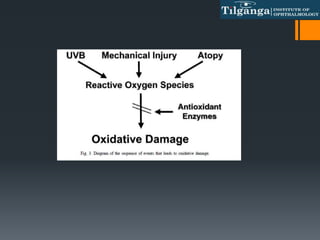
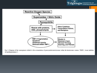
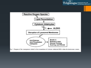
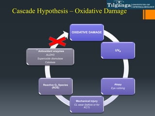
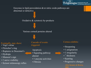
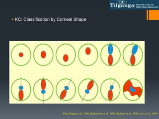
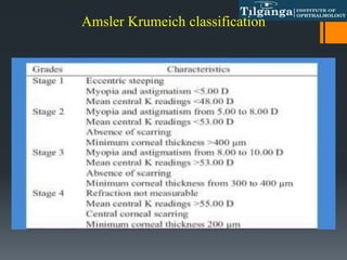
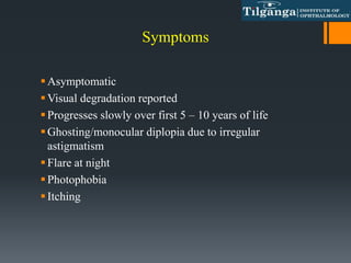
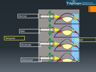
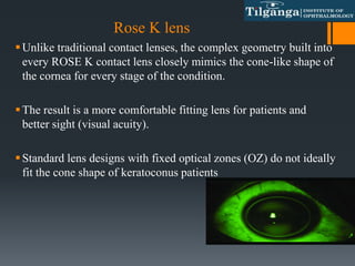
This document discusses ectatic disorders of the cornea, focusing on keratoconus. It defines keratoconus as a non-inflammatory thinning of the cornea that results in a protrusion and irregular astigmatism. The document covers the cascade hypothesis of oxidative damage in keratoconus pathogenesis, classification systems for keratoconus severity, signs and symptoms, and non-surgical management approaches like spectacles, contact lenses, and RGP fitting philosophies. The goal of management is to eliminate irregularities and provide optimal vision correction while minimizing further corneal damage.
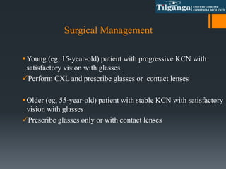
This presentation describes all the clinical aspects of keratoconus management
You can watch the illustrated presentation in this link :
https://www.youtube.com/watch?v=pYxwZPGm7e4&list=PLZ_mM13I_TrhWavjTmE9NjW1O5bGxkONO&index=13
This presentation describes all the clinical aspects of keratoconus management
You can watch the illustrated presentation in this link :
https://www.youtube.com/watch?v=pYxwZPGm7e4&list=PLZ_mM13I_TrhWavjTmE9NjW1O5bGxkONO&index=13
Corneal Cross Linking: Protocols and Literature Review
For sharing:
-- If you found our videos valuable, give us a like --
-- If you know someone who needs to see it, share it:
-- Leave a comment below with your thoughts --
-- Add it to a playlist if you want to watch it later again --
Link: https://youtu.be/rZSjEk3pgco
-- If you like the idea of my channel stay tuned and subscribe--
-- If you would like to check for more information about myself follow
the links on Linkedin and Researchgate:
Linkedin: https://at.linkedin.com/in/tukezban-h...
Researchgate: https://www.researchgate.net/profile/
Instagram: Eye_dr_Tuti
Twitter: https://twitter.com/Eye_DrTuti
Management of Keratoconus
for more information about icourses
https://www.facebook.com/i.courses.ophthalmology/
https://wa.me/201092909418
https://www.youtube.com/channel/UChSK-t5QtUa7Y6ct889ql7Q?reload=9&
https://t.me/icoursesophthalmology
https://www.instagram.com/i.courses.ophthalmology/
https://www.linkedin.com/in/ahmed-hamdy-626527188/
These simplified slides by Dr. Sidra Arshad present an overview of the non-respiratory functions of the respiratory tract.
Learning objectives:
1. Enlist the non-respiratory functions of the respiratory tract
2. Briefly explain how these functions are carried out
3. Discuss the significance of dead space
4. Differentiate between minute ventilation and alveolar ventilation
5. Describe the cough and sneeze reflexes
Study Resources:
1. Chapter 39, Guyton and Hall Textbook of Medical Physiology, 14th edition
2. Chapter 34, Ganong’s Review of Medical Physiology, 26th edition
3. Chapter 17, Human Physiology by Lauralee Sherwood, 9th edition
4. Non-respiratory functions of the lungs https://academic.oup.com/bjaed/article/13/3/98/278874
- Video recording of this lecture in English language: https://youtu.be/lK81BzxMqdo
- Video recording of this lecture in Arabic language: https://youtu.be/Ve4P0COk9OI
- Link to download the book free: https://nephrotube.blogspot.com/p/nephrotube-nephrology-books.html
- Link to NephroTube website: www.NephroTube.com
- Link to NephroTube social media accounts: https://nephrotube.blogspot.com/p/join-nephrotube-on-social-media.html
Lung Cancer: Artificial Intelligence, Synergetics, Complex System Analysis, S...Oleg Kshivets
RESULTS: Overall life span (LS) was 2252.1±1742.5 days and cumulative 5-year survival (5YS) reached 73.2%, 10 years – 64.8%, 20 years – 42.5%. 513 LCP lived more than 5 years (LS=3124.6±1525.6 days), 148 LCP – more than 10 years (LS=5054.4±1504.1 days).199 LCP died because of LC (LS=562.7±374.5 days). 5YS of LCP after bi/lobectomies was significantly superior in comparison with LCP after pneumonectomies (78.1% vs.63.7%, P=0.00001 by log-rank test). AT significantly improved 5YS (66.3% vs. 34.8%) (P=0.00000 by log-rank test) only for LCP with N1-2. Cox modeling displayed that 5YS of LCP significantly depended on: phase transition (PT) early-invasive LC in terms of synergetics, PT N0—N12, cell ratio factors (ratio between cancer cells- CC and blood cells subpopulations), G1-3, histology, glucose, AT, blood cell circuit, prothrombin index, heparin tolerance, recalcification time (P=0.000-0.038). Neural networks, genetic algorithm selection and bootstrap simulation revealed relationships between 5YS and PT early-invasive LC (rank=1), PT N0—N12 (rank=2), thrombocytes/CC (3), erythrocytes/CC (4), eosinophils/CC (5), healthy cells/CC (6), lymphocytes/CC (7), segmented neutrophils/CC (8), stick neutrophils/CC (9), monocytes/CC (10); leucocytes/CC (11). Correct prediction of 5YS was 100% by neural networks computing (area under ROC curve=1.0; error=0.0).
CONCLUSIONS: 5YS of LCP after radical procedures significantly depended on: 1) PT early-invasive cancer; 2) PT N0--N12; 3) cell ratio factors; 4) blood cell circuit; 5) biochemical factors; 6) hemostasis system; 7) AT; 8) LC characteristics; 9) LC cell dynamics; 10) surgery type: lobectomy/pneumonectomy; 11) anthropometric data. Optimal diagnosis and treatment strategies for LC are: 1) screening and early detection of LC; 2) availability of experienced thoracic surgeons because of complexity of radical procedures; 3) aggressive en block surgery and adequate lymph node dissection for completeness; 4) precise prediction; 5) adjuvant chemoimmunoradiotherapy for LCP with unfavorable prognosis.
The prostate is an exocrine gland of the male mammalian reproductive system
It is a walnut-sized gland that forms part of the male reproductive system and is located in front of the rectum and just below the urinary bladder
Function is to store and secrete a clear, slightly alkaline fluid that constitutes 10-30% of the volume of the seminal fluid that along with the spermatozoa, constitutes semen
A healthy human prostate measures (4cm-vertical, by 3cm-horizontal, 2cm ant-post ).
It surrounds the urethra just below the urinary bladder. It has anterior, median, posterior and two lateral lobes
It’s work is regulated by androgens which are responsible for male sex characteristics
Generalised disease of the prostate due to hormonal derangement which leads to non malignant enlargement of the gland (increase in the number of epithelial cells and stromal tissue)to cause compression of the urethra leading to symptoms (LUTS
Ethanol (CH3CH2OH), or beverage alcohol, is a two-carbon alcohol
that is rapidly distributed in the body and brain. Ethanol alters many
neurochemical systems and has rewarding and addictive properties. It
is the oldest recreational drug and likely contributes to more morbidity,
mortality, and public health costs than all illicit drugs combined. The
5th edition of the Diagnostic and Statistical Manual of Mental Disorders
(DSM-5) integrates alcohol abuse and alcohol dependence into a single
disorder called alcohol use disorder (AUD), with mild, moderate,
and severe subclassifications (American Psychiatric Association, 2013).
In the DSM-5, all types of substance abuse and dependence have been
combined into a single substance use disorder (SUD) on a continuum
from mild to severe. A diagnosis of AUD requires that at least two of
the 11 DSM-5 behaviors be present within a 12-month period (mild
AUD: 2–3 criteria; moderate AUD: 4–5 criteria; severe AUD: 6–11 criteria).
The four main behavioral effects of AUD are impaired control over
drinking, negative social consequences, risky use, and altered physiological
effects (tolerance, withdrawal). This chapter presents an overview
of the prevalence and harmful consequences of AUD in the U.S.,
the systemic nature of the disease, neurocircuitry and stages of AUD,
comorbidities, fetal alcohol spectrum disorders, genetic risk factors, and
pharmacotherapies for AUD.
ARTIFICIAL INTELLIGENCE IN HEALTHCARE.pdfAnujkumaranit
Artificial intelligence (AI) refers to the simulation of human intelligence processes by machines, especially computer systems. It encompasses tasks such as learning, reasoning, problem-solving, perception, and language understanding. AI technologies are revolutionizing various fields, from healthcare to finance, by enabling machines to perform tasks that typically require human intelligence.
3. “Ectasia” as defined in most medical dictionaries refers
to a dilation or distention of a tubular structure. *
“Ectasia progression” is defined by a consistent change
in at least 2 of the following parameters :
1. Steepening of the anterior corneal surface
2. Steepening of the posterior corneal surface
3. Thinning and/or an increase in the rate of corneal
thickness change from the periphery to the thinnest
point.”*
Gomes JA, Rapuano CJ, Belin MW, Ambrósio Jr R. Global consensus on keratoconus diagnosis. Cornea. 2015
Dec 1;34(12):e38-9.
Corneal Ectasia
4. As opposed to “thinning disorders” the following are
classified under “ectatic diseases”
Keratoconus
PMD
Keratoglobus
Postrefractive surgery progressive corneal ectasia
Gomes JA, Rapuano CJ, Belin MW, Ambrósio Jr R. Global consensus on keratoconus diagnosis. Cornea. 2015 Dec
1;34(12):e38-9
6. Introduction
Non-inflammatory, progressive thinning of the cornea that results
in apical protrusion (ectasia) resulting in a high degree of irregular
myopic astigmatism with observable structural changes appearing
in later stages.
The prevalence of keratoconus varies widely depending upon the
geographic location and diagnostic criteria used*
The reported incidence of keratoconus is 1 in 2000 individuals.*
The reports of two surveys in the UK indicated a prevalence 4.4
and 7.5 times greater for Asian (Indian, Pakistani, and
Bangladeshi) subjects compared with white subject.*
Gokhale NS. Epidemiology of keratoconus. Indian journal of ophthalmology. 2013 Aug;61(8):382
Georgiou T, Funnell CL, Cassels-Brown A, O’Connor R. Influence of ethnic origin on the incidence of keratoconus and associated atopic diseases in Asian and
white patients. Eye (Lond) 2004;18:379–83
11. Pearson AR, Soneji B, Sarvananthan N, Sandforth-Smith JH. Does ethnic origin influence the incidence or severity of keratoconus? Eye
(Lond) 2000;14:625–8
Rabinowitz YS. Keratoconus. Surv Ophthalmol 1998;42(4):297–319.
Edwards M, McGhee CN, Dean S. The genetics of keratoconus. Clin Exp Ophthalmol 2001;29(6):345–51.
7. Keratoconus and ectatic corneal diseases have been
recognized for more than 150 years.*
Posterior corneal elevation abnormalities must be
present to diagnose mild or subclinical keratoconus.*
Krachmer JH, Feder RS, Belin MW. Keratoconus and related non inflflammatory corneal thinning disorders.
Surv Ophthalmol. 1984;28: 293–322.
Rabinowitz YS. Keratoconus. Surv Ophthalmol. 1998;42:297–319.
Gomes JA, Rapuano CJ, Belin MW, Ambrósio Jr R. Global consensus on keratoconus diagnosis. Cornea. 2015
Dec 1;34(12):e38-9.
8. No gender predisposition
More common in hot, dry climates
Zadnik K, Barr JT, Edrington TB, et al. Baseline findings in the Collaborative Longitudinal Evaluation of Keratoconus
(CLEK) Study. Invest Ophthalmol Vis Sci 1998;39(13):2537-46.
9.
10. 1. Characterization of the abnormal matrix
The keratoconus corneas had decreased levels of
fibronectin, laminin, entactin, type IV collagen and type
XII collagen associated with the epithelial basement
membranes.
There were also increased levels of fibrosis-associated
extracellular matrices such as type III collagen,
tenascin-C, and fibrillin-1 in the regions of anterior
stromal scars and disrupted Bowman’s layer
11. 2.Enzyme and inhibitor abnormalities
Keratoconus corneas had elevated levels of gelatinase activity.
It is likely that keratoconus corneas have an imbalance
between the corneal MMP-2 and its inhibitors, tissue inhibitors
of metalloproteinases (TIMPs)
Keratoconus corneas have decreased levels of enzyme
inhibitors and increased enzyme activities that can degrade the
various extracellular matrices within the keratoconus corneas.
The inhibitor–enzyme imbalance undoubtedly plays a major
role in the stromal thinning and Bowman’s layer/basement
membrane breaks that are characteristic of keratoconus
corneas.
12.
13. 3. Apoptosis in keratoconus corneas
The keratoconus corneas have increased apoptosis and
this phenomenon of cell death may be important in its
pathogenesis.
Factors that cause apoptosis include chronic epithelial
cell damage, increased levels of LAR(of leukocyte
common antigen related protein) and decreased levels of
TIMP-1.
14. 4. Abnormal regulation and signal transduction
Keratoconus corneas have increased levels of Sp1, a
transcription factor that can down-regulate proteinase
inhibitor.
Phosphotyrosine phosphatase enzyme (LAR)(function
of the phosphotyrosine phosphatase is removal of the
phosphates from the tyrosine molecules) that was found
in keratoconus but lacking in normal corneas
15. 5. Oxidative damage in keratoconus corneas
Keratoconus corneas have increased oxidative damage
compared to normal .They lack the necessary enzyme
components (ALDH3, superoxide dismutase) to process the
reactive oxygen species that occur.
With an accumulation of the reactive oxygen species, there
is a resultant deposition of cytotoxic by-products
(malondialdehyde and peroxynitrites) that can damage the
corneal tissues.
20. Enzymes in lipid peroxidation &/or nitric oxide pathways are
abnormal or defective
Oxidative & cytotoxic by-products
Various corneal proteins altered
Cascade of events
triggered:
Apoptosis
• Signaling pathway
altered
• ↑ enzyme activities
• Fibrosis
Cornea exhibits:
• Steepening
• ↑ astigmatism
• ↑ irregularity
• ¯ thickness
• Scarring
• Progressive changes
After Kenney & Brown,
2003
Eventually, may show:
• Vogt’s striae
• Fleischer’s ring
• Ruptures in Descemet’s
• Hydrops
• Munson’s sign
• ↑ nerve visibility
• Scissor retinoscopy reflex
21. Mechanical factors : Floppy eyelid syndrome ,eye
rubbing associated with atopy or vernal keratoconjunctivitis
Connective tissue disorders(Marfan syndrome, Ehlers–
Danlos syndrome).
Positive family history(The prevalence of keratoconus in
first degree relatives is 3.34%, which is 15–67 times higher
than the general population.)
Down syndrome(0.5-15% of patients)
Ethnic factors (eg, Asian and Arabian)
22. • Mild: <45 D
• Moderate: 45 – 52 D
• Advanced: 52 – 62 D
• Severe: > 62 D
Booysen, 2003
KC: Classification by Corneal Curvature
32. Later sign :
Advanced cone formation seen in profile
Apical scar formation
Munson’s sign on lower lid
Fleischer’s iron ring
↑ visibility of corneal nerve fibres
Vogt’s striae
Non-uniform red reflex with ophthalmoscopy
Rupture of Descemet’s membrane (corneal hydrops)
↓ IOP
33. Additional Sign in Advanced KCN
Thinning
Stromal edema
Splits/tears in the endothelium/
Descemet’s membrane
Endothelial cells may be elongated
Corneal scarring
34. KISA%: Rabinowitz Criteria
Corneal power (K) [>47.2 D]
Inferior-Superior dioptric asymmetry (I-S) [>1.2 D]
Sim K Astigmatism (Ast) >1.5 D
Skewed Radial Axis (SRAX) [>21°]
If KISA% > 60%, case is a KC suspect
KISA = (Central K) × (I−S) × (AST) × (SRAX) × 100/300
36. Non surgical Management
Verbal guidance to the patient regarding the importance
of not rubbing eyes and use of topical lubricants (in case
of ocular irritation) to decrease the impulse to rub eyes.
Use of topical antiallergic medication(ie, antihistamines,
mast cell stabilizer, antiinflammatory) in patients with
allergy.
“Use of eye drops without preservatives is preferable in
keratoconus patients.”
37. Does spectacle works in Keratoconus?
Mild keratoconus in early stage can be corrected with
spectacles.
As the cornea steepens and becomes more irregular,
glasses not capable of providing adequate visual
improvement.
38. Soft contact lens
Suited to early stage progression only as it does have any tear lens
formation doesn't correct the irregularities.
In cases of GP CL intolerance
Usually, astigmatic component corrected with over-spectacles
Contact Lens
39. RGP contact lens
Eliminate corneal irregularities with pre-corneal tear lens
formed by these lenses thus provides better visual
correction
41. APICAL CLEARANCE
The central pooling or no apical
touch and the bearing is towards the
periphery
Reduces the risk of scarring
Tightening at the periphery may
result in sealing the tear exchange
42. APICAL BEARING
The central bearing or apical touch and the
pooling is towards the periphery resulting in
good visual acuity
Results in the corneal scarring and
intolerance
43. 3 POINT or DIVIDED SUPPORT
Divided support or three point touch • An apical contact area of two
to three millimetres, a intermediate clearance zone and mid
peripheral contact annulus with conventional edge clearance at the
periphery.
CL weight distributed over larger area.
Woodward EG. Contact lenses in abnormal ocular conditions—keratoconus. In: Phillips AJ, Speedwell L, eds.
44. So, what if patient cannot tolerate
RGP lenses??
Piggy back system
Hybrid lens system
45. Piggyback CLs
Rigid lens fitted over a hydrogel lens increases comfort resulting
in adequate wearing time with good vision
SCL acts as a ‘carrier’ for the overlying GP CL
Used to ↑ comfort & ↓ risk of epithelial abrasion by a GP CL
Ultra-thin SCL (usually disposable)
46. Potential problems Piggyback CLs????
Handling and care of two different types of lenses.
Difficulty in obtaining centration of the rigid lens.
47. Better options than Piggy back !
One way to overcome the problems with piggy-
back lenses, yet have the optics of a rigid lens with
the comfort of a hydrogel, is to fuse a soft rim onto
a hard central portion
Hybrid Lens system
48. Combine the benefits of rigid lens optics, including
better lens centration and decreased aberrations, along
with the comfort of a soft lens Potential
Complications…
Flexure of the GP centre lens (leading to astigmatism
and decreasing visual results),
Difficulties with insertion and removal of the lenses,
and
Tearing at the GP lens and hydrogel skirt junction.
50. Scleral Lens
Cornea is completely vaulted
Creating an equal and opposite keratoconic surface
ultimately restoring uniform optical lens and elimination
of astigmatism.
This result in less ghosting and much crisper vision
Mini-Scleral – 12.5mm to 14.5mm
Medium Range Scleral– 15.0mm to
18.0mm
Full Scleral – 18.5mm to 28.0mm
52. Rose K lens
Unlike traditional contact lenses, the complex geometry built into
every ROSE K contact lens closely mimics the cone-like shape of
the cornea for every stage of the condition.
The result is a more comfortable fitting lens for patients and
better sight (visual acuity).
Standard lens designs with fixed optical zones (OZ) do not ideally
fit the cone shape of keratoconus patients
53. Keratoconus show high flattening between the very
steep apex of the cone and the flat periphery.
Need to have very steep lenses in the center that get
flatter quickly at periphery.
54. Fig shows a standard lens that will yield unwanted
pooling at the base of the cone and peripheral
bearing that can seal off and cause corneal
problems
55. Fig demonstrates the benefits of a smaller optical zone to
fit the cone contour. The design results in little tear
pooling at the base of the cone and shows an even
distribution of tears under the lens.
56.
57. Key points:
Use UV protection in the contact lenses and glasses
Improve patient comfort in order to minimize eye
rubbing
Non-steroidal anti-inflammatory medications (NSAIDs)
Preservative-free artificial tears
Allergy medications
Use properly fit contact lenses
Contact and scleral lenses are extremely important for
visual rehab. in patients with keratoconus.
59. Corneal collagen cross-linking (CXL)
First described in 1998 by Spoerl et al. as a
modality for increasing the corneal biomechanical
strength to halt disease progression
First data presented in 2003 by Wollensak et al.
In April 2016, the U.S. FDA gave approval to
corneal collagen cross-linking to treat progressive
keratoconus and post-LASIK ectasia.
Spoerl E, Huhle M, Seiler T. Induction of cross-links in corneal tissue. Exp Eye Res. 1998;66:97–103
G.Wollensak, E. Spoerl, and T. Seiler, “Riboflavin/ultraviolet-ainduced ollagen crosslinking for the treatment of
keratoconus,” The American Journal of Ophthalmology, vol. 135, no. 5, pp. 620– 627,
60. Corneal collagen cross-linking (CXL)
In CXL, riboflavin (vitamin B2) is administered in
conjunction with ultraviolet A (UVA, 365 nm).
–The interaction of riboflavin and UVA leads to the
formation of reactive oxygen species, which leads to
the formation of additional covalent bonds between
collagen molecules, with consequent biomechanical
stiffening of the cornea
61. Surgical Management
Young (eg, 15-year-old) patient with progressive KCN with
satisfactory vision with glasses
Perform CXL and prescribe glasses or contact lenses
Older (eg, 55-year-old) patient with stable KCN with satisfactory
vision with glasses
Prescribe glasses only or with contact lenses
62. Patient with stable severe KCN with unsatisfactory vision with
glasses and contact and scleral lenses? This patient has moderate
anterior corneal scarring but no evidence of previous corneal
hydrops
Perform DALK
Patient with stable severe KCN with unsatisfactory vision with
glasses and contact and scleral lenses? This patient has moderate
anterior and deep corneal scarring with evidence of previous
corneal hydrops
PK alone
63. As for PK, the most important factor in considering
keratoplasty in keratoconus is when
Significant corneal scarring (eg, posthydrops) is present.
Contact lens intolerant or is not keen on wearing contact
lenses; other surgical strategies fail, or are
contraindicated; the cornea is very thin (200 um); and
K eratoconus is deemed to be severe and at a potential
risk of acute hydrops.
64. TAKE HOME MESSAGE
A proper assessment of corneal topography is required
Careful Skilled fluorescein-pattern interpretation is the
cornerstone of successful CL fitting